Introduction
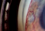
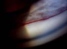
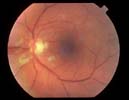
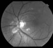
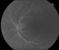
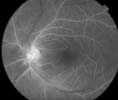
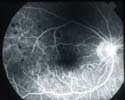
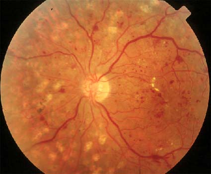
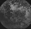
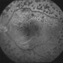
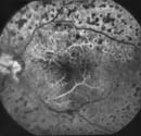
Ocular ischemic syndrome (OIS) is a rare but important cause of blindness. It is a constellation of signs arising from a reduction in perfusion pressure to the globe (mean blood pressure minus intraocular pressure [IOP]), leading to anterior and posterior segment ischemia (Slide 1A, Slide 1B, Slide 2A, Slide 2B, Slide 3A, Slide 3B, Slide 4, Slide 5, Slide 6A, Slide 6B, and Slide 7). Orbital structures supplied by the ophthalmic artery can also be affected. This reduction in perfusion pressure is usually due to atherosclerotic carotid artery disease. Ophthalmic artery occlusion can also lead to OIS. Other contributing factors must also be considered, as many patients with occlusive carotid artery disease do not develop OIS. An important consideration is the combination of small and large vessel disease, as suggested by the higher incidence of diabetes mellitus in OIS patients compared to the general population.
Clinical Characteristics
The prospective study by Mizner and colleagues has outlined many of the significant clinical characteristics of patients with OIS. 1 The average age of patients with OIS is 68 years. Relative to age-matched controls in the general population, these patients are more likely to have diabetes mellitus, systemic hypertension, coronary artery disease, and cerebrovascular disease. Patients with OIS typically present with sudden vision loss (41%), gradual visual loss (28%), or amaurosis fugax (15%). More than 60% of eyes have visual acuity of less than 20/400. The primary clinical feature in eyes with OIS is ocular neovascularization: 87% have iris neovascularization; 59% have angle neovascularization; 13% have disc neovascularization; and 3% have retinal neovascularization. Other findings include episcleral injection, corneal edema, cataracts, optic disc pallor, mid-peripheral retinal hemorrhages (hypotensive retinopathy), retinal circulatory stasis, and, in most cases, inducible or spontaneous retinal arterial pulsations. Dilated episcleral vessels may reflect the presence of external carotid artery collateral vessels supplying the ophthalmic artery by retrograde flow. IOP at presentation is variable. In Mizner’s report, 56% of eyes had an IOP less than 22 mm Hg, though IOPs ranged from 4 mm Hg to 60 mm Hg. Most patients have severe ipsilateral carotid artery disease: 60% have complete occlusion and an additional 15% have 80% to 99% stenosis. Up to 22% of patients will present with bilateral OIS.
Management
Eyes with hypotensive retinopathy in OIS must be distinguished from other clinical entities, such as central retinal vein occlusion or diabetic retinopathy, that typically have normal retinal arterial pressures. Patients must also be carefully evaluated for other rarer causes of OIS including giant cell arteritis or carotid artery dissection.
Fluorescein angiography in OIS eyes usually demonstrates delayed choroidal filling (> 5 seconds) and retinal arteriovenous circulation times (> 11 seconds). Late staining of arterioles and veins, and capillary nonperfusion (especially in the mid-periphery) can also be seen. Carotid artery ultrasound can detect occlusive carotid artery disease which is usually 90% or more in eyes with OIS. Color Doppler imaging (CDI) of the retrobulbar circulation can also provide additional diagnostic information. Relative to patients who have severe carotid stenosis (> 70% occlusion) without OIS, patients with OIS are more likely to demonstrate bilateral occlusive carotid artery disease, high-grade carotid stenosis, and reversed ophthalmic artery flow. The use of CDI, however, may be limited by difficulties in reliably reproducing orbital blood flow measurements. Electroretinography usually reveals diminished b- and a-waves consistent with inner and outer retinal ischemia.
Treatment of eyes with OIS consists of controlling IOP and the complications of retinal ischemia, including neovascular glaucoma (NVG). Generally, panretinal photocoagulation (PRP) should be performed in eyes with capillary nonperfusion on fluorescein angiography and in eyes with ocular neovascularization. For reasons that remain elusive, PRP reduces iris neovascularization regression in only one-third of eyes with OIS. The presence of uveal ischemia without concomitant retinal ischemia may be one factor and some authorities have suggested that PRP be performed only in eyes with angiographic evidence of capillary nonperfusion. This would minimize complications of PRP such as pain or visual field loss. In eyes with uncontrolled NVG, filtering surgery with mitomycin C, tube shunt procedures, and ciliary body ablation with cryotherapy or diode laser can also be used.
Also, it is important to manage any associated systemic disease, as cardiovascular and cerebrovascular diseases are the leading causes of death in patients with OIS.
Although carotid endarterectomy lowers the risk of stroke in patients with symptomatic carotid stenosis, it is unclear whether this procedure impacts the visual prognosis in eyes with OIS because no randomized controlled studies have compared the surgical results to the natural history of the disease. However, there have been reports of stabilization of visual acuity and, rarely, improvement of visual acuity in eyes with OIS after carotid endarterectomy. In one series of 60 eyes with OIS that received carotid endarterectomy, 7% had visual improvement, 33% had no change, and 60% worsened. A smaller series of 11 eyes, five of which had reversed ophthalmic artery flow measured by CDI, reported improved antegrade ophthalmic artery flow in all eyes postoperatively; visual acuity improved in five eyes and stabilized in six. In most cases, a carotid endarterectomy does not cause regression of iris neovascularization.
The best predictor of final visual prognosis is initial visual acuity: eyes with visual acuity of 20/80 or better usually retain their baseline vision; similarly, eyes with visual acuity less than 20/400 tend to remain the same or worsen. The 5-year mortality rate in patients with OIS is 40%.
References
- Mizner JB, Podhajsky P, Hayreh SS. Ocular ischemic syndrome. Ophthalmology. 1997;104:859-864.
Bibliography
Brown GC, Magargal LE, Simeone FA, et al. The ocular ischemic syndrome. Clinical, fluorescein angiographic and carotid angiographic features. Int Ophthalmol. 1988;11:239-251.
Costa VP, Kuzniec S, Molnar LJ, et al. Clinical Findings and hemodynamic changes associated with severe occlusive carotid artery disease. Ophthalmology. 1997;104:1994-2002.
Hayreh SS. Vasospasm and intraocular blindness. N Engl J Med. 1992;326:837-838.
Hayreh SS. Chronic ocular ischemic syndrome in internal carotid artery disease: Controversy on "venous stasis retinopathy." In: Bernstein EF, ed. Amaurosis Fugax. New York: Springer-Verlag; 1988:135-158.
Ino-ue M, Azumi A, Kajiura-Tsukahara Y, et al. Ocular ischemic syndrome in diabetic patients. Japan J Ophthalmol. 1999;43:31-35.
Kawaguchi S, Sakaki T, Morimoto T, et al. Effects of bypass on ocular ischemic syndrome caused by reversed flow in the ophthalmic artery. Lancet. 1999;354:2052-2053.
Kawaguchi S, Okuno S, Sakaki T, et al. Effect of carotid endarterectomy on chronic ocular ischemic syndrome due to internal carotid artery stenosis. Neurosurgery. 2001;48:328-333.
Sivalingam A, Brown GC, Magargal LE. The ocular ischemic syndrome. III. Visual prognosis and the effect of treatment. Int Ophthalmol. 1991;15:15-20.
Sivalingham A, Brown GC, Magargal LE, Menduke H. The ocular ischemic syndrome II. Mortality and systemic morbidity. Int Ophthalmol. 1989;13:187-191.