Case Presentation
A 60-year-old man was followed by the New England Eye Center glaucoma service since 1993. He was first seen by an ophthalmologist at the age of 18 months because he was noted to have "crossed eyes." Despite wearing glasses, patching his left eye, and using a drop in "one of his eyes," he developed amblyopia in his right eye. He has since followed with several different ophthalmologists for routine eye care.
The patient had no known allergies and his past medical history was unremarkable. He past ocular history was as above. He took no medications. His family history was unremarkable and he used neither alcohol nor cigarettes.
On examination, visual acuity was 20/200 OD, 20/30 OS. Manifest refraction was +3.75 +2.25 x 90 OD, +1.75 +1.00 x 10 OS. Pupils were equally reactive without an afferent papillary defect. Extraocular motility was normal and the patient was orthophoric near and distance fixation. Applanation tonometry measured intraocular pressures of 16 mm Hg OU. Visual fields showed an enlarged blind spot in the left eye (Slide 1A and Slide 1B).
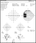
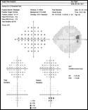
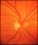
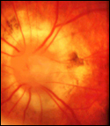
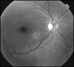
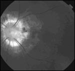
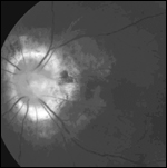
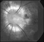
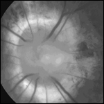
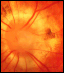
The slit lamp examination was unremarkable. Examination of the fundus showed an anomalous disc appearance (Slide 2A and Slide 2B). Red free photographs revealed a relatively healthy nerve fiber layer in each eye and demonstrated the morning glory disc anomaly (Slide 3A, Slide 3B, Slide 4A, Slide 4B and Slide 4C). Slide 5A, Slide 5B compares the patient’s anomalous optic nerve head with the morning glory flower for which the anomaly was named.

Comment
Handmann first described morning glory disc anomaly in 1929. However, the name morning glory disc anomaly was not given to this entity until 1970, when Kindler published his findings of an abnormal optic nerve appearance in 10 patients.1 His definition of morning glory disc anomaly was:
- An enlarged optic disc
- A funnel-shaped, excavated peripapillary region surrounded by a wide elevated annulus of chorioretinal pigment disturbance
- Central overlying white, fluffy tissue
- Retinal vessels appearing at the edge of the disc as multiple narrow branches.
Since this description, many reports of this disease have appeared in the literature. It has been associated with a number of anomalies2 including cleft lip and palate, basal encephalocele, neurofibromatosis type 2,3 absent corpus callosum, moyamoya disease,4 retinal arteriovenous communication,5 renal abnormalities,6 and endocrine abnormalities.7 Morning glory disc anomaly is typically unilateral and occurs equally among men and women. The prevalence and incidence of this disorder is unknown. Patients typically present in childhood because of strabismus, poor vision, or leukocoria. The eye with morning glory disc anomaly will usually have poor vision (90% with 20/200 or worse8) on the basis of either amblyopia or retinal detachment. However, patients will occasionally have good vision in the affected eye.
Histology and Pathophysiology
The cause of morning glory disc anomaly is speculative to date. Some support the hypothesis that morning glory disc anomaly is caused by a mesenchymal abnormality causing faulty closure of the posterior scleral wall and lamina cribrosa. This idea stems from the association of morning glory disc anomaly with craniofacial abnormalities, which also derive from mesenchymal tissue. Others believe that the pathology is similar to optic nerve coloboma, with deficiency of closure of the embryonic fissure as the cause of morning glory disc anomaly. Yet others9 believe that the defect is a spectrum of persistent hyperplastic primary vitreous (PHPV) because of the whitish tissue overlying the optic disc.
Manschot10 performed a histopathologic examination of one eye with morning glory disc anomaly. In this eye, he found a 4-mm deep staphyloma with a diameter of 5 mm, covered by a layer of normal retinal pigment epithelium. The nerve head was retrodisplaced and smooth muscle cells were noted within the outer scleral layer. The whitish tissue overlying the nerve head consisted of folded retina with glial fibrous and pigment epithelial proliferation. The retinal vessels seen at the head of the optic disc were noted to be the second order vessels of the retinal artery, with the first order vessels displaced into the nerve. Other histopathologic examinations have supported these findings.
Differential Diagnosis
The entities most often mistaken for morning glory disc anomaly are optic nerve pit, optic nerve coloboma, and Bergmeister’s papilla. Bergmeister’s papilla is a tuft of whitish tissue overlying the optic disc. This is believed to be in the spectrum of PHPV. There are several important differences between optic pit, coloboma, and morning glory disc anomaly (Table 1).11-14
Complications of Morning Glory Disc Anomaly
The main complication of morning glory disc anomaly is visual loss in the affected eye, either on the basis of amblyopia or retinal detachment. Eyes with morning glory disc anomaly tend to be myopic relative to the fellow unaffected eye. Thus, in the absence of vision screening, children with morning glory disc anomaly tend to use their better eye (typically the unaffected eye, but occasionally the eye with morning glory disc anomaly). As a result, amblyopia develops. This complication can be avoided to some extent by appropriate amblyopic therapy.
Retinal detachment in morning glory disc anomaly is often difficult to treat. Detachments in this setting are serous in nature. One series of 30 patients15 found that 11 had serous retinal detachments throughout the course of their follow-up (mean 10 years). The source of the fluid in these serous retinal detachments is unknown.
One theory is that there is an abnormal communication between the subretinal space and the subarachnoid space.16 Another is that a defect in the intermediary tissue of Kuhnt allows communication between the subretinal space and the vitreous cavity.17 Repair of retinal detachment in this condition has been approached in a variety of ways. Chang and associates16 reported anatomic success with optic nerve sheath fenestration and subretinal fluid drainage in a 14-year-old girl. Harris and colleagues described reattachment of the retina in a 14-year-old boy via pars plana vitrectomy, subretinal fluid drainage, fluid-gas exchange, and laser photocoagulation.18 Coll and colleagues17 describe reattachment using vitrectomy, laser, and silicone oil in a 69-year-old man. However, treatment of retinal detachments associated with morning glory disc anomaly is often difficult. Surgical repair is often tempered by prior vision in the affected eye, predicted postoperative visual acuity, and the possibility of spontaneous reattachment, which has been reported.15
References
- Kindler P. Morning glory syndrome: Unusual congenital optic disk anomaly. Am J Ophthalmol. 1970;69(3):376-384.
- Traboulsi E, O’Neill J. The spectrum in the morphology of the so-called "morning glory disc anomaly." J Pediatr Ophthalmol Strabismus. 1988;25(2):93-98.
- Brodsky H, Landau K, et al. Morning glory disc anomaly in neurofibromatosis type 2. Arch Ophthalmol. 1999;117:839-841.
- Bakri S, Siker D, et al. Ocular malformations, moyamoya disease and midline cranial defects: A distinct syndrome. Am J Ophthalmol. 1999;127(3):356-357.
- Brodsky M. Retinal arteriovenous communications in the morning glory disc anomaly. Arch Ophthalmol. 1995;133:410-411.
- Dureau P, Attie-Bitach T, et al. Renal coloboma syndrome. Ophthalmology. 2001;108:1912-1916.
- Eustis HS, Sanders M, Zimmerman T. Morning glory syndrome in children: association with endocrine and central nervous system anomalies. Arch Ophthalmol. 1994;112:204-207.
- Lit E, D’Amico D. Retinal manifestations of morning glory disc syndrome. Int Ophthalmol Clin. 2001;41(1):131-138.
- Brown G, Gonder J, Levin A. Persistence of the primary vitreous in association with the morning glory disc anomaly. J Pediatr Ophthalmol Strabismus. 1984;21(1):5-7.
- Manschot W. Morning glory syndrome: A histopathological study. Br J Ophthalmol. 1990;74:56-58.
- Apple D, Rabb M, Walsh P. Congenital anomalies of the optic disc. Surv Ophthalmol. 1982;27(1):3-41.
- Brodsky M. Congenital optic disk anomalies. Surv Ophthalmol. 1994;39(2);89-112.
- Brodsky M. Congenital optic disc anomalies. In: Yanoff M, Duker J, eds. Ophthalmology. St. Louis: Mosby-Yearbook; 1999:11.4.1-11.4.4.
- Brown G. Coexistent optic nerve and macular abnormalities. In: Yanoff M, Duker J, eds. Ophthalmology. St. Louis: Mosby-Yearbook; 1999:8.35.1-8.35.5.
- Haik B, Greenstein S, et al. Retinal detachment in the morning glory anomaly. Ophthalmology. 1984;91:1638-1647.
- Chang S, Haik B, et al. Treatment of total retinal detachment in morning glory syndrome. Am J Ophthalmol. 1984;97:596-600.
- Coll G, Chang S, et al. Communication between the subretinal space and the vitreous cavity in the morning glory syndrome. Graefe’s Arch Clin Exp Ophthalmol. 1995;233:441-443.
- Harris M, De Bustros S, et al. Treatment of combined traction-rhegmatogenous retinal detachment in the morning glory syndrome. Retina. 1984;4:249-252.