Introduction
Many methods have been used to shunt aqueous out of the anterior chamber. Wicks or tubes made of various materials have been described since the early 1900s; however, none proved safe and effective. In 1969, Molteno described the first modern glaucoma drainage device,1 which was an acrylic tube connected to an acrylic plate fixated subconjunctivally near the limbus. His goal was to shunt fluid from the anterior chamber and create a permanent, functioning filtration bleb of consistent size. This arrangement led to large blebs causing discomfort, dellen formation, and plate extrusion, as well as hypotony. Moving the plate further posterior on the globe addressed these difficulties and laid the foundation for current devices.
Device Design
Continued research and development has ensued over the past 30 years and today numerous drainage devices are available (Slide 1). All have in common the presence of a tube, inserted either into the anterior chamber or through the pars plana, leading to a variably sized plate explant that is fixated to the sclera and covered with conjunctiva. The plate serves as a focus for fibrous encapsulation, forming a reservoir with an overlying filtration bleb. Devices differ by materials used, shape and surface area of the plate, and presence or absence of a flow-restricting valve. Recently, double-plate models of some devices have been developed to increase the surface area for encapsulation and, theoretically, to decrease intraocular pressure (IOP).
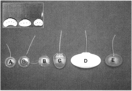
Commonly used nonrestrictive devices include the single- and double-plate Molteno shunts and the Baerveldt shunts. The Molteno consists of a silicone tube connected to a round polypropylene plate with a 134-mm2 surface area. A second plate is attached to the first by a connector tube in the double-plate model, thus doubling the surface area. The Baerveldt devices are available in three sizes — 200 mm2, 350 mm2, and 425 mm2. These silicone plates are curved and somewhat wide and, unlike other plates, require isolation of the bounding rectus muscles during fixation of the plate to the globe. Another nonvalved device, the Schocket, employs an encircling silicone band, similar to that used in scleral buckling surgery, as the explant. The Schocket is not commonly used today.
Common flow-restrictive implants include the single- and double-plate Ahmed valve and the Krupin valve. The Ahmed consists of an ovoid, polypropylene plate with an internal pair of silicone elastomer membranes at the proximal end. These membranes function as a pressure-dependent valve that requires a force of 8 mm Hg to 12 mm Hg to separate the leaves of the membrane. The surface area of the single-plate device is 184 mm2 and approximately doubles with addition of a secondary, nonvalved plate. The Krupin valve consists of a silastic tube connected to a silastic disk and has a surface area of 183 mm2 (alternatively, the tube can be connected to a #220 Silastic band as with a Schocket procedure). The valve in this device is formed by a series of horizontal and vertical slits in the sealed distal end of the tube, requiring a force of approximately 9 mm Hg to 11 mm Hg for aqueous flow through the device. Additional flow-restrictive devices include the Joseph, Optimed, and White shunts, which are used less often than those previously discussed.
Function and Physiology of Drainage Devices
With the exception of modifications designed to restrict flow (e.g., valved devices), all tube shunts function in a similar manner (Table 1). The tube, inserted translimbally into the anterior chamber or through the pars plana into the vitreous cavity after complete vitrectomy, serves as a permanent sclerostomy and is capable of remaining patent despite the presence of anterior synechiae which may otherwise obstruct a simple sclerostomy. The tube connects to an explant plate that serves as a focus of fibrous encapsulation, a process that progresses over several weeks to months.2
Authors have demonstrated a relatively acellular, collagenous capsule forming a smooth internal wall around the plate, with minimal signs of inflammation.3-5 Similarly, little inflammation and no significant fibrosis have been associated with the intracameral portion of the tubes. Minckler and colleagues2 have found the capsular wall to be the primary site of resistance to aqueous flow in these devices, with passive, hydrostatic movement of fluid across the capsule. Thus, the thickness of the capsular wall and the surface area available for encapsulation should be the limiting factors in IOP reduction.
Capsule thickness cannot be easily controlled; however, surface area is easily altered with larger or multiple plates. Heuer and associates6 compared 132 patients randomized to either a single- or double-plate Molteno, finding 2-year life-table success of 46% and 71%, respectively. These findings suggest that increased surface area correlates with a drop in IOP and surgical success. However, this effect of plate size may yield diminishing results as the surface area continues to increase. Lloyd and colleagues7 compared 73 patients undergoing implantation of either a 350-mm2 Baerveldt device or a 500-mm2 Baerveldt device, with 18-month life-table success rates of 93% and 88%, respectively. Although efficacy was not improved in the 500-mm2 Baerveldt device, complications, notably choroidal effusions and strabismus, were more common with the larger plate.
Indications and Selection of Device
Aqueous drainage devices are generally indicated for patients with uncontrolled IOP despite being treated with maximum tolerated medical therapy and often after one or more failed filtration procedures. The devices are useful in both open-angle and closed-angle glaucoma in adults and are increasingly being used in congenital and juvenile glaucoma. Tube shunts are particularly useful in patients with secondary glaucoma, such as inflammatory, neovascular (NVG), aphakic, and postkeratoplasty glaucomas, who are at high risk for failure with traditional filtration surgery. Implantation of these devices may be indicated as a primary procedure in the presence of severe angle neovascularization, extensive peripheral synechiae, or limbal scarring.
The choice of device depends on personal preference, as little supporting data exist comparing outcomes among shunts. Typically, the choice is based upon the degree of IOP reduction required (plate size and number), quadrants available for plate placement, desire for a valved or nonvalved system, and a surgeon’s comfort/experience with each device.
Researchers have attempted to compare efficacy of different tube shunts. Smith and colleagues8 compared the double-plate Molteno with a Schocket procedure. With at least 6 months’ follow-up, the mean IOP was 14.4 mm Hg with the Molteno and 15.1 mm Hg with the Schocket. The mean number of required medications was 0.95 in the Molteno group and 0.43 in the Schocket group. Stability of visual acuity and complication rates were similar between groups. Smith later compared the double-plate Molteno with the 350-mm2 Baerveldt device.9 With an average follow-up of 1 year, the mean IOP was 13.4 mm Hg for the Molteno device and 13.1 mm Hg for the Baerveldt device. The mean number of required medications was 0.8 in the Molteno group and 1.3 in the Baerveldt group. Stability of visual acuity and complication rates were similar between groups.
Surgical Results
Evaluations of tube shunt efficacy are complicated by a number of factors, including authors’ definitions of success, surgical technique, duration of follow-up, study design, and type and number of plates. Such differences between studies lead to difficulty in comparison of outcomes. Most studies indicate overall success rates between 50% and 80%. As previously noted, devices with larger plate surface area may have greater success in maintaining adequate IOP control. The majority of data exist for the Molteno device, as it has been in use the longest.
Mills and colleagues10 conducted a retrospective review of 77 eyes undergoing single- or double-plate Molteno implantation. Single plates were placed in 39 eyes and double plates in 38 eyes, with tubes in the anterior chamber in 58 eyes and through the pars plana in 19 eyes. Success was defined as IOP higher than 22 mm Hg with or without medications. The overall success rate was 57%, and the most common reasons for failure were progression to phthisis, loss of light perception, and lack of IOP control. Success was greatest in patients with uveitic glaucoma (75%) and traumatic glaucoma (63%), and poorest in patients with neovascular glaucoma (50%). The reason for failure was most commonly lack of IOP control; however, in the neovascular group, 60% failed due to progression to no light perception, whereas only 10% failed by IOP criteria.
Broadway and colleagues11 evaluated 119 consecutive cases of patients undergoing single or double-plate Molteno implantation, using definitions of success and failure similar to those of Mills, with an average follow-up of 43 months. Overall success was 60.5%, with primary open-angle glaucoma and chronic angle-closure glaucoma faring best with 69.2% and 70.0% success rates, respectively. Patients with NVG did poorly, with 52.6% success. NVG cases were least likely to maintain a 30% target IOP reduction.
As NVG is a common indication for glaucoma drainage device implantation, Mermoud and colleagues12retrospectively reviewed 60 eyes undergoing single-plate Molteno for NVG. Patients had at least light perception vision preoperatively. Glaucoma was secondary to diabetes mellitus in 53% and central retinal vein occlusion in 40%. Success rates were 62.1% at 1 year, 43.1 % at 3 years, and 10.3% at 5 years. Patients with diabetes had statistically significantly better outcomes than those with vein occlusions. Patients older than 55 years had better results than younger patients, who were more likely to develop thickened encapsulations and higher IOP. Vision remained stable or improved in 38% of eyes, whereas 48% of eyes lost light perception. Of the eyes that lost light perception, 76% were due to progression of retinal disease and 24% due to progressive glaucoma.
Da Mata and colleagues13 reviewed 21 eyes undergoing single-plate Ahmed valve implantation for uveitic glaucoma. Diagnoses included juvenile rheumatoid arthritis, herpetic uveitis, HLA-B27-associated uveitis, sarcoidosis, congenital syphilis, and idiopathic uveitis. Eyes had undergone an average of 1.8 previous surgeries. Cumulative probability of success was 94% at 1 year, with a mean decrease in IOP from 35.1 ± 10.4 to 11.6 ± 3.8 (P<.0001).
Role of Antimetabolites
Many studies have demonstrated improved success rates of trabeculectomy with the intraoperative use of the antimetabolites 5-fluorouracil and mitomycin-C.14-16 Given that the degree of plate encapsulation determines the permeability of the bleb, it seems logical that these agents may effect similar improvements in outcome with tube shunts, as well. The results of studies addressing this issue have been variable.
Lee and colleagues17 evaluated 49 eyes undergoing single-plate Molteno implantation with adjunctive mitomycin-C, 0.5 mg/mL for 3 to 5 minutes, and compared them with 51 historic controls. There was a trend toward increased success with mitomycin; however, surgical survival, IOP reduction, and mean number of medications required were not significantly different among groups. The incidence of hypotony was greater with mitomycin (63.3%) versus controls (45.1%).
In a 1-year, randomized, masked, prospective trial, Cantor and colleagues18 evaluated 25 eyes undergoing Molteno tube shunt surgery with adjunctive mitomycin-C, 0.4 mg/mL for 2 minutes, versus controls. They found no statistically significant differences in postoperative IOP, percent decrease of IOP, or number of postoperative medications required.
Perkins and colleagues19 retrospectively compared 21 eyes undergoing double-plate Molteno implantation with 0.5 mg/mL of mitomycin administered for five minutes with 21 controls. Eighty-five percent of eyes that received mitomycin and only 20% that did not receive mitomycin maintained IOP between 6 and 21 mm Hg, which is a significant difference. Seven percent of patients who received mitomycin versus 40% who did not receive mitomycin required postoperative medications to achieve these pressures. The incidence of complications relative to overfiltration and hypotony was higher in patients who received mitomycin.
Complications and Their Management
Implantation of tube shunts involves many of the same complications as traditional filtration surgery, as well as a number of issues specific to these devices. Complications can generally be considered as intraoperative, early postoperative, and late postoperative. (Table 2).
Hypotony is one of the most common potentially serious complications that can lead to choroidal detachment, flat anterior chamber, corneal decompensation, suprachoroidal hemorrhage, vitreous hemorrhage, hyphema, and retinal detachment. Significant effort has been expended developing methods, both intrinsic and extrinsic to tube shunt design, to limit the occurrence of hypotony and its sequelae.
A number of techniques have been described to limit initial flow through the earlier available, nonvalved devices. The tube can be occluded during surgery with a 3-0 nylon or polypropylene suture, the so-called "rip cord" suture, the end of which is left in the subconjunctival space to be released if necessary once plate encapsulation has developed.20 Occlusion of the tube with other materials, such as collagen plugs, has been attempted but found less reliable.21 The tube can be ligated subconjunctivally or intracamerally with an absorbable or nonabsorbable suture that can be released, if necessary, by laser.20,22 Staged procedures can be performed whereby the plate is implanted and allowed to encapsulate with the tube in the subconjunctival space, followed by intracameral placement of the tube several weeks to months later. The Molteno implant has recently been modified with a ridge on the superior surface of the plate, which is meant to limit immediate postoperative flow. Gerber and colleagues23 found that six of 15 eyes with the pressure ridge Molteno implant developed hypotony with shallow or flat anterior chambers.
These difficulties lead to the development of valved implants such as the Ahmed and Krupin devices, which offer the benefit of immediate IOP control with limited risk of serious hypotony. They have been designed to function as true valves, with relatively consistent opening and closing pressures. A number of authors have evaluated the validity of this function.
In 159 eyes receiving single-plate Ahmed valves, Huang and colleagues24 found transient postoperative hypotony in 8%. Ayyala and colleagues25 reported hypotony in 9.4% of 85 eyes with single-plate Ahmed valves on postoperative day 1. Suprachoroidal hemorrhage occurred in 4.7%.
Eisenberg and colleagues26 developed a test apparatus and protocol to characterize the flow properties of drainage devices. They found that Ahmed devices demonstrated reliable valve performance with a mean opening pressure of 13.64 mm Hg, closing pressure of 6.1 mm Hg, and flow facility of 1.2 µL/min/mm Hg. In contrast, patients implanted with Baerveldt devices, as expected, demonstrated no opening or closing pressure, with a flow facility of 7.56 µL/min/mm Hg. Other authors have found greater variability in the function of these devices with similar testing.27,28
Suprachoroidal hemorrhage (SCH) is a potentially serious, vision-threatening complication of intraocular surgery. Hypotony is a known risk factor for SCH and, as previously discussed, hypotony is a common complication of tube shunt surgery. Tuli and colleagues29 reviewed 2,285 filtration procedures performed by one surgeon over a 14-year period. There were 66 cases (2.9%) of delayed SCH. These occurred in 1.5% of patients who underwent trabeculectomy without antimetabolite, 2.4% of trabeculectomies with antimetabolites, 2.8% of valved tube shunts procedures, and 7.1% of nonvalved tube shunts procedures. Of numerous risk factors evaluated, severe hypotony was the only significant factor.
Paysse and colleagues30 reviewed 197 cases of eyes receiving either a single- or double-plate Molteno. Delayed SCH occurred in 6%. Eight cases occurred within the first 24 hours, three more during the first 5 postoperative days. No intraoperative hemorrhages were reported. Significant risk factors included history of angle closure, rapid decrease in IOP, duration of hypotony, and number of previous surgeries.
Motility disturbances and diplopia have also been reported after tube shunt procedures.31,32 These are often transient, and may be associated with perioperative inflammation and edema. Motility disorders, however, may become chronic, possibly associated with a large bleb crowding a small orbit, scarring of Tenon’s capsule, or fat adhesion. Problems may develop with any type of implant; however, they have been reported most commonly with Baerveldt models. Early reports of difficulty with these plates led to modifications in an attempt to limit overall bleb elevation. Generally, it is recommended that plates be placed temporally when possible, with the superotemporal quadrant as the usual first choice.
Corneal decompensation and graft failure are well-known complications of glaucoma drainage device implantation. The mechanisms of endothelial cell damage have not been fully elucidated. Possible causes include direct tube-endothelium contact, intermittent tube-endothelium contact with blinking, eye rubbing or squeezing, or non-mechanical factors, possibly immunologic or inflammatory. In a study evaluating long-term survival of Molteno implants, Mills and colleagues10 found an 18% rate of corneal decompensation or graft failure. Broadway and colleagues11 found a 10% rate of corneal failure in a similar study.
Kaynak and colleagues33 evaluated 17 eyes undergoing pars plana vitrectomy with tube shunt implantation through the pars plana for intractable glaucoma. Five eyes developed corneal decompensation despite the lack of a tube in the anterior chamber. Gandham and colleagues33 noted seven eyes with corneal edema in a similar analysis. Although the eyes that had significant disease and, in some cases, had undergone multiple previous procedures may have been prone to corneal edema, these studies suggest that a factor other than direct tube-corneal contact may play a role in corneal failure after tube shunt implantation.
References
Molteno AC. New implant for drainage in glaucoma. Clinical trial. Br J Ophthalmol. 1969;53:606-615.
Minckler DS, Shammas A, Wilcox M, Ogden TE. Experimental studies of aqueous filtration using the Molteno implant. Trans Am Ophthalmol Soc. 1987;85:368-392.
Cameron JD, White TC. Clinico-histopathologic correlation of a successful glaucoma pump-shunt implant. Ophthalmology. 1988;95:1189-1194.
Philipp W, Klima G, Miller K. Clinicopathological findings 11 months after implantation of a functioning aqueous-drainage silicone implant. Graefe's Arch Clin Exp Ophthalmol. 1990;228:481-486.
Rubin B, Chan CC, Burnier M, Munion L, Freedman J. Histopathologic study of the Molteno glaucoma implant in three patients. Am J Ophthalmol. 1990;110:371-379.
Heuer DK, Lloyd MA, Abrams DA, Baerveldt G, Minckler DS, Lee MB, Martone JF. Which is better? One or Two? A randomized clinical trial of single-plate versus double-plate Molteno implantation for glaucomas in aphakia and pseudophakia. Ophthalmology. 1992;99:1512-1519.
Lloyd MA, Baerveldt G, Fellenbaum PS, Sidoti PA, Minckler DS, Martone JF, LaBree L, Heuer DK. Intermediate results of a randomized clinical trial of the 350- versus 500-mm2 Baerveldt implant. Ophthalmology. 1994;101;1456-1463.
Smith MF, Sherwood MB, McGorray SP. Comparison of the double-plate Molteno drainage implant with the Schockett procedure. Arch Ophthalmol. 1992;110:1246-1250.
Smith MF, Doyle JW, Sherwood MB. Comparison of the Baerveldt glaucoma implant with the double-plate Molteno drainage implant. Arch Ophthalmol. 1995;113:444-447.
Mills RP, Reynolds A, Emond MJ, Barlow WE, Leen MM. Long-term survival of Molteno glaucoma drainage devices. Ophthalmology. 1996;103:299-305.
Broadway DC, Jester M, Schulzer M, Douglas GR. Survival analysis for success of Molteno tube implants. Br J Ophthalmol. 2001;85:689-695.
Mermoud A, Salmon JF, Alexander P, Straker C, Murray AND. Molteno tube implantation for neovascular glaucoma. Ophthalmology. 1993;100:897-902.
Da Mata A, Burk SE, Netland PA, Baltatzis S, Christen W, Foster CS. Management of uveitic glaucoma with Ahmed glaucoma valve implantation. Ophthalmology. 1999;106:2168-2172.
The Fluorouracil Filtering Surgery Study Group. Three-year follow-up of the Fluorouracil Filtering Surgery Study. Am J Ophthalmol. 1993;115:82-92.
Jampel HD. Impact of adjuvant chemotherapy on glaucoma filtration surgery. J Glaucoma. 1993;2:58-63.
Chen C-W. Enhanced intraocular pressure controlling effectiveness of trabeculectomy by local application of MMC. Trans Asia-Pacific Acad Ophthalmol. 1983;9:172-177.
Lee D, Shin DH, Birt CM, Kim C, Krupin TH, Oliver MMG, Khatan AK, Reed SY. The effect of adjunctive mitomycin C in Molteno implant surgery Ophthalmology. 1997;104:2125-2135.
Cantor L, Burgoyne J, Sanders S, Bhavnani V, Hoop J, Brizendine E. The effect of mitomycin C on Molteno implant surgery: A 1 year randomized, masked prospective study. J Glaucoma. 1998;7:240-246.
Perkins TW, Cardakli UF, Eisele JR, Kaufman PL, Heatly GA. Adjunctive mitomycin C in Molteno implant surgery. Ophthalmology. 1995;102:91-97.
Hoare Nairne JE, Sherwood D, Jacob JS, Rich WJ. Single stage insertion of the Molteno tube for glaucoma and modification to reduce postoperative hypotony. Br J Ophthalmol. 1988;72:846-51.
Stewart W, Feldman RM, Gross RL. Collagen plug occlusion of Molteno tube shunts. Ophthalmic Surg. 1993;24:47-48.
el-Sayyad F, el-Maghraby A, Helal M, Amayem A. The use of releasable sutures in Molteno glaucoma implant procedures to reduce postoperative hypotony. Ophthalmic Surg. 1991;22:82-84.
Gerber SL, Cantor LB, Sponsel WF. A comparison of postoperative complications from pressure-ridge Molteno implants versus Molteno implants with suture ligation. Ophthalmic Surg Lasers. 1997;28:905-910.
Huang MC, Netland PA, Coleman AL, Siegner SW, Moster MR, Hill RA. Intermediate-term clinical experience with the Ahmed Glaucoma Valve implant. Am J Ophthalmol. 1999;127:27-33.
Ayyala RS, Zurakowski D, Smith JA, Monshizadeh R, Netland PA, Richards DW, Layden WE. A clinical study of the Ahmed glaucoma valve implant in advanced glaucoma. Ophthalmology. 1998;105:1968-1976.
Eisenberg DL, Koo EY, Hafner G, Schuman JS. In vitro flow properties of glaucoma implant devices. Ophth Surg Lasers. 1999;30:662-667.
Porter JM, Krawczyk CH, Carey RF. In vitro flow testing of glaucoma drainage devices. Ophthalmology. 1997;104:1701-1707.
Prata JA, Mermoud A, LaBree L, Minckler DS. In vitro and in vivo flow characteristics of glaucoma drainage implants. Ophthalmology. 1995;102:894-904.
Tuli SS, WuDunn D, Ciulla TA, Cantor LB. Delayed suprachoroidal hemorrhage after glaucoma filtration procedures. Ophthalmology. 2001;108:1808-1811.
Paysse E, Lee PP, Lloyd MA, Sidoti PA, Fellenbaum PS, Baerveldt G, Minckler DS, Heuer DK. Suprachoroidal hemorrhage after Molteno implantation. J Glaucoma. 1996;5:170-175.
Smith SL, Starita RJ, Fellman RL, Lynn JR. Early clinical experience with the Baerveldt 350-mm2 glaucoma implant and associated extraocular muscle imbalance. Ophthalmology 1993;100:914-918.
Coats DK, Paysse EA, Orengo-Nania S. Acquired Pseudo-Brown’s syndrome immediately following Ahmed valve glaucoma implant. Ophthalmic Surg Lasers. 1999;30:396-397.
Kaynak S, Tekin NF, Durak I, Berk AT, Saatci AO, Soylev MF. Pars plana vitrectomy with pars plana tube implantation in eyes with intractable glaucoma. Br J Ophthalmol. 1998;82:1377-1382.
Gandham SB, Costa VP, Katz LJ, Wilson RP, Sivalingam A, Belmont J, Smith M. Aqueous tube shunt implantation and pars plana vitrectomy in eyes with refractory glaucoma. Am J Ophthalmol. 1993;116:189-195.