Reprinted with permission from Lee P. Epidemiology and socioeconomic impact of glaucoma. In: Sassani J, ed. Ophthalmic Fundamentals: Glaucoma. 2000:1.
Introduction
Glaucoma is the leading cause of blindness among adult African-Americans and the second leading cause of blindness among adult Americans ages 18 to 65.1 An estimated 80,000 Americans are legally blind from glaucoma (best visual acuity of worse than 20/200 or best visual field of less than 20°).1 It has been estimated that the costs of caring for glaucoma including the indirect costs of transfer payments for those disabled due to glaucoma in federal and state government programs exceeded $2.4 billion in 1992 and are likely to increase significantly in the future.2,3 Nevertheless, it has also been estimated that one-half of all patients with glaucoma do not know that they have it and are therefore not receiving treatment that may prevent vision loss.1
To determine if glaucoma is a significant health concern, we must clearly understand several issues:
- The number of people who have glaucoma, which depends in large part on how one defines glaucoma.
- The natural history of glaucoma, treated and untreated.
- Does everyone with glaucoma need treatment?
- The factors that influence how best to deliver treatment for glaucoma if it is warranted.
Defining Glaucoma
Glaucoma has become a descriptive term, like congestive heart failure, that identifies a group of clinical conditions, as opposed to a specific disease entity with a specific etiology. The term glaucoma today encompasses a large number of disparate conditions united by a final common pathway of damage to the optic nerve.4 Yet, traditionally, glaucoma has been organized not by mechanisms of damage to the optic nerve but by the mechanisms that cause an elevation of intraocular pressure (IOP). Thus, there are two major categories based upon where obstruction to aqueous outflow occurs (open-angle or closed-angle, also called angle-closure), further subdivided by whether the cause is primary or secondary. Understanding the differences between the traditional and the newer terminology of glaucoma is essential to placing the epidemiological and socioeconomic data about glaucoma in the proper context.
Under the traditional system, angle-closure glaucoma consists of conditions in which the iris occludes the trabecular meshwork, thus impeding outflow. Although pupillary block is the major mechanism underlying primary angle-closure glaucoma (PACG), there are other conditions in which the iris secondarily occludes the meshwork. Damage to the optic nerve in angle-closure glaucoma often results from extreme elevations in IOP causing direct or indirect injury to the optic nerve. Open-angle glaucoma refers to conditions in which the iris does not occlude the meshwork and the presumed primary difficulty is impediment to outflow in the trabecular meshwork.
In a break with the traditional categorization of the glaucomas, the new American Academy of Ophthalmology (AAO) definition of primary open-angle glaucoma (POAG) states that the disease is multifactorial in origin but that it primarily affects the optic nerve. IOP is an important element of the group of putative causes of damage to the optic nerve, but it is not the only one. As such, the new definition of POAG has no IOP criterion. It requires only optic nerve damage that is consistent with the clinical syndrome to be present for a diagnosis of glaucoma to be made.4 Characteristic findings include thinning of the neuroretinal rim, notching, asymmetry, progressive thinning of the neuroretinal rim, or nerve fiber layer defects. Furthermore, by definition, early glaucoma has no associated visual field loss. Instead, the presence of any characteristic glaucomatous field loss automatically makes the glaucoma of at least moderate severity. This definition has since been adopted by other organizations including Prevent Blindness America; its screening program based on perimetric screening is targeted toward detecting moderately advanced or worse glaucoma.1 Several authors have proposed organizing concepts to encompass the range of potential factors that may affect the development and progression of abnormal loss of optic nerve structure and function.
The rationale for the new definition of glaucoma centers on three important clinical observations. First, optic nerve changes precede visual field damage in the majority of patients with glaucoma.5,6 Second, all previous studies using IOP criteria are subject to significant measurement error for not including adjustments for corneal thickness, which can introduce significant artifact relative to the accurate measurement of IOP.7 The entire concept of a normal IOP is based on a statistical distribution of IOP among studied populations.8 Third, studies have repeatedly shown that the IOP at any one reading is within the normal range in 30% to 50% of patients with demonstrated glaucoma by optic nerve and visual field criteria.8
Thus, the major focus today is not on the drainage pathways of the eye but on injury to the optic nerve and how best to prevent or even reverse such injury. The definitions and terminology used in glaucoma most likely will change over the next few years.
Determining People Currently under Care for Glaucoma
One method of determining how many people have glaucoma is to ascertain the number of people being treated for glaucoma in the United States. Data from the RAND Eye Care Workforce study, based on analyses from representative samples of the physicians and institutions providing care maintained by the National Center for Health Statistics, indicate that approximately 2 million people in 1994 had glaucoma of any type (including suspects) and received care.9
Use of this approach can further identify the proportion of the different forms of glaucoma in five broad categories similar to the overall clinical categories noted above:
1. POAG
2. Secondary open-angle glaucoma (SOAG)
3.
Angle-closure (both primary and secondary) glaucoma
4. Congenital
glaucoma
5. Suspect glaucoma
As seen in Table 1, the proportions clearly indicate that POAG is the predominant form of glaucoma in the United States, in terms of the work demanded of eye care providers.9
This approach is unsatisfying for several reasons. First, we cannot tell how each case is being identified by the treating practitioner. Previous work by Lichter and Varma has identified a high degree of variability in the interpretation of optic nerve head appearance, a critical feature in every diagnostic definition of glaucoma.10,11 As a result, many patients being seen by eye care providers could either be mistakenly diagnosed as having glaucoma or as being free from glaucoma when they actually have glaucoma. Second, using the estimates of individuals currently receiving treatment ("demand") is particularly concerning in understanding the "glaucomas" because of the high degree of unidentified cases not receiving treatment or monitoring by eye care providers at all.1 Thus, it would be much better to use estimates based on population-based epidemiological studies of the rates and numbers of people with glaucoma rather than those currently being cared for with glaucoma. Clinicians should evaluate studies carefully as to whether reported rates reflect those under treatment or those ascertained on the basis of population-based methods.
Population-Based Estimates of the Prevalence of Glaucoma
POAG
Fortunately, data from several
population-based studies provide varying estimates of the numbers of people
with POAG in diverse populations. Unfortunately, each of these studies - Beaver
Dam in Wisconsin12; the Baltimore Eye Survey13; and
studies in other countries, such as Blue Mountains in Australia,14
Rotterdam in the Netherlands,15 and St. Lucia in the
Caribbean16 - use different definitions to identify a case of
glaucoma. In addition, these studies generally have insufficient sample sizes
to draw reliable conclusions about the numbers of people with secondary forms
of glaucoma. Thus, any such population-based estimates generally are based on
extrapolations from the POAG data. Finally, these estimates are further muddied
by the recent change in definition of open-angle glaucoma in the AAO Preferred
Practice Pattern (PPP) for POAG noted earlier.
The essential implication of the new definition of glaucoma is that the rates of glaucoma reported in the published population studies must be reassessed, because all case definitions of POAG used in those studies required the presence of optic nerve, visual field, and, in some, IOP abnormalities. When the published data are reanalyzed to allow for either optic nerve or visual field criteria to be sufficient, the rates of POAG in the population increase significantly. Indeed, it has been estimated that over 15 million Americans in 1993 would be classified as having glaucoma if optic nerve head criteria alone were used, as they are in the new PPP.17
Table 2 summarizes the prevalence rates from several published population-based studies on the different forms of glaucoma and the definitions used in each study. As noted earlier, every study except aspects of the Baltimore Eye Study uses a definition of POAG that requires optic nerve damage, however defined, and another element, either IOP or visual field. In these studies, the prevalence rate of glaucoma would be significantly higher utilizing the new definition of glaucoma. For example, the prevalence rate of the Beaver Dam population would increase 31%, whereas the already tremendously high rate among blacks noted in the Barbados Eye Study would be increased by 55%.18 Thus, if one were to use the new definition of glaucoma, the published studies on population-based rates of POAG underestimate prevalence rates of glaucoma by at least one third, and perhaps even more, depending upon the exact optic nerve or visual field defects one would include as being characteristic of glaucoma.
PACG
Far fewer population-based studies have
examined the rates of angle-closure glaucoma, as indicated in
Table 2. In these studies, case identification
depended upon the gonioscopic identification of occludable drainage angles
together with either elevated IOP or characteristic optic nerve/visual field
findings of glaucoma. This definitional specificity is vital because the
prevalence rates of potentially occludable angles alone would be expected to be
significantly greater than if elevated IOP or glaucomatous damage are also
required for the diagnosis of angle-closure glaucoma.19 Furthermore,
one must keep in mind that even glaucoma specialists may disagree as to what is
an occludable angle in a given patient.
There might be far different rates of PACG if the definition were one based on biometric measurements of anterior chamber depth or angle configuration on ultrasound assessment.20 Finally, care must also be taken to distinguish studies based on true population-based ascertainment methods as opposed to those that rely upon physician diagnoses in office settings.
The relationship of PACG to refractive error and axial length has been well defined; eyes that are hyperopic or shorter are more likely to have PACG, whereas eyes that are myopic are far less likely to have an angle configuration susceptible to PACG. As such, population-based studies must be carefully interpreted relative to the distribution of refractive error in that population. This fact is of critical significance in studies outside of Europe and North America, where rapid economic development has transformed the population distribution of refractive error from one of hyperopic predominance to myopic dominance within a generation in a country such as Taiwan.21 Thus, studies reporting higher rates of PACG among those above age 50 in such populations may not be valid in the future as today's younger, far more myopic adults reach those age thresholds.
Risk Factors for POAG and PACG
Regardless of how
strictly one defines POAG, there are several important risk factors for the
development of POAG that have been documented in virtually every study. First,
older age is a tremendous risk for developing glaucoma in every study that has
examined it. Comparing patients in their 40s to those in their 80s, the
Baltimore Eye Study found that rates increased in whites nearly 2.5-fold, from
0.92% to 2.16%, while that of blacks increased more than 10-fold, from 1.23% to
11.26%.13 The Rotterdam Study found over a 16-fold increase, from
0.2% among those 55 to 59 years of age to 3.3% among those in their late
80s.15 Finally, the Blue Mountains Study in Australia found almost a
30-fold increase in POAG, from 0.4% among patients in their 50s to 11.4% among
those in their 80s.14
Age plays a similar risk factor for the development of PACG, with older individuals much more likely to have PACG than younger cohorts. Among native populations in North America and Africa, the risk is as much as eight times greater in those older than 70 years compared to those in their 40s.22
Second, race is also an important glaucoma risk factor. Blacks and those with darker pigmentation are more likely to develop POAG and to do so at an earlier age. Data from a direct comparison in the same geographic area as the Baltimore Eye Study demonstrate a four- to five-fold increase among blacks.13 Similarly, the rates in the Barbados Eye Study showed a nearly nine-fold difference between whites and blacks, with an intermediate rate for the mixed race subpopulation.18
Ethnic or racial differences similarly appear to exist for PACG. Native populations of Asian descent are reputed to have greater rates of PACG than whites; however, such differences may have more to do with socioeconomic contexts than an underlying racial predisposition. As economic development has increased educational opportunities in Asia, the population distribution of refractive error has shifted from hyperopia to myopia,21 so that future rates of PACG may be no greater than that of whites. Indeed, over 80% of young adults in Taiwan and other newly developed countries are myopic, perhaps supporting the finding that biometric measurements of anterior chamber depth and axial length among residents of Taiwan do not differ significantly from white means.20
Third, patients with diabetes have higher rates of POAG than those without the disease, and those with glaucoma have higher rates of diabetes than those who do not have glaucoma. Although the initial report from the Baltimore Eye Study found no relationship between POAG and self-reported history of diabetes,23 three other population-based studies, which were based on laboratory tests of diabetes, found a 2-fold increase in both directions.24-26 No similar finding has been found among those with PACG.
Fourth, there is a suggestion that men may be more likely than women to have glaucoma. Although most population-based studies have found no difference by gender, both the Rotterdam Study and the Barbados Study found a greater likelihood of glaucoma in men.15,18
Finally, IOP remains a strong risk factor for the development of POAG. Mathematical models suggest that at IOPs of 28 to 30 mm Hg or greater, the probability of having or developing glaucoma exceeds 50%.8,27-30 Further, population-based studies have repeatedly identified IOP as a significant risk factor in the subsequent development of glaucomas according to the case criteria used in those studies.
Although most of these glaucoma risk factors are probably so significant that they would persist even with the recent changes in the case definition, the reader should be attuned to potential changes due to reanalysis of study results. Indeed, one should be particularly alert to potential new risk factors arising from new case definitions altering the case identification within even these completed studies.
The Natural History of Glaucoma - Treated or Untreated?
POAG
Little data currently exist on the natural
history of POAG followed in a scientifically rigorous fashion without
treatment.4 The only controlled trial to date is the Collaborative
Normal-Tension Glaucoma Study Group (CNTGSG) results which indicate that
therapy initiated with the intent to lower IOP by 30% from baseline was
associated with a reduction of 50% in the rate of progression of visual field
loss over 5 years.31,32 However, such a survival benefit was found
only after removing the effect of cataracts from the analyses. The performance
of surgery to lower the IOP sufficiently was associated with a higher rate of
cataract progression, which adversely affected visual acuity endpoints. As
such, significant questions remain to be answered. Additional knowledge is
currently being gained as part of a no-treatment arm in a randomized controlled
trial of the benefits and side effects of the medical treatment of early POAG
in Scandinavia.
Instead, knowledge of the history of glaucoma is derived from anecdotal experiences of providers (e.g., in published consecutive series cases) or from trials (e.g., few randomized, controlled) of the efficacy of different kinds of interventions. In addition, reanalyses of existing data sets such as the Baltimore Eye Study and the Olmstead County Registry in Minnesota are providing important new insights.33
POAG, even under treatment, historically has been associated with measurable rates of progression of visual field loss. Published studies indicate that between 3% and 8% of patients under care by an ophthalmologist may suffer progressive field damage on an annual basis.34-36 In a study of patients followed longitudinally in Olmstead County in Minnesota, who were cared for at least 20 years, 27% became legally blind in at least one eye and 9% became legally blind in both eyes.37 Such rates of progression of disease, even under treatment, have led some to wonder whether treatment is even effective for POAG and whether these rates represent the natural history of glaucoma.
Evidence collected by the AAO in preparing its PPP, however, indicates that progression rates over 5 years rise with higher IOP levels, such that more than 60% of those who have IOPs of 20 mm Hg or greater after surgery progress, compared to less than 16% of those with IOPs of 14 mm Hg or less.1 Thus, it appears that the clinical course of even the treated history of POAG may be marked by progressive loss of vision in a significant portion of patients.
In contrast, analysis of progression rates from many studies show that the rates of progression to POAG from glaucoma suspects are significantly lower, and that less than 1% of ocular hypertensives have any visual field progression each year.38,39 In contrast, the above data should be compared to the rate of blindness in the Olmstead County data, which would average to more than 1% per year of blindness incidence.37 What is the true rate?
The answer is that both rates are probably correct. When one considers that the low rates are based on a population study of ocular hypertensives and suspects, and the higher rates are based on care provided generally at academic referral centers, the difference can most likely be explained on the basis of population biases in the populations being reported upon. The lower rate indicates that most suspects do well over time. In comparison, the published literature clearly identifies a subset of treated patients who do very poorly over the long term and have significantly higher rates of progression. In addition, many of the published rates of progression represent older paradigms of treatment. One would expect that, given the newer modalities of treatment and the more aggressive stance of glaucoma surgical intervention encoded in the new AAO PPP, current progression rates should be lower than the historical rates. Such a supposition, of course, is based on the predicate that treatment for POAG is effective.
PACG
In contrast to POAG, the natural history of
untreated PACG is well known from historical antecedents. Untreated, this
bilateral condition is likely to cause blindness in both eyes, often through
painful acute attacks, but also commonly through quiet, relatively
asymptomatic, intermittent episodes. Studies have demonstrated that up to 75%
of individuals with acute PACG in one eye will suffer a similar PACG attack in
the fellow eye over 5 to 10 years, even under treatment with miotic
drops.40
The current standard of treatment is to perform definitive surgery to relieve the anatomic predisposition for such attacks through the creation of an iridotomy or iridectomy, through either laser or incisional surgery. Such therapy definitively relieves the mechanism of PACG, though residual damage to the trabecular meshwork from previous angle-closure attacks may result in a secondary form of glaucoma that requires continuing care.
Does Everyone with Glaucoma Need Treatment?
The clear implication of the studies on disease progression are that not every patient with glaucoma, however defined, may require treatment. As indicated in Slide 1, modified from figures by Spaeth, Quigley, and others, many patients will die before they have significant visual field loss. In a recent analysis, Quigley demonstrated that although visual field progression can be as high as 8% per year on a population basis, most patients will not go blind even at this rate of progression.35 We also now have preliminary data on what amounts of visual field loss are required to create significant effects on patient quality of life and vision-related functioning. In other words, there are at least two distinct parts to answering this question. First, how likely is any particular person to develop loss of vision functioning, defined as visual field, visual acuity, contrast sensitivity, or some other physiological variable? Second, how much loss of physiological functioning is required before an individual begins to have an associated loss of visual abilities or overall quality of life?
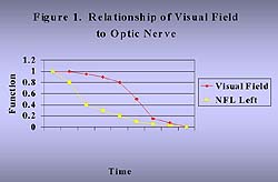
Understanding the Likelihood of Loss of Vision Functioning
In Slide 1, we can see that the loss of optic nerve fibers must occur for a period of time before measurable visual field deficits are detected.6 Newer visual field techniques such as blue-on-yellow perimetry (short wavelength automated perimetry, or SWAP) may identify visual field loss at an earlier stage of disease.41 Once loss has occurred, though, additional nerve fiber degeneration is likely to be associated with an accelerating course of visual field loss for the same absolute amount of nerve fiber loss. This fact can easily be understood by the mathematical notion that each equivalent additional quantitative amount of nerve fiber layer loss is a greater proportionate or percentage amount of the remaining tissue (see Slide 1). Thus, a nerve fiber layer loss of 5000 with 50,000 remaining fibers (10%) is likely to have less devastating consequences than the same 5000 nerve fiber loss at 15,000 remaining fibers (33%). This simple mathematical relationship may account for the clinical observations that underlie the axiom that the greater amount of nerve damage, the more susceptible the nerve is to additional damage. If the mechanisms of damage are unaltered, one might expect the same amount of nerve fiber loss per unit of time, but the relative effects of such loss would be progressively more pronounced as the remaining amount of nerve tissue declines.
At this time, we cannot identify the subgroup of patients who are likely to have steeper slopes of nerve loss, and thus are likely to progress to significant vision loss or blindness prior to death. As such, our goal is to treat every person so that that the smallest possible minority of patients lose vision prior to death. Such a philosophy is consonant with medical therapeutics in other fields of medicine, especially systemic hypertension.42 Without natural history data on an untreated population basis, it is currently not practical to model the relative risks and benefits of treatment. Thus, it should be among our highest priorities to identify factors that would allow us to better differentiate high risk individuals who should be treated from lower risk individuals who may be able to be followed without treatment until progression suggestive of misclassification occurs.
We do have glimpses at some potential methods of identifying those at greater risk. First, the extent of fluctuation of IOP in an eye may indicate those eyes at higher risk.43 Larger fluctuations in IOP can be found in those with glaucoma rather than those without glaucoma.44 Such findings can be understood in the context that connective and other tissues in the human body are possibly more susceptible to injury by dynamic stress rather than static loads.
Similarly, those with vasculopathic diseases, such as diabetes and hypertension, have also been found in some studies to be at higher risk of developing glaucoma. The apparent conflict among studies regarding hypertension may have to do with the inadequate definitions of hypertension and with its characterization (e.g., the need to use mean arterial pressure), and to confounding effects of medications on systemic blood pressure, as well as to the potential effects of hypotension.45,46
Third, we already know that there are certain IOP levels that are so high that intervention is warranted. In neovascular glaucoma or acute angle-closure glaucoma (primary or secondary), blindness can result in weeks or months if the pressure and underlying causes are not adequately treated. In congenital glaucomas, untreated elevations in pressure lead to blindness in all cases over time. Experience and mathematical models indicate that those with IOPs of higher than 28 mm Hg to 30 mm Hg have a 50% probability of having glaucoma, defined using older clinical paradigms (which, as noted above, have a higher threshold for diagnosis).28-30 From these situations, it is clear that significantly elevated IOP levels are associated with vision loss and that intervention, therefore, is warranted.
If we are better able to broaden this ability to characterize those who suffer faster rates of progression from those who suffer much slower rates of progression of nerve fiber loss (and hence visual field and other performance measures), then we can begin to tailor both the need for and the intensity of treatment. Even a relative risk of 3 or 4 could help identify those at greatest need for intensive treatment and follow-up. Thus, much more research is warranted to better characterize these subpopulations of patients with glaucoma.
Quality of Life Impact of Visual Loss in Glaucoma
Impaired vision is associated with increased rates of falls, hip fractures, and decreased physical mobility.47,48 Visual symptoms, such as trouble seeing or blurred vision, are associated with measurable decrements in general quality of life measures such as the SF-36 and instrumental activities of daily living.49,50 Self-rated quality of vision has a clear relationship to general health functioning. Indeed, vision is a vital aspect of our lives, and decrements in vision can be directly related to decrements in general well being and quality of life. Nevertheless, merely having a disease such as cataract or glaucoma is not independently associated with general quality of life measures, once the appropriate adjustments for demographic factors and medical and ocular comorbidities are included.51,52
Decrements in vision also are directly related to difficulties in performing vision-related or vision-specific tasks.48,53,54 Not surprisingly, such relationships are stronger and more easily seen relative to vision-specific quality of life than general quality of life measures. As in the analyses of general quality of life, adjustments for demographic differences, visual acuity, and comorbid medical and ocular conditions need to be made. In the case of visual fields, such adjustments reduce but do not eliminate the statistical or clinical significance of visual field decrements in vision-related functioning, as measured by validated instruments.52,55
The critical query then becomes one of how much visual field loss needs to occur before a clinically significant decrement in vision-related functioning can be measured. Such a seemingly simple inquiry is complicated, however, by important questions of how we can best quantitate and classify visual field loss, if field loss should be measured binocularly or can be imputed through the visual field loss in the better eye (with the worse eye having more and deeper field loss), and what the method of field measurement should be (central fields or full fields). Such questions will need to be addressed and techniques further modified in future investigations.
Using the information we do have available, however, we can begin to assess the impact of field loss on vision functioning using two common field grading systems - the Advanced Glaucoma Intervention Study (AGIS) grading scale and the Esterman binocular visual field grading system.52,55 In a pilot study using Esterman fields, Parrish found that there appeared to be a linear relationship between field loss and decrements in the National Eye Institute - Visual Functioning Questionnaire (VFQ).52 Nevertheless, analysis of many of the scatter plots of results suggests that perhaps not all of the VFQ subscales are directly linear in nature, but additional investigation is needed. In a multi-site study, Gutierrez et al found that better eye AGIS scores were similarly linearly related to VFQ scores.55 Although the data presented in this study more strongly support a linear relationship, evaluation of the results suggests that many if not most of the VFQ subscales have steeper rates or slopes of loss with more severely affected fields, suggesting a potentially non-linear but inverse parabolic relationship. Put together, these results suggest that it is the loss of field in the better eye that generally drives vision functioning, and that even early loss in the better eye may not be associated with truly significant clinical decrements in quality of life. In other words, individuals probably do not notice a meaningful impact on their lives until relatively noticeable visual field loss has occurred in both eyes, excluding special situations (e.g., the professional athlete's or professional truck driver's peripheral vision).
Implications for Treating Glaucoma
Current standards of care call for treatment of all patients who have glaucoma. Clearly, all patients with PACG or who are susceptible to PACG should be treated, given the natural history of those without treatment. The AAO PPP for POAG clearly enables eye care providers and other physicians to diagnose and treat individuals with glaucoma who have no field loss and no elevations in IOP, but do have characteristic optic nerve or nerve fiber layer loss. Such characteristic loss includes: defects in the nerve fiber layer, asymmetry between the two eyes, asymmetry above and below in the same optic nerve, notches in the optic nerve rim, optic nerve hemorrhages, or enlarged cups. In addition, individuals with elevated IOP beyond a "safe" range, generally 28 mm Hg or higher, also usually are treated. As estimated from the Baltimore Eye Study results, such a treatment protocol could potentially place upward of 15 million Americans on treatment for POAG.17
The evidence cited here suggests that this strategy could be modified significantly if additional data could be generated in the areas of risk factors analyses for progression (as opposed to risk factors for having glaucoma) and pace of progression. Additional data also need to be generated to better identify the degree of physiological dysfunction arising from glaucoma that is associated with noticeable patient functioning defects. When these two elements are put together, we can then identify not only those who are at higher risk of developing loss that interferes with their quality of vision and quality of life but also those individuals for whom we should intervene before noticeable decrements occur in functioning.
How Can We Best Deliver Glaucoma Care?
Glaucoma of all forms is a leading cause of impaired vision and blindness around the world. Even in the United States, 19% of blindness among blacks is caused by glaucoma.1 Such vision loss and blindness has large and significant effects and costs on both the individuals affected and our society. In order to assess this burden, we need to address two questions:
- Should we screen for glaucoma?
- How can we ensure access to health care for those who are diagnosed as having glaucoma?
Screening for Glaucoma
All current preventive care guidelines and screening strategies uniformly state that mass glaucoma screening is not warranted and should not be conducted.56 Using traditional means of IOP evaluation, optic disc examination, or traditional visual field testing, insufficient sensitivity, specificity, predictive values, and cost-effectiveness have been found.56,57 One study in the United Kingdom estimated screening would be worthwhile if the value of detecting a case (in terms of savings) would exceed $850 per case, using existing resources and staff.58 Furthermore, the open questions about the effectiveness of treatment for POAG militate against aggressive screening, because the benefits of treatment once cases are identified are as yet not completely resolved.
The recent development of the frequency-doubled perimeter (FDP), however, suggests that the test performance of the perimeter as a screening instrument may finally be sufficient for use on a population screening basis.59 With sensitivities and specificities in excess of 95%, the posterior probabilities of a positive test result are sufficiently high as to justify its use. Together with the new definition of POAG and the finding of any visual field defect defines one as at least a moderate case of glaucoma, the FDP offers the possibility of economically feasible screening for glaucoma. To the extent that the prior probability of glaucoma is enhanced by use of a second screening technique (such as risk factor analysis), the performance of the FDP will only be more accurate.
Nevertheless, efforts to provide more accurate and reliable methods of screening still leave unanswered questions surrounding the value of treatment once the diagnosis of glaucoma has been made. No screening strategy would be cost-effective if no effective treatment existed for cases so identified. Thus, the final endorsement for glaucoma screening necessarily awaits the results of the ongoing randomized, controlled trials with a no-treatment arm to confirm the findings of the CNTGSG results.
Access to Care
Care Patterns by Ophthalmologists
Assuming that
treatment is found to be effective, significant issues still exist about
whether people receive appropriate care for glaucoma. Although the survey of
the membership of the AAO conducted for the RAND Workforce Study suggests that
the average patient with POAG should be seen four times per year,9
data from the Medicare Provider (Part B) files indicate that only a minority of
patients with any eye condition are seen at least this many times per
year.60 Furthermore, although patients cared for in an academic
setting appear to receive care in accordance with the recommendations of the
AAO PPP,61 patients receiving care in the community setting often
receive care not in line with the recommendations of the PPP for
POAG.62 Thus, for all patients, significant attention needs to be
paid to how care is provided.
Differential Access by Groups
This concern is even
more troubling relative to care provided to minorities and the poor in the
United States. Data clearly indicate that black Americans do not access the
health care system in proportion to the rates of glaucoma that have been
detected on the basis of population studies. Although black Americans undergo
glaucoma surgery twice as often as white Americans, this rate is still 50% less
than what would be expected.63 Similarly, the homeless and the poor
do not access health care in a manner similar to those in better socioeconomic
situations.64 Finally, blacks and the elderly do not have glaucoma
treatment initiated at rates that might otherwise be expected.65 The
problem does not appear to be the care provided by physicians once patients
enter the care system, unlike the situation with many other areas of medical
care. Nevertheless, the lack of expected access raises important questions for
providers to address.66 Why is this the case?
Traditional approaches to this issue have cited the lack of income and the lack of sufficient insurance coverage for the groups just cited. Yet, even among Medicare beneficiaries, where these influences are far less, one sees the same patterns of care.66 As such, additional factors or obstacles to care need to be examined. One important new finding is that black Americans trust the health care system and health care providers much less than white Americans.67 The result of such lower trust may be a decrease in the likelihood of seeking care or of complying with provider recommendations once care is sought. Thus, access may be decreased on the basis of the patient's conscious decisions and behaviors.
Second, loss of vision may be seen culturally as part of the aging process, so that loss of vision in one eye is not cause for alarm. Similarly, there may be other cultural barriers to receiving care for a chronic condition.68 Although this may appear to be an educational issue on the surface, there may be hidden subtexts that severely diminish the returns from traditional educational efforts. For example, among Latino women with diabetes, the cultural norms of their gender roles severely hamper their ability to care for themselves, both in terms of diet and in their being able to attend regular medical follow-up.68 Such scenarios also are likely to be present for minorities relative to glaucoma care. Thus, it is clear that a wider set of factors needs to be evaluated in enhancing access to care.
Conclusion
When we examine the population prevalence of POAG and PACG, we can easily see that these estimates are strongly dependent on how one defines each condition and the quality of the screening and examination technique employed.69,70 Nevertheless, it is clear that many patients who might benefit from treatment are not currently receiving it. Yet, several outstanding questions remain.
First, is treatment effective in altering the natural history of POAG? We know that treatment does alter the natural history of PACG favorably, and we have evidence that it also does so for POAG (though not yet definitive evidence). Second, can we identify those at greatest risk for the most rapid progression of damage to vision? We know that visual field loss has a measurable effect on vision-related quality of life, so targeting those at greatest risk for developing such field loss prior to its onset clearly would be beneficial. Third, can we enhance the likelihood that those who do have glaucoma will receive adequate care? This obligation involves not only increasing access to those at greatest risk of developing severe glaucoma but also optimizing the quality of care that all patients with glaucoma receive. Each of these tasks is a major undertaking that will challenge our profession and ourselves.
References
- American Academy of Ophthalmology. Preferred practice pattern: Primary open-angle glaucoma. San Francisco, Calif: American Academy of Ophthalmology; 1992.
- Guzman G, Javitt J, Glick H, Tielsch J, McDonald R. Glaucoma in the United States population: The economic burden of illness. Invest Ophthalmol Vis Sci. 1992;33(suppl):759.
- Lee P. Economic concerns in glaucoma management in the 21st century. J Glaucoma. 1993;2:148-151.
- American Academy of Ophthalmology. Preferred practice pattern: primary open-angle glaucoma. San Francisco, Calif: American Academy of Ophthalmology; 1996.
- Caprioli J. Clinical evaluation of the optic nerve in glaucoma. Trans Am Ophthalmol Soc. 1994;92:589-641.
- Quigley JA, Addicks EM, Green WR. Optic nerve damage in human glaucoma. III: quantitative correlation of nerve fiber loss and visual field defect in glaucoma, ischemic neuropathy, papilledema, and toxic neuropathy. Arch Ophthalmol. 1982;100:135-146.
- Herndon LW, Choudhri SA, Cox T, et al. Central corneal thickness in normal, glaucomatous, and ocular hypertensive eyes. Arch Ophthalmol. 1997;115:1137-1141.
- Sommer A, Tielsch JM, Katz J, et al. Relationship between intraocular pressure and primary open angle glaucoma among white and black Americans. The Baltimore Eye Survey. Arch Ophthalmol. 1991;109:1090-1095.
- Lee PP, Jackson CJ, Relles DA. Estimating Eye Care Provider Supply and Workforce Requirements. Santa Monica, Calif: RAND, MR-516-AAO; 1995.
- Lichter PR. Variability of expert observers in evaluating the optic disc. Trans Am Ophthalmol Soc. 1977;74:532-572.
- Varma R, Steinmann WC, Scott IU. Expert agreement in evaluating the optic disc for glaucoma. Ophthalmology. 1992;99:215-221.
- Klein BE, Klein R, Sponsel WE, et al. Prevalence of glaucoma. The Beaver Dam Eye Study. Ophthalmology. 1992;99:1499-1504.
- Tielsch JM, Sommer A, Katz J, et al. Racial variations in the prevalence of primary open-angle glaucoma. The Baltimore Eye Survey. JAMA. 1991;266:369-374.
- Mitchell P, Smith W, Attebo K, Healey PR. Prevalence of open-angle glaucoma in Australia. The Blue Mountains Eye Study. Ophthalmology. 1996;103:1661-1669.
- Dielemans I, Vingerling JR, Wolfs RC, et al. The prevalence of open-angle glaucoma in a population-based study in the Netherlands. The Rotterdam Study. Ophthalmology. 1994;101:1851-1855.
- Mason RP, Kosoko O, Wilson MR, et al. National survey of the prevalence and risk factors of glaucoma in St. Lucia, West Indies. Part I. Prevalence findings. Ophthalmology. 1989;96:1363-1368.
- Quigley HA. Medical progress: Open-angle glaucoma (review article). N Engl J Med. 1993;328:1097-1106.
- Leske MC, Connell AM, Schachat AP, Hyman L. The Barbados Eye Study. Prevalence of open angle glaucoma. Ophthalmology. 1994;112:821-829.
- Congdon N, Wang F, Tielsch JM. Issues in the epidemiology and population-based screening of primary angle-closure glaucoma. Surv Ophthalmol. 1992;36:411-423.
- Congdon NG, Youlin Q, Quigley H, et al. Biometry and primary angle-closure glaucoma among Chinese, white, and black populations. Ophthalmology. 1997;104:1489-1495.
- Wen L-Y, Myopic choroidal degenerations. In: Tso MOM, ed. Retinal Diseases: Biomedical Foundations and Clinical Management. New York, NY: JB Lippincott Co; 1988;242-246.
- Arkell SM, Lightman DA, Sommer A, et al. The prevalence of glaucoma among Eskimos of northwest Alaska. Arch Ophthalmol. 1987;105:482-485.
- Tielsch JM, Katz J, Quigley HA, Javitt JC, Sommer A. Diabetes, intraocular pressure, and primary open-angle glaucoma in the Baltimore Eye Survey. Ophthalmology. 1995;102:48-53.
- Mitchell P, Smith W, Chey T, Healey PR. Open-angle glaucoma and diabetes: The Blue Mountains Eye Study, Australia. Ophthalmology. 1997;104:712-728.
- Dielemans I, de Jong PT, Stolk R, et al. Primary open-angle glaucoma, intraocular pressure, and diabetes mellitus in the general elderly population. The Rotterdam Study. Ophthalmology. 1996;103:1271-1275.
- Klein BE, Klein R, Jensen SC. Open-angle glaucoma and older-onset diabetes. The Beaver Dam Eye Study. Ophthalmology. 1994;101:1173-1177.
- Hollows FC, Graham PA. Intra-ocular pressure, glaucoma, and glaucoma suspects in a defined population. Br J Ophthalmol. 1996;50:570-586.
- Davanger M. Low-pressure glaucoma and the concept of the IOP tolerance distribution curve. Acta Ophthalmol. 1989;67:256-260.
- Davanger M, Ringvold A, Blika S. The frequency distribution of the glaucoma tolerance limit. Acta Ophthalmol. 1991;69:782-775.
- Davanger M, Ringvold A, Blika S. The probability of having glaucoma at different IOP levels. Acta Ophthalmol. 1991;69:565-568.
- Comparison of glaucomatous progression between untreated patients with normal-tension glaucoma and patients with therapeutically reduced intraocular pressures. Collaborative Normal-Tension Glaucoma Study Group. Am J Ophthalmol. 1998;126(4):487-497.
- The effectiveness of intraocular pressure reduction in the treatment of normal-tension glaucoma. Collaborative Normal-Tension Glaucoma Study Group. Am J Ophthalmol. 1998;126(4):498-505.
- Erie JC, Hodge DO, Bray DT. The incidence of primary angle-closure glaucoma in Olmsted County, Minnesota. Arch Ophthalmol. 1997;115:177-181.
- Kass MA, Kolker AE, Becker B. Prognostic factors in glaucomatous visual field loss. Arch Ophthalmol. 1976;94:1274-1276.
- Quigley HA, Tielsch J, Katz J, et al. Rate of progression in open-angle glaucoma estimated from cross-sectional prevalence of visual field damage. Am J Ophthalmol. 1996;122:355-363.
- Mao LK, Stewart WC, Shields MB. Correlation between intraocular pressure control and progressive glaucomatous damage in primary open-angle glaucoma. Am J Ophthalmol. 1991;111:51-55.
- Hattenhauer MG, Johnson DH, Ing HH, et al. The probability of blindness from open-angle glaucoma. Ophthalmology. 1998;105(11):2099-2104.
- Hart WM. The epidemiology of primary open-angle glaucoma and ocular hypertension. In: Ritch R, Shields MB, Krupin T, eds. The Glaucomas. Baltimore, Md: CV Mosby Co; 1989;790-791.
- Leske MC. The epidemiology of open-angle glaucoma: a review. Am J Epidemiology. 1983;118:166-191.
- Ritch R, Lowe RF, Reyes A. Therapeutic overview of angle-closure glaucoma. In: Ritch R, Shields MB, Krupin T, eds. The Glaucomas. Baltimore, Md: CV Mosby Co; 1989;860.
- Sample PA, Bosworth CF, Weinreb RN. Short-wavelength automated perimetry and motion automated perimetry in patients with glaucoma. Arch Ophthalmol. 1997;115:1129-1133.
- Joint National Committee on Prevention, Detection, Evaluation and Treatment of High Blood Pressure. The sixth report of the Joint National Committee on Prevention, Detection, Evaluation and Treatment of High Blood Pressure. Arch Int Med. 1997;157:2413-2446.
- Zeimer RC, Wilensky JT, Gieser DK, Viana MA. Association between intraocular pressure peaks and progression of visual field loss. Ophthalmology. 1991;98:64-69.
- Sacca SC, Rolando M, Marletta A, et al. Fluctuations of intraocular pressure during the day in open-angle, normal-tension glaucoma and normal subjects. Ophthalmologica. 1998;212(2):115-119.
- Tielsch JM, Katz J, Sommer A, Quigley HA, Javitt JC. Hypertension, perfusion pressure, and primary open-angle glaucoma. Arch Ophthalmol. 1995;113:216-221.
- Hayreh SS, Zimmerman MB, Podhajsky P, Alward WLM. Nocturnal arterial hypotension and its role in optic nerve head and ocular ischemic disorders. Am J Ophthalmol. 1994;117:603-624.
- Tinetti ME, Sppechley M, Ginter SF. Risk factors for falls among elderly persons living in the community. N Engl J Med. 1988;319:1701-1707.
- Mangione CM, Lee PP, Hays RD. Measurement of visual functioning and health-related quality of life in eye disease and cataract surgery. In: Spilker B, ed. Quality of Life and Pharmacoeconomics in Clinical Trials. 2nd ed. Philadelphia, Pa: Lippincott-Raven Publishers; 1996:1045-1051.
- Kington R, Rogowski R, Lillard L, Lee PP. Functional associations of "trouble seeing." J Gen Int Med. 1997;12:125-128.
- Lee PP, Spritzer K, Hays R. The impact of blurred vision on functioning and well-being. Ophthalmology. 1997;104:390-396.
- Fryback DG, Dasbach EJ, Klein R, et al. The Beaver Dam Health Outcomes Study: Initial catalog of health-state quality factors. Med Decis Making. 1993;13:90-102.
- Parrish RK. Visual impairment, visual functioning, and quality of life assessments in patients with glaucoma. Trans Am Ophthalmol Soc. 1996;94:919-1028.
- Javitt JC, Brenner MH, Curbow B, et al. Vision change and quality of life in the elderly: response to cataract surgery and treatment of other chronic ocular conditions. Arch Ophthalmol. 1993;111:680-685.
- Steinberg EP, Tielsch JM, Schein OD, et al. The VF-14: An index of functional impairment in patients with cataract. Arch Ophthalmol. 1994;112:630-638.
- Gutierrez P, Wilson MR, Johnson C, et al. Influence of glaucomatous visual field loss on health-related quality of life. Arch Ophthalmol. 1997;115:777-784.
- Canadian Task Force on the Periodic Health examination. Periodic health examination, 1995, update: 3. Screening for visual problems among elderly patients. Can Med Assoc J. 1995;152(8):1211-1222.
- Tielsch JM, Katz J, Singh K, et al. A population-based evaluation of glaucoma screening: the Baltimore Eye Survey. Am J Epidemiol. 1991;134:1102-1110.
- Tuck MW, Crick RP. The cost-effectiveness of various modes of screening for primary open angle glaucoma. Ophthalmic Epidemiol. 1997;4:3-17.
- Sponsel WE, Ritch R, Stamper R, et al. Prevent Blindness America Visual Field Screening Study. Am J Ophthalmol. 1995;120:699-708.
- Ellwein LB, Friedlin V, McBean AM, Lee PP. Use of eye-care services among the 1991 Medicare population. Ophthalmology. 1996;103:1732-1743.
- Albrecht K, Lee PP. Conformance with Preferred Practice Patterns in glaucoma care. Ophthalmology. 1994;101:1668-1671.
- Hertzog LH, Albrecht KG, LaBree L, Lee PP. Glaucoma care and conformance with preferred practice patterns: examination of the private, community-based ophthalmologist. Ophthalmology. 1996;103:1009-1013.
- Javitt JC, McBean AM, Nicholson GA, et al. Undertreatment of glaucoma among black Americans. N Engl J Med. 1991;325:1418-1422.
- Ho JH, Change RJ, Wheeler NC, Lee DA. Ophthalmic disorders among the homeless and nonhomeless in Los Angeles. J Am Optom Assoc. 1997;68:567-573.
- Glynn RJ, Gurwitz JH, Bohn RL, et al. Old age and race as determinants of initiation of glaucoma therapy. Am J Epidemiol. 1993;138:395-406.
- Wang F, Javitt JC, Tielsch JM. Racial variations in treatment for glaucoma and cataract among Medicare recipients. Ophthalmic Epdiemiol. 1997;4(2):89-100.
- Corbie-Smith G, Thomas SB, Duncan T, et al. Racial differences in perceptions of physicians and research: results of a national telephone survey. J Gen Int Med. 1998;13(suppl):63.
- Van Boemel GB, Lee PP. Adherence to traditional female sex role as a hidden barrier in preventable eye disease: a case history. J Vis Impair & Blindness. In press.
- Bathija R, Gupta N, Zangwill L, Weinreb RN. Changing definition of glaucoma. J Glaucoma. 1998;7:165-169.
- Kahn HA, Milton RC. Alternative definitions of open-angle glaucoma. Effect on prevalence and associations in the Framingham eye study. Arch Ophthalmol. 1980;98:2172-2177.
- Wensor MD, McCarty CA, Sttanislavsky YL, Livingston PM, Taylor HR. The prevalence of glaucoma in the Melbourne Visual Impairment Project. Ophthalmology. 1998;105:733-739.
- Bonomi L, Marchini G, Marraffa M, et al. Prevalence of glaucoma and intraocular pressure distribution in a defined population. The Egna-Neumarkt Study. Ophthalmology. 1998;105:209-215.
- Cedrone C, Culaasso F, Cesareo M, et al. Prevalence of glaucoma in Ponza, Italy: A comparison with other studies. Ophthalmic Epidemiol. 1997;4:59-72.
- Foster PJ, Baasanhu J, Alsbirk PH, et al. Glaucoma in Mongolia. A population-based survey in Hovsgol province, northern Mongolia. Arch Ophthalmol. 1996;114:1235-1241.