Diabetic retinopathy accounts for 12% of all new cases of blindness each year in the United States and is the leading cause of blindness in patients between 20 and 64 years old. Vision loss is most often attributed to macular edema, nonresolving vitreous hemorrhage, traction retinal detachment, and macular ischemia. The disease is often best treated before symptoms develop, making screening ophthalmic examinations important. Landmark studies have been performed to elucidate the natural history of this condition as well as the benefits of early intervention with laser surgery to prevent visual loss.
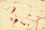
The prevalence of diabetic retinopathy increases with the age of the patient and the duration of diabetes. After 20 years of diabetes, 99% of insulin-dependent diabetics and 60% of non-insulin-dependent diabetics show some degree of retinopathy. Other factors increasing the risk for development of retinopathy include pregnancy, hypertension, renal disease, anemia, chronic hyperglycemia, and hyperlipidemia. Although the exact cause of retinopathy is not known, it is accepted that microvascular compromise results from endothelial cell damage due to extended hyperglycemia. Pathologic specimens (Slide 1) have shown loss of pericytes and basement membrane thickening in the retinal vasculature.
Clinical Findings
Diabetic retinopathy is subdivided into nonproliferative (NPDR) and proliferative (PDR) forms with macular edema occurring at any stage. NPDR (Slide 2 and 3) is characterized by microaneurysms, intraretinal hemorrhages, hard exudates, and nerve fiber layer infarcts. More advanced findings include venous caliber changes, intraretinal microvascular abnormalities (IRMA), and retinal capillary nonperfusion (Slide 4) . Microaneurysms represent abnormal outpouchings of retinal capillaries. Hemorrhages reflect the retinal layer in which they are located. Nerve fiber layer hemorrhages assume a flame shape, whereas deeper hemorrhages appear as dots or blots. Retinal hypoxia results in nerve fiber layer infarcts and venous caliber changes such as beading and dilation. IRMAs most likely represent preexisting vessels that act as shunts through areas of nonperfusion.
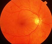
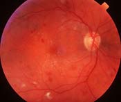
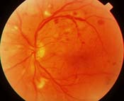
Macular edema is a common cause of visual loss in diabetic retinopathy (Slide 5). The Early Treatment Diabetic Retinopathy Study (ETDRS) defined clinically significant macular edema (CSME) for which a 50% reduction in moderate visual loss was reported with laser photocoagulation. CSME was defined as any one of the following identified on clinical examination:
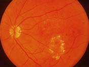
- Retinal thickening at or within 500 µm from the center of the macula
- Hard exudates at or within 500 µm from the center of the macula, if associated with thickening of the adjacent retina
- A zone of retinal thickening larger than 1 disc area located within 1 disc diameter from the center of the macula
PDR is heralded by neovascularization of the retina. Increased retinal ischemia results in the production of vasoproliferative factors resulting in endothelial cell proliferation. New vessels appear at or near the optic disc (Slide 6) (neovascularization of optic disc [NVD]) or elsewhere in the retina (neovascularization elsewhere [NVE]). Retinal neovascularization is most often seen at the junction of perfused and nonperfused retina. The posterior hyaloid serves as a scaffold for proliferation. Neovascular tissue is fragile, with a propensity to bleed and produce the sight-threatening complication of vitreous hemorrhage. This tissue also has a propensity to become fibrotic, resulting in other sight-threatening complications including macular traction/heterotopia, macular edema, and traction retinal detachment. Contraction of fibrovascular tissue may also produce retinal breaks, resulting in rhegmatogenous retinal detachment. The Diabetic Retinopathy Study defined high-risk characteristics when panretinal photocoagulation (PRP) was shown to reduce severe visual loss by 50%. High-risk PDR was defined by any one of the following clinical findings:
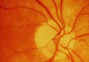
- Mild NVD (25% to 34% disc area) with vitreous hemorrhage
- Moderate to severe NVD with or without vitreous hemorrhage
- Moderate NVE (50% disc area) with vitreous hemorrhage
With severe ischemia, neovascularization can also involve the anterior segment at the iris and anterior chamber angle (Slide 7). Obstruction of aqueous outflow may result in neovascular glaucoma, another sight-threatening complication.
The ETDRS studied the progression from NPDR to PDR and developed the 4:2:1 rule to define NPDR. Severe NPDR is characterized by any one of the following manifestations:
- 4 quadrants of diffuse intraretinal hemorrhages and microaneurysms
- 2 quadrants of venous beading
- 1 quadrant of intraretinal microvascular abnormalities
Very severe NPDR showed a 15% rate of progression to high-risk PDR within 1 year. Severe NPDR was defined as any two of the above characteristics and showed a 45% rate of progression to high-risk PDR within 1 year.
Management
Regardless of the stage of retinopathy, control of blood sugar is correlated with a better prognosis. The Diabetes Control and Complications Trial demonstrated that intensive glycemic control delays the onset of new retinopathy and slows the progression of existing retinopathy in insulin-dependent diabetics. These patients also showed a reduced incidence of macular edema and decreased need for panretinal and focal photocoagulation.
In patients who require laser photocoagulation, focal treatment of macular edema is recommended prior to PRP when possible. Peripheral scatter treatment may exacerbate macular edema, justifying a 6- to 8-week delay of PRP in patients with clinically significant macular thickening who are approaching high-risk characteristics. Similarly, treatment of macular edema is recommended prior to cataract surgery.
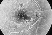
Although the ETDRS showed a 50% reduction in moderate visual loss with focal laser photocoagulation for macular edema, certain risk factors predispose to a poor visual outcome. These include diffuse macular edema, macular ischemia (Slide 8A and 8B), foveal hard exudate, and significant cystoid macular edema.
Complications of laser photocoagulation for diabetic retinopathy are low relative to the visual benefit. For macular edema, focal laser treatment may cause mild scotomata or visual field defects. For retinal neovascularization, PRP may cause a decrease in night vision, color vision, and/or peripheral vision. Visual acuity loss of 1 or 2 lines has been reported in some patients. PRP may also exacerbate macular edema as discussed above. Intense treatment may result in choroidal effusion. Accidental foveal burn is a potentially devastating complication of all retinal laser therapies.
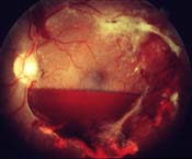
When vitreous hemorrhage fails to resolve spontaneously or fibrovascular proliferation causes traction that threatens the macula, surgery is usually indicated (Slide 9). Pars plana vitrectomy with or without membrane dissection is performed in these cases. The Diabetic Retinopathy Vitrectomy Study studied the role of vitrectomy for PDR. It clearly demonstrated a benefit of early vitrectomy for severe vitreous hemorrhage in insulin-dependent diabetics. No such benefit was found for non-insulin-dependent diabetics. More recent advances in microsurgery may improve the overall prognosis in operated eyes.
Current prevention and treatment of diabetic retinopathy involve strict glycemic control coupled with regular screening examinations to detect disease when it is asymptomatic and most effectively treated. Prompt application of laser photocoagulation and judicious use of vitrectomy can improve the ultimate prognosis relative to the natural history of disease. Future modalities may involve pharmacologic interventions that have targets such as angiogenic factors in preventing or treating the complications of retinopathy.