Reprinted from Crouch ER Jr, Crouch ER. Management of Traumatic Hyphema: Therapeutic Options. J Pediatr Ophthalmol Strabismus. 1999;36(5):238.
Introduction
The accumulation of blood in the anterior chamber is one of the most challenging clinical entities encountered by the ophthalmologist. Even a small hyphema can be a sign of major intraocular trauma with associated damage to other intraocular tissues. The lack of an ideal therapeutic program, the potential for secondary hemorrhage, and the secondary onset of glaucoma threaten to transform an eye with an initially good visual prognosis into a complex therapeutic problem with poor final visual result. Traumatic hyphema potentially can cause significant visual reduction, particularly if secondary hemorrhage occurs. There are a host of therapeutic options in traumatic hyphema, including the preferred medical management of traumatic hyphema and the indications for surgical intervention.
Before examining the treatment options, let us examine the complications directly related to traumatic hyphema. In particular, elevated intraocular pressure (IOP) occurs in 24% to 32% of all traumatic hyphemas.1 In total hyphemas, the incidence of elevated IOP is higher than 95%. Treatment modalities should be directed at controlling the IOP, reducing the incidence of secondary hemorrhage, and reducing the associated visual complications of corneal blood staining and optic atrophy.
The types of glaucoma in hyphema have a variety of mechanisms. The pathogenesis of the glaucoma related to erythrocytes in the anterior chamber includes a trabecular blockage with normal or sickle erythrocytes. Pupillary block glaucoma occurs when the entire anterior chamber is filled with clotted blood. Glaucoma not related to the erythrocytes in the anterior chamber includes angle-recession glaucoma, involving greater than 180° of the trabecular meshwork and peripheral anterior synechiae that occur after 9 days of traumatic hyphema.2
Major complications directly related to the hyphema are posterior synechiae, peripheral anterior synechiae, corneal blood staining, and optic atrophy.1,3 In two perspective studies of 196 patients, we observed 11 patients (5.6%) with corneal blood staining. Predisposing factors of corneal blood staining include hyphemas lasting more than 6 days, hyphemas occupying more than half of the anterior chamber, corneal epithelial dysfunction, and elevated IOP. Frequently, a combination of one or more of these factors contributes to corneal blood staining.
Optic atrophy can occur as a result of an initial blunt trauma or secondary to elevations of IOP. Optic atrophy occurred in 10 of 196 patients (5.1%). Five of these patients had pressures higher than 50 mm Hg for 5 days, three patients had pressures of higher than 35 mm Hg for 7 days, two of the patients had positive sickle cell trait or SA hemoglobin with IOP of 35 mm Hg to 39 mm Hg for 2 to 4 days.
Evaluation
The patient with traumatic hyphema should have a thorough ophthalmologic evaluation. The time of onset and type of injury should be recorded accurately. The object producing the injury and the method by which it produced the injury give additional insight into the amount of ocular damage. If several days have elapsed since the injury, the onset of diminished visual acuity or an increase in ocular pain is significant, as they may indicate secondary hemorrhage, elevated IOP, or both. Any other history of ocular problems including amblyopia should be recorded.
The ocular examination should be thorough and include the surrounding tissues. Resist the temptation to abandon the examination when one cannot see beyond the anterior chamber. Search the periorbital skin for areas of hypesthesia or anesthesia. Blowout fractures frequently accompany hyphemas.4 Examine the lids and conjunctiva carefully for laceration. Regard disproportionate conjunctival edema as a possible indication of scleral rupture until proven otherwise (Slide 1). Proptosis may indicate retrobulbar hemorrhage; enophthalmos and/or restrictions in extraocular muscle rotations suggest a blowout fracture. Record visual acuity of both eyes with best optical correction. If the acuity of the injured eye is markedly reduced, light projection and color sense should be tested, including evaluations of consensual reaction of the pupil of the uninjured eye. Inappropriately poor visual acuity suggests concomitant optic nerve or macular damage.
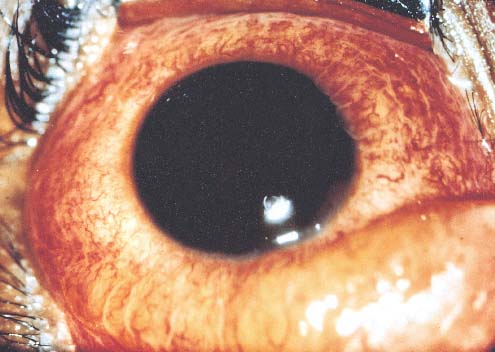
The cornea should be examined for damage or preexisting disease, because any compromise of the endothelium increases its susceptibility to blood staining. Record the amount and character of the hyphema as it first presents. Careful drawing of the shape of the clot or of the level of free cells aids in evaluation of secondary hemorrhage or resolution of the hyphema. Note iris and lens details that are visible beyond the hyphema (i.e., pupillary size and shape, sphincter tears, iridodialysis, and dislocation of the lens or other injuries). Examination of the fundus should be attempted but may be difficult until the hyphema has resolved.
The pupil should be dilated if one suspects intraocular foreign body, rupture or perforation of the globe, or retinal tears or detachment. In most instances, a more favorable fundus evaluation is possible later, with a clear media following resolution of the hyphema. B-scan ultrasonography should be performed when fundus details are markedly obscured.
IOP should be recorded by applanation tonometry. Topical anesthesia and facial akinesia may be required if lid squeezing prevents adequate examination. Avoid gonioscopy until day 7 to avoid a secondary hemorrhage.
Many patients with hyphema appear somnolent. The exact mechanism for this reaction is unclear. If associated head trauma is a possibility, obtain neurological consultation and a magnetic resonance image. Laboratory data should include platelet count, prothrombin time, and partial thromboplastin time. All African American patients require a sickle cell screening and, if the test is positive, hemoglobin electrophoresis.
Classification and Characteristics
Traumatic hyphema is most often encountered in children. Most reports indicate a prevalence of 70% or greater in the pediatric population.5 The agent producing a hyphema is usually a projectile that strikes the exposed portion of the eye. Sources of trauma have included balls, rocks, air-gun or BB pellets, paint-ball pellets, and the human fist.6,7
With the increase of child abuse, fists and belts have begun playing a prominent role. Males are involved in 75% of cases.1,5 U.S. studies indicate that hyphemas in the pediatric population occur in approximately 20 per 100,000 people per year.8
Sports injuries from soccer, hockey, baseball, and basketball account for up to 60% of traumatic hyphemas. In many cases, traumatic hyphemas are related to a lack of protective eye gear. A greater awareness in the community regarding eye protection safety and use of protective gear would likely decrease the incidence of traumatic hyphema in the pediatric patient population.
We prefer the following grading system for traumatic hyphemas. Grade 1 hyphema consists of layered blood occupying less than one-third of the anterior chamber. Grade 2 hyphema consists of blood filling one-third to one-half of the anterior chamber, and grade 3 hyphema is layered blood filling one-half to less than total. Grade 4 hyphema includes total clotted hyphemas, often referred to as black-ball or eight-ball hyphema.
The majority of hyphemas fill less than one-third of the anterior chamber (Slide 2). When hyphemas are divided into four groups according to the amount of filling of the anterior chamber, hyphemas involving less than one-third of the anterior chamber comprise 58%, one-third to one-half of the anterior chamber, 20%; one-half to less than total, 14%; and total hyphemas, 8% (Slide 3). Slightly fewer than half of all hyphemas settle inferiorly to form a level; approximately 40% form a definite clot, usually adherent to the iris stroma; 10% have a dark clot in contact with the endothelium.l
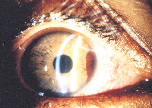
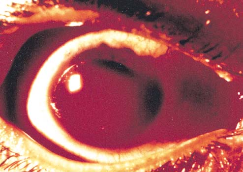
The source of anterior chamber hemorrhage in contusion injuries is believed to be related to the displacement of tissues posteriorly or to the resultant fluid wave in the aqueous and vitreous, which stretches the limbal vessels and displaces the iris and lens. This usually results in a tear at the iris or ciliary body, usually at the angle structures.9 A tear at the anterior aspect of the ciliary body is the most common site of bleeding and occurs in about 71% of cases.1 The blood exits from the anterior chamber via the trabecular meshwork and Schlemm's canal or the juxtacanalicular tissue.
Rarely, spontaneous hyphemas may occur and be confused with traumatic hyphemas. Spontaneous hyphemas are secondary to neovascularization, ocular neoplasms (retinoblastoma), and vascular anomalies (juvenile xanthogranuloma). Vascular tufts that exist at the pupillary border have been implicated in spontaneous hyphema.10
The usual duration of the uncomplicated hyphema is 5 to 6 days. The mean duration of elevated IOP is 6 days.
Elevated IOP
Increased IOPs (above 24 mm Hg) may accompany hyphemas of any size and may be anticipated in approximately 32% of all patients with hyphema at some time during their hospitalization.1 Higher, more prolonged elevations of IOP are more commonly associated with near total or total hyphemas.
These highly elevated IOPs occur during the acute phase of the hyphema and are separate from those related to angle recession.2 In patients with pressure elevations, abnormal tonometric readings are frequently detected during the first 24 hours after injury. This initial period of elevated IOP is often followed by a period of normal or below normal pressure from the second to the sixth day.
The early period of elevated IOP is probably the result of trabecular plugging by erythrocytes and fibrin. The following period of reduced pressure is most likely due to reduced aqueous production and may actually increase the chances of secondary hemorrhage. This period of hypotony is commonly followed by a subsequent rise in IOP with the recovery of the ciliary body. Intraocular hypertension then subsides with recovery of the trabecular meshwork and disappearance of the hyphema.
Exceptions to this are patients with hyphema occupying more than 75% of the anterior chamber and total hyphema, when pressure elevation frequently has its onset simultaneously with the initial hyphema and remains continually elevated until the hyphema has had considerable resolution. When large segments of the anterior chamber angle are irreparably damaged or when organization of the fibrin or clot produces extensive peripheral anterior synechiae, the intraocular hypertension continues, becoming intractable glaucoma.
Ghost-cell glaucoma with hyphema and vitreous hemorrhage may cause elevation of IOP 2 weeks to 3 months after initial injury.11 There is gradual clearing of the hyphema with erythrocytes losing hemoglobin and becoming "ghost cells" in the vitreous cavity. The ghost cells then press forward into the anterior chamber, with resultant later elevation of IOP.
Secondary Hemorrhage
Secondary bleeding into the anterior chamber results in a markedly worse prognosis. Eventual visual recovery to an acuity of 20/50 (6/15) or better occurs in approximately 64% of patients with secondary hemorrhage as compared with 79.5% of those in whom no rebleeding occurred.1,3,7 True secondary bleeding into the anterior chamber is indicated by an obvious increase in the amount of blood in the anterior chamber. Secondary hemorrhage occurs in approximately 22% of all patients with hyphema with a range of 7% to 38%.1,3,7,12 The incidence of secondary hemorrhage is higher in grades 3 and 4.5 With total to near total hyphemas, in which the blood is dark and clotted, bright red blood often begins to appear at the periphery of the clot on the fourth to sixth day. This is probably the result of early dissolution of the clot and does not necessarily indicate a secondary hemorrhage.
A large proportion of patients younger than 6 have secondary hemorrhages; the likelihood decreases with age. One-third of all secondary hemorrhages proceed to total hyphema. Secondary hemorrhage usually occurs on the third or fourth day, but may occur from the second to the seventh day after trauma.1,12
Several studies documented that secondary hemorrhage occurs more frequently in African American patients. Spoor and colleagues observed secondary hemorrhage in 24.2% of African American and in only 4.5% of Caucasian patients.13 Three other studies demonstrated greater secondary hemorrhage rates in African American patients that are highly significant (P<.05.).7,14,15 In our recent topical aminocaproic acid (ACA) study, the control group had 54 patients. Twenty percent of the controls had positive sickle cell trait by hemoglobin electrophoresis. Twelve patients suffered secondary hemorrhage in the control group16: 10 (83%) were African American.
Ten (32%) of 31 African American patients in the control group had secondary hemorrhage and four (40%) of the 10 patients with secondary hemorrhage had positive sickle cell trait or SA hemoglobin. Other reports have observed a higher incidence of complications and secondary hemorrhage in this population.1,7,13,17-19 In our previous series, three of eight (37%) such patients with positive SA hemoglobin had secondary hemorrhage.7
Secondary hemorrhage is probably due to lysis and retraction of the clot and fibrin aggregates that have occluded the initially traumatized vessel.1 The secondary bleeding may result in increased IOP and corneal staining and is associated with a poorer visual prognosis. Studies have indicated the incidence of glaucoma following secondary hemorrhage is at least 50%.20,21 Other predictive factors for secondary hemorrhage are included in (Table 1).22
Ocular Findings in Hyphema
Angle recession, separation of the longitudinal and circular muscle fibers, produces a cleft in the ciliary muscle. The angle recession generally does not correlate with an acute rise in IOP. However, extensive angle recession is associated with a high incidence of late-onset (sometimes months later) glaucoma, which occurs in 6% to 10% of patients with hyphema. Traumatic mydriasis occurs in more than 10% of patients and is secondary to sphincter muscle tears or paralysis. Most commonly, miosis occurs with a severe traumatic iritis. Iridodialysis occurs in fewer than 10% of cases.
Corneal abrasions are also quite common after blunt trauma. Blunt trauma may potentially cause a rupture to the globe, which must be considered when evaluating pediatric patients with hyphema. Generally, the corneoscleral rupture will occur near the limbus just behind the rectus muscle insertions perpendicular to the limbus. Chemosis, subconjunctival hemorrhage, and deepening of the anterior chamber depth are important associated findings (Slide 1). Corneal edema may occur related to the initial blunt injury and may lead to early or late corneal edema.
Anterior segment injuries associated with poor prognosis include corneal blood staining, iridodialysis, lens subluxation, cataract, and traumatic mydriasis.23 Macular edema, epiretinal membrane, choroidal rupture, and retinal hemorrhage are all significant posterior segment injuries associated with poor visual outcome. High IOP and low vision on initial examination are associated with poor visual outcome.24 Final visual acuity worse than 20/30 is associated with macular hole, traumatic cataract, and macular degeneration.25 Optic atrophy may result as a direct result of trauma to the optic nerve or following elevated IOP. In patients with sickle cell trait or disease, pressures of 35 mm Hg to 39 mm Hg between 48 and 96 hours may result in significant optic atrophy.1,7,16
Complications of Hyphema
Four complications of traumatic hyphema that may be directly attributed to the retention of blood in the anterior chamber include posterior synechiae, peripheral anterior synechiae, corneal blood staining, and optic atrophy.1,18 Optic atrophy may result from either acute, transiently elevated IOP or chronically elevated IOP. The occurrence of each of these was studied in a series of patients with hyphema in an attempt to identify predisposing factors1,3,18; the results are discussed below.
Posterior synechiae may form in patients with traumatic hyphema. This is secondary to iritis or iridocyclitis. However, they are relatively rare complications in patients treated medically and occur more frequently in patients who have had surgical evacuation of the hyphema. (It is possible that patients requiring surgery had the worse hyphemas and might have developed posterior synechiae even if surgery had not been performed; alternatively, this may be a combined postsurgical-posttraumatic result.)
Peripheral anterior synechiae occur frequently in medically treated patients in whom the hyphema has remained in the anterior chamber for a prolonged period (9 days or more). The pathogenesis of peripheral anterior synechiae (PAS) in traumatic hyphema is not definitely established. They may be due to a prolonged iritis associated with the initial trauma and/or chemical iritis resulting from blood in the anterior chamber. Alternatively, the clot in the chamber angle may subsequently organize, producing a fibrosis that closes the angle. Both mechanisms may be involved.1,3
Corneal Blood Staining
Corneal blood staining occurs primarily in patients who have a total hyphema and associated elevation of IOP. The following factors may increase the likelihood of corneal blood staining: the initial state of the corneal endothelium (decreased viability resulting from trauma; surgical trauma to the endothelium; a large amount of formed clot in contact with the endothelium; and prolonged elevation of IOP. All these factors affect endothelial integrity. Corneal blood staining may occur with low or normal IOPs; on rare occasions it may also occur in hyphemas that are less than total. However, these latter two instances can probably be anticipated only in eyes with a severely damaged or compromised endothelium. Corneal blood staining has a greater potential for occurrence in patients who have a total hyphema that remains for at least 6 days with concomitant, continuous IOPs above 25 mm Hg.1,3 Corneal blood staining may require several months to 2 years to clear (Slide 4).
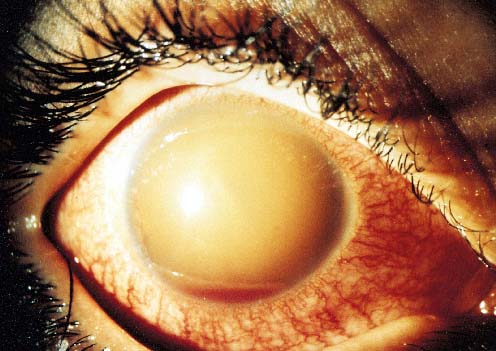
Optic Atrophy
Nonglaucomatous optic atrophy in hyphema patients may be due either to the initial trauma or to transient periods of markedly elevated IOP. Diffuse optic pallor (not glaucomatous cupping) is the result of transient periods of markedly elevated IOP, and occurs with constant pressure of 50 mm Hg or higher for 5 days, or 35 mm Hg or higher for 7 days.1,7 However, a number of patients with sickle cell trait developed a nonglaucomatous optic atrophy with relatively small elevations of IOP of 35 mm Hg to 39 mm Hg for 2 to 4 days1,7,16 (Slide 5). Despite maximum medical therapy, final visual acuity was less than 20/400 in all patients. We continue to observe optic atrophy in patients with sickle cell trait referred to our institution who have not had vigorous control of IOP and/or delay in paracentesis.
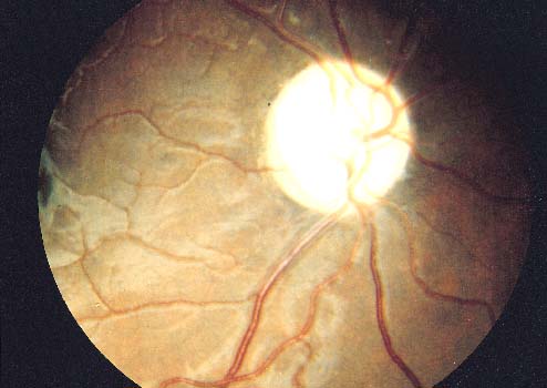
Other studies indicate that patients with sickle cell hemoglobinopathies and anterior chamber hyphemas have a greater ratio of sickled erythrocytes in their anterior chambers than in their circulating venous blood.19 The sickled erythrocytes obstruct the trabecular meshwork more effectively than normal cells and with a consequential elevation of IOP to greater levels with lesser amounts of hyphema. Systemic hypotensive agents may not always be successful in reducing the IOP. They may, in fact, be contraindicated in high or repeated dose regimens, due to their possible contribution to intravascular hemoconcentration and increased microvascular sludging, both of which are detrimental in sickle cell hemoglobinopathy. Possibly, the increased IOP is tolerated less in these patients because of the increased susceptibility to impaired vascular perfusion within the optic nerve and retina. Indeed, moderate elevation of IOP in patients with sickle cell hemoglobinopathy may produce rapid deterioration of visual function due to profound reduction of central retinal artery and posterior ciliary artery perfusion.25
For these African American patients, the prevention of secondary hemorrhage is a critical factor.
Prognosis and Treatment
It is important to recognize that the prognosis for visual recovery is directly related to three factors:
- The amount of associated damage to other ocular structures (i.e., choroidal rupture or macular scarring)
- Occurrence of secondary hemorrhage
- Occurrence of complications of glaucoma, corneal blood staining, or optic atrophy
Treatment modalities should be directed at reducing the incidence of secondary hemorrhage and reducing the risk of corneal blood staining and optic atrophy. The success of hyphema treatment, as judged by recovery of visual acuity, is good in approximately 75% of patients. Approximately 80% of hyphema patients with less than one-third filling of the anterior chamber regain visual acuity of 20/40 (6/12) or better. Of these, 60% of those with greater than half and less than total hyphema regain 20/40 or better; of those with an initial total hyphema, only 35% have good visual results. Sixty percent of hyphema patients younger than 6 years have good visual results; older age groups have progressively higher percentages of good visual recovery.
The severity of the trauma is frequently related to the final visual outcome. Lens opacities, choroidal rupture, vitreous hemorrhage, angle-recession glaucoma, and retinal detachment are commonly associated with traumatic hyphema, compromising the final visual result.
Fourteen percent of patients have poor visual results from associated trauma (i.e., glaucoma, vitreous hemorrhage, retinal detachment, or scleral rupture). Poor visual outcome in traumatic hyphema can be attributed directly to the hyphema in 11% of patients,3,18 usually resulting from secondary hemorrhage associated with optic atrophy or corneal blood staining.
Medical Management
Several studies provide evidence that no statistically significant difference exists in most areas of comparison among patients treated with bed rest, bilateral patches, and sedation, and those treated with ambulation, a patch and shield on the injured eye only, and no sedation.1,3,17 We recommend ambulation and a patch and shield for the injured eye. Sedation is recommended only in the apprehensive or hyperactive pediatric patient. If analgesics are required for the relief of pain, we prefer acetaminophen, codeine, or both, depending on the severity of the pain. The antiplatelet effect of aspirin tends to increase the incidence of rebleeding in traumatic hyphema and should be avoided.26 Nonsteroidal anti-inflammatory drugs with analgesic activity, such as mefenamic acid, also share this antiplatelet effect.
In any therapeutic regimen, the injured globe requires adequate protection with a patch and shield. Elevating the head of the bed to 30° to 45° facilitates settling of the hyphema in the inferior anterior chamber and aids in classifying the hyphema. Inferior settling facilitates more rapid improvement of visual acuity, earlier evaluation of the posterior pole, and greater clearing of the anterior chamber angle. Also, a better estimate of the decrease or increase in the amount of blood in the anterior chamber is possible during subsequent biomicroscopic examinations.
Various topical medications have been recommended for the treatment of traumatic hyphema, including cycloplegics for the traumatic iridocyclitis and miotics to increase the surface area of the iris to enhance resorption of the hyphema.27-29 Topical corticosteroids and estrogens have been recommended with contradictory results.29 A summary of medical management is included in Table 2.
Most topical medications are not necessary because of the lack of definite evidence of therapeutic advantages.1,20 Atropine sulfate 1% is indicated for iritis and to break the pupillary block.27 Topical use of steroids after the fourth or fifth day of retained hyphema may be advantageous to decrease the associated iridocyclitis and to prevent or deter the development of PAS or posterior synechiae.
One of our double-masked studies indicates the value of systemic administration of ACA in the prevention of recurrent hemorrhages.7 If secondary hemorrhages are the result of lysis and retraction of a clot that has produced an occlusion of the traumatized vessel, then prevention of normally occurring clot lysis for 5 or 6 days should be advantageous to allow the injured blood vessel to repair its integrity more completely.1,7,16 The antifibrinolytic activity of ACA given systemically has been demonstrated to decrease the incidence of secondary hemorrhage in other areas of the body.
ACA retards clot lysis by preventing plasmin from binding to lysine molecules in the fibrin clot. ACA, a lysine analogue, competitively inactivates plasmin by occupying the lysine-binding site on plasmin that would normally bind to fibrin. In addition, ACA binds to plasminogen, so that when activated to plasmin, it cannot attach to fibrin. These effects stabilize the clot-vessel-wall interface, decreasing the potential for secondary hemorrhage.1,7,16
Based on the work of Ablondi and colleagues30 and Alkjaersig and colleagues,31 a concentration of ACA of 10 mg/mL to 32.5 mg/mL is required to produce antifibrinolytic effects. These concentrations are achieved in aqueous humor after systemic administration.32
When ACA was administered in a dosage of 100 mg/kg every 4 hours, orally, for 5 full days, a statistically significant reduction in the incidence of rebleeding of traumatic hyphemas was observed.7,33
For systemically administered ACA to be effective, it must penetrate into the anterior segment in sufficient concentration to retard fibrinolysis. To directly determine the concentration of ACA in aqueous humor following systemic administration, using an animal model we compared plasma and aqueous humor concentrations of ACA following intravenous (IV) administration of 50 mg/kg and 100 mg/kg, as well as after constant infusion of 25 mg/kg/hr.32 Plasma levels after administration were 10-fold higher than in the aqueous humor. Antifibrinolytic activity correlated directly with ACA concentration in plasma or aqueous humor. The time to clot dissolution was greatest (2.5 times control) when the ACA concentration in the aqueous humor reached 30 mg/dL to 35 mg/dL, which thus became our target concentration to achieve with topical therapy.
Systemic ACA should be used in patients with hyphemas that occupy 75% or less of the anterior chamber since the clot may persist in the anterior chamber for an increased period during administration of the drug. The continued retention of the clot in the anterior chamber would be a disadvantage with larger, grade 4 hyphemas.
In our prospective study, as well as in two additional studies, patient groups treated with ACA and placebo were randomized and double-masked.7,14,33 In the ACA-treated group, the incidence of secondary hemorrhage varied between 3% and 4%.7,14,33 In the placebo-treated group, the incidence was 28% to 33%. ACA in a dosage of 50 mg/kg every 4 hours is equally as effective as 100 mg/kg every 4 hours, orally, for 5 days.14 The total dosage of ACA should not exceed 30 g per day, and should not be used in pregnant patients or those with renal or hepatic impairment.
Because systemic ACA significantly reduces the incidence of secondary hemorrhage, a topical preparation should decrease the incidence of adverse effects. By concentrating the medication in the aqueous humor, a topical preparation would decrease the systemic concentration of ACA associated with many of the adverse effects. Our long-term goal is to improve the treatment of hyphema by decreasing the incidence of secondary hemorrhage using topical-drug therapy that is more effective, less toxic, and better accepted by both patients and ophthalmologists than the currently available oral therapy with ACA.
Seven preparations containing ACA were studied to assess which could deliver the required amount of ACA into the aqueous humor.34 The greatest ACA concentrations were obtained by using carboxypolymethylene (CPM) 58 mg/dL. Using an experimental model for hyphema, ACA in CPM was applied topically every 6 hours for 6 days or until a secondary hemorrhage occurred.35 Compared with no treatment or the administration of a placebo (e.g., vehicle without ACA), topical application of ACA significantly decreased the incidence of rebleeds (P<.05.). No ocular side effects were noted after topical application of either formulation.
Additional studies have been performed to optimize the concentration of vehicle and drug.36 The optimum combination is 30% ACA to 2% CPM. This combination, however, did not lead to an increase in the duration of action using hyaluronic acid or collagen shields as a depot.37
We established a prospective, multicenter, double-masked, randomized clinical trial comparing oral and topical ACA.16 Sixty-four patients with traumatic hyphema treated with topical or systemic ACA were compared with 54 control patients with hyphema. Compared with the control group, topical and systemic ACA were statistically significant in preventing secondary hemorrhage. Only 3% (2/64) of the patients who received topical or systemic ACA had secondary hemorrhage compared with 22% (12/54) of the control group (P=.002). Final visual acuity was 20/40 or better in 30 patients (86%) in the topical group, compared with 23 patients (43%) in the control group (P=.001). Final visual acuity was 20/40 or better in 20 patients (69%) in the systemic ACA group, compared with 23 patients (43%) in the control group (P=.04). The topical ACA group had a final visual acuity of 20/40 or better in 86% of patients, compared with 69% of patients in the systemic group.
Topical ACA appears to be a safe, effective treatment to prevent secondary hemorrhage in traumatic hyphema. It is as effective as systemic ACA in reducing secondary hemorrhage. No systemic side effects were observed with topical use. Topical ACA provides an effective outpatient treatment for traumatic hyphemas.
Although not approved for ophthalmic use in the United States, another lysine analog, tranexamic acid, also has antifibrinolytic properties. In a series of children treated with tranexamic acid, 25 mg/kg/day, the incidence of secondary hemorrhage was significantly reduced.38 Like ACA, tranexamic acid has been associated with nausea, vomiting, and hypotension. Unlike ACA, tranexamic acid is associated with visual abnormalities, which could complicate ophthalmologic evaluation of the patient. In addition, some patients in this study were also treated with other drugs including topical steroids.
Other investigators have suggested that systemic steroids decrease the incidence of secondary hemorrhage. Initial studies supporting this claim were neither randomized nor double-masked.28,39 A randomized, double-masked, prospective study by Spoor and colleagues showed a secondary hemorrhage of 20% in controls and 13% in treated patients, not statistically significant.40 Farber and colleagues compared treatment of oral ACA with oral prednisone in a well-controlled trial.41 Their study suggested that both drugs decrease the incidence of secondary hemorrhage by a similar amount, albeit by different mechanisms.
Due to the small number of rebleeds, the confidence limits were large and may have masked a real difference. The major difficulty with this study was that there were no controls. The lack of a true control population is unfortunate in comparing the two groups. In addition, the study excluded all patients with sickle-cell trait. These patients should be considered for topical or systemic ACA or systemic corticosteroid treatment. In addition, patients with gastric ulcer or diabetes mellitus, those who were intoxicated, and those who had bleeding were excluded.
The pharmacologic mode of action of the prednisone is unclear and may be related to an anti-inflammatory influence on traumatized blood vessels with reduced engorgement and a propensity for rebleeding. Additional randomized studies with controls would be helpful to determine whether a significant reduction of secondary hemorrhage with systemic prednisone occurred in comparison with systemic ACA.
Some studies have investigated the application of intracameral tissue plasminogen activator (t-PA) in the management of traumatic hyphema.42 However, these studies have been neither large nor randomized. A potential risk with t-PA is the associated risk of developing rebleeding of the initial wound. The application has been considered in resolving hyphemas that either fail to clear spontaneously or are associated with malignant IOP.43 The actual timing of t-PA administration from initial injury has yet to be determined.
Generally, medical management seems to produce the best visual result for patients with less than 75% hyphema. Surgical intervention is rarely indicated for hyphemas that occupy less than half of the anterior chamber; these lesser hyphemas (either primary or secondary) usually resolve spontaneously under medical regimen and require no surgical intervention. In two prospective series totaling 196 patients, no corneal blood staining or optic atrophy was noted in hyphemas of 50% or less.1,3,7 Corneal blood staining occurs primarily in patients with hyphemas that are larger than 75% at some time during their course.
The results of surgical evacuation to improve secondary glaucoma in the small hyphemas (75% or less) are disappointing. The ocular hypertension in these instances results more frequently from damage to the trabecular structures than from plugging by red cells and fibrin. Surgical evacuation in these instances may produce only temporary postsurgical hypotony, with a rapid return to preoperative IOP. Topical antiglaucomatous medications may achieve lowering of IOP. Beta-blockers and topical carbonic anhydrase inhibitors are started followed by other topical agents.
If IOP is still uncontrolled, systemic medication should be given during the acute phase of the hyphema. Acetazolamide, 20 mg/kg/day, may be administered in four divided doses for IOP higher than 22 mm Hg. Acetazolamide can increase the concentration of anterior chamber ascorbate. It lowers the pH of human plasma and exacerbates sickling of erythrocytes. Therefore, methazolamide 10 mg/kg/day administered in four divided doses is preferred in pediatric patients with sickle-cell trait or sickle-cell disease.7,19 Osmotic agents (preferably mannitol) should be considered for pressures higher than 35 mm Hg. Orally administered glycerol is effective; however, nausea and vomiting are often associated with its administration in patients with elevated IOP. Mannitol is given intravenously, 1.5g/kg (usually in a 10% solution), over a period of approximately 45 minutes. This may be given two times a day (or every 8 hours in patients with extremely high IOP) in an attempt to keep the IOP below 35 mm Hg. Renal output, blood urea nitrogen, and electrolyte values should be monitored in all patients in whom such therapy is continued for several days.
Outpatient Care vs Hospitalization
With increasing emphasis on cost containment, outpatient management of hyphema has become more popular in recent years. Several studies have demonstrated no significant difference in final visual acuities in patients treated at home versus those treated in a hospital.1,16,44-46 Indications for hospital admission are shown in Table 2.16
Patients with traumatic hyphema with microhyphemas can be treated on an outpatient basis with systemic ACA and other topical or systemic medical regimens. If the hyphema occupies greater than 50% of the anterior chamber or the IOP is elevated, or both, hospitalization is recommended. The decision to hospitalize also depends on the cooperation of the patient, family members, and extent of ocular injury. Daily ocular examinations should be performed and should include an evaluation of the amount of hyphema and IOP. Daily ophthalmic sketches are helpful in estimating the amount and rate of resolution or rebleeding. Applanation tonometry must be performed at least once daily, twice daily in patients with elevated IOPs.
Daily corneal examination should include careful inspection with the biomicroscope for corneal blood staining. Minimal blood staining is often difficult to detect against a background of blood in the anterior chamber. Under such circumstances, the cornea often assumes a yellowish cast, which is reflected from the yellowish fibrinous coagulum in the anterior chamber. The most typical early sign of corneal blood staining is the presence of tiny yellowish granules that initially appear in the posterior third of the corneal stroma. An additional finding is a lack of definition or a blurred appearance of the ordinarily sharply defined fibrillar structure of the involved corneal stroma. The latter is independent of the yellowish color transmitted to the stroma by the contents of the anterior chamber.
We have found this sign useful in recognizing the very early stages of corneal blood staining. These biomicroscopic signs of corneal blood staining usually precede gross staining by only 24 to 36 hours. Surgical treatment in this early stage may prevent gross staining, and the cornea may clear in 4 to 6 months. However, once grossly visible staining develops, many months may elapse before clearing is complete.
Surgical Intervention
We believe that most hyphemas, including total hyphema, should be treated medically for the first 4 days. Spontaneous resolution of the hyphema occurs rapidly during this period, and these cases have the best prognosis. In one series of 20 eyes with total hyphemas, four of the 20 eyes (20%) cleared sufficiently by day 4 to rule out surgery.18 An additional four eyes resolved spontaneously on medical treatment over a longer period. Generally, with the exception of patients with sickle-cell trait or anemia, surgery is seldom indicated for hyphema less than 50%. Our indications for surgical intervention are shown in (Table 3).1,17,18
It is best not to delay surgery if the IOP remains elevated at 50 mm Hg or higher for 4 days. One study noted optic atrophy in 50% of patients with total hyphemas when surgery was delayed. Corneal blood staining occurred in 43% of patients.47
Patients with sickle-cell hemoglobinopathies and even those with sickle-cell trait, require surgical intervention if IOP is not controlled within 24 hours.7,16,19 Surgery for hyphema should be cautiously approached. In two series involving 196 patients, surgery was performed in only 14 patients (7.1%).1,3 Risks of surgery include damage to corneal endothelium, lens, and/or iris, prolapse of intraocular contents, rebleeding, and increased synechiae formation. With the exception of patients with sickle-cell trait, no patients in our series required surgery if the hyphema occupied less than 50% of the anterior chamber.
Historically, various surgical methods have been recommended to remove the hyphema. Paracentesis and enzymatic digestion of the clot with fibrinolysin or urokinase have been advocated.48-49 A 90° to 120° limbal incision with manual expression of the clot also has been recommended.50 The recommended time for manual expression of the clot has varied from the time of onset of the total hyphema and secondary glaucoma to the fourth day following the eight-ball hyphema. As to the recommended time for surgical intervention, histologic studies of the clot from an eight-ball hyphema suggest and support intervention on the fourth day of a total hyphema.51 Lysis of the clot periphery at this time favors removal on the fourth day to avoid organization of the periphery of the clot to anterior chamber structures. Total hyphema evacuation by vitrectomy instrumentation and peripheral iridectomy procedures have also been recommended.52-55
Generally, we recommend the type of surgical intervention with which the surgeon is most familiar. The four major approaches are listed in Table 4. Hyphema surgery should be preceded by IV acetazolamide and mannitol, if the IOP is elevated above 35 mm Hg. The procedure should be performed under general anesthesia in all patients if medically feasible.
The technique that is preferred at present is evacuation of the hyphema with vitrectomy instrumentation. The initial clear corneal incision is made with a diamond blade. To avoid the iris and lens, the blade is oriented and pushed into the anterior chamber in such a manner that it is parallel to the plane of the iris. A 20-gauge instrument attached to an infusion line of balanced salt solution plus is then fitted into the anterior chamber. The balanced salt solution bottle should be between 30 cm and 40 cm above the eye to maintain normal IOP.
With the vitrectomy cutting port half open and the infusion line in place, it is possible to irrigate and aspirate free blood from the formed clot. The aspiration mode is initially set at 200 mm Hg vacuum and the cutting speed set at 200 cycles per minute for the procedure. Aspiration mode is increased to 400 mm Hg as needed. Extreme care is required to avoid contact with any iris, lens, or corneal endothelium. This operative procedure is used to remove the central portion of the clot. It is not necessary to remove the entire clot in the periphery of the anterior chamber.
If a secondary hemorrhage occurs during the operative procedure, we recommend tamponade of the bleeding by elevation of the infusion bottle to approximately 70 cm above the eye for several minutes. If the bleeding continues, it is helpful to fill the anterior chamber with sodium hyaluronate after evacuating the clot to tamponade the bleeding site. If bleeding persists, bimanual bipolar diathermy is helpful when the bleeding site is visible.56
At the end of the surgical procedure, fill the anterior chamber with an air bubble. This will also help control any secondary bleeding. Close the corneal incision with two 10-0 nylon sutures. The response in lowering IOP with the vitrectomy instrumentation has been successful. Each eye operated on with this technique has shown an initial decrease in IOP associated with the surgery.
Paracentesis causes little surgical trauma and relieves the elevated IOP. Paracentesis is especially beneficial in patients with sickle-cell trait or disease. However, the decrease in IOP may be transient, and there may be no appreciable reduction in the amount of the formed clot.
Irrigation by a single- or double-needle technique has the advantage of a small incision. We prefer entry at the 1 o'clock position in the right eye and at the 11 o'clock position in the left eye with a diamond blade. The entry should be through clear cornea. The irrigating needle should then extend just through the corneal endothelium and a slow push-pull maneuver with the single-needle technique washes out the erythrocytes from the anterior chamber clot, often leaving the fibrin matrix. To reduce the likelihood of rebleeding during the operative procedure, care should be undertaken not to produce violent alterations in the anterior chamber pressure. If rebleeding does occur, sodium hyaluronate can be effectively introduced for tamponade. After a 5-minute wait, irrigation maneuvers can be resumed.
Using a one- or two-needle technique, the surgeon must be particularly careful to have direct visualization of the anterior chamber, but this technique has some disadvantages. Maintaining the position of the needle tip in the anterior chamber may be difficult during the procedure. A hazardous situation is created when the collar-button type of formed clot occupies both the anterior and posterior chambers. This produces pupillary block with anterior displacement of the iris-lens diaphragm.50
A fourth technique is clot removal with trabeculectomy.1,16,47,57 Generally, trabeculectomy is not used in smaller hyphemas. Trabeculectomy with anterior chamber washout and peripheral iridectomy is a beneficial primary surgical treatment for traumatic hyphema unresponsive to medical management with antiglaucomatous agents. Trabeculectomy provides a means to keep IOP lowered while the remaining blood is cleared from the anterior chamber. In patients with total hyphema, trabeculectomy with peripheral iridectomy should be considered. Trabeculectomy is performed with gentle irrigation of the anterior chamber hyphema. It is relatively safe and should be performed early for cases of total hyphema unless the elevated IOP is controlled medically and resolution of the hyphema is clearly imminent.
We use this technique in patients with total hyphema at day 4 and find this superior to clot evacuation. Several patients referred to our institution have had attempts at clot evacuation. One patient sustained complete iridodialysis related to attempted clot evacuation. In addition, we have had other patients who have been referred after optic atrophy developed with total hyphemas. When the trabeculectomy surgery is performed, we use a 3 x 3 mm scleral trap door, hinged at the limbus. Superficial episcleral vessels are coagulated with the bipolar cautery. A superficial lamellar flap is developed through one-third scleral thickness creating the trap door hinged at the limbal area. Peripheral iridectomy is performed followed by gentle irrigation of the clot in the area of the trabeculectomy site. Two 10-0 nylon sutures are used to close the trabeculectomy site. The conjunctiva is closed with a running 7-0 or 8-0 vicryl suture. Once the conjunctiva has healed, the nylon suture(s) can be lasered to open the trabeculectomy site when necessary. This technique has been invaluable in the difficult total hyphema cases. Each of these surgical procedures has its own set of complications. The surgeon should approach each patient with caution and individualize the surgical approach.
References
- Crouch ER Jr, Williams PB. Trauma: Ruptures and bleeding. In: Tasman W, Jaeger EM, eds. Duane's Clinical Ophthalmology. Philadelphia, Pa: JB Lippincott; 1993;4:1-18.
- Blanton FM. Anterior chamber angle recession and secondary glaucoma: A study of the after effects of traumatic hyphemas. Arch Ophthalmol. 1964;72:39-44.
- Read JE, Goldberg MF. Traumatic hyphema: Comparison of medical treatment. Trans Am Acad Ophthalmol Otolaryngol. 1974;78:799-815.
- Milauskas AT, Fueger GF. Serious ocular complications associated with blowout fractures of the orbit. Am J Ophthalmol. 1966:62:670-672.
- Edwards WC, Layden WE. Traumatic hyphema. A report of 184 consecutive cases. Am J Ophthalmol. 1973;75:110-116.
- Thach AB, Ward TP, Hollifield RD, et al. Ocular injuries from paintball pellets. Ophthalmology. 1999;106:533-537.
- Crouch ER Jr, Frenkel M. Aminocaproic acid in the treatment of traumatic hyphema. Am J Ophthalmol. 1976;81:355-360.
- Parver LM, Dannenberg AL, Blacklow B, Fowler CJ, Brechner RJ, Tielsch, JM. Characteristics and causes of penetrating eye injuries reported to the National Eye Trauma System Registry, 1985-1991. Public Health Rep. 1993;108:625-630.
- Hoskins HD. Secondary glaucoma. In: Heilman K, Richardson KT, eds. Glaucoma: Conceptions of a Disease, Pathogenesis, Diagnosis, Therapy. Philadelphia, Pa: WB Saunders; 1978:376-389.
- Podolsky M, Srinivasan B. Spontaneous hyphema secondary to vascular tuft of pupillary margin of the iris. Arch Ophthalmol. 1979;97:301-302.
- Campbell D. Ghost cell glaucoma following trauma. Ophthalmology. 1981;88:1151-1158.
- Pilger IS. Medical treatment of traumatic hyphema. Surv Ophthalmol. 1975;20:28-34.
- Spoor TC, Kwitko GM, O'Grady JM, Ramocki JM. Traumatic hyphema in an urban population. Am J Ophthalmol. 1990;109:23-27.
- Palmer DJ, Goldberg MF, Frenkel M, Fiscella R, Anderson, RJ. A comparison of the two dose regimens of epsilon aminocaproic acid in the prevention and management of secondary traumatic hyphemas. Ophthalmology. 1986;93:102-108.
- Kutner B. Fourman S. Brein K, et al. Aminocaproic acid reduces the risk of secondary hemorrhage patients with traumatic hyphema. Arch Ophthalmol. 1987;105:206-208.
- Crouch ER Jr, Williams PB, Gray MK, Crouch ER, Chames M. Topical aminocaproic acid in the treatment of traumatic hyphema. Arch Ophthalmol. 1997;115:1106-1112.
- Fourman S. Topical aminocaproic acid in the treatment of patients with traumatic hyphema. Arch Ophthalmol. 1998;116:395-396.
- Read J. Traumatic hyphema: Surgical vs medical management. Ann Ophthalmol. 1975;7:659-670.
- Goldberg MF. The diagnosis and treatment of sickled erythrocytes in human hyphemas. Trans Am Ophthalmol Soc. 1978;76:481-501.
- Crouch ER. Traumatic hyphema. J Pediatr Ophthalmol Strabismus. 1986;23:95-97.
- Darr JL, Passmore JW. Management of traumatic hyphema. Am J Ophthalmol. 1967;63:134-136.
- Fong LP. Secondary hemorrhage in traumatic hyphema. Predictive factors for selective prophylaxis. Ophthalmology. 1994;101:1583-1588.
- Cho J, Jun BK, Lee YJ, Uhm KB. Factors associated with the poor final visual outcome after traumatic hyphema. Korean J Ophthalmol. 1998;12:122-129.
- Rahmani B, Jahadi HR, Rajaeefard A. An analysis of risk for secondary hemorrhage in traumatic hyphema. Ophthalmology. 1999;106:380-285.
- Michelson PE, Pfaffenbach D. Retinal arterial occlusion following ocular trauma in youths with sickle-trait hemoglobinopathy. Am J Ophthalmol. 1972;74:494-497.
- Crawford JS, Lewandowski RL, Chan W. The effect of aspirin on rebleeding in traumatic hyphema. Am J Ophthalmol. 1975;80:543-545.
- Gilbert HD, Jensen AD. Atropine in the treatment of traumatic hyphema. Ann Ophthalmol. 1973;5:1297-1300.
- Yasuna E. Management of traumatic hyphema. Arch Ophthalmol. 1974;91:190-191.
- Spaeth GL, Levy PM. Traumatic hyphema: Its clinical characteristics and failure of estrogens to alter its course. A double-blind study. Am J Ophthalmol. 1966;62:1098-1106.
- Ablondi FB, Hagan JJ, Philips M, DeRenzo EC. Inhibition of plasmin, trypsin and the streptokinase-activated fibrinolytic system by e-ACA. Arch Biochem Biophys. 1959;82:153-159.
- Alkjaersig N, Fletcher P, Sherry S. E-ACA: An inhibitor of plasminogen activation. J Biol Chem. 1959;234:832-837.
- Loewy DM, Williams PB, Crouch ER Jr, Cooke WJ. Systemic aminocaproic acid reduces fibrinolysis in the aqueous humor. Arch Ophthalmol. 1987;105:272-276.
- McGetrick JJ, Jampol LM, Goldberg MF, Frenkel M, Fiscella RG. Aminocaproic acid decreases secondary hemorrhage after traumatic hyphema. Arch Ophthalmol. 1983;101:1031-1033.
- Allingham RR, Williams PB, Crouch ER Jr, Loewy DM, Demkowski HC. Topically applied aminocaproic acid concentrates in the aqueous humor of the rabbit in therapeutic levels. Arch Ophthalmol. 1987;105:1421-1423.
- Allingham RR, Crouch ER Jr, William PB, Catlin JC, Loewy DM, Jacobson J. Topical aminocaproic acid significantly reduces the incidence of secondary hemorrhage in traumatic hyphema in the rabbit model. Arch Ophthalmol. 1988;106:1436-1438.
- Ehlers WH, Crouch ER Jr, Williams PB, Riggs PK. Factors affecting therapeutic concentration of topical aminocaproic acid in traumatic hyphema. Invest Ophthalmol Vis Sci. 1990;31:2389-2394.
- Mattox C, Williams PB, Crouch ER Jr, et al. Aqueous humor concentrations after use of reservoir systems for topical delivery of aminocaproic acid. Invest Ophthalmol Vis Sci. 1991;32:1293.
- Deans R. Noel LP, Clarke WN. Oral administration of tranexamic acid in the management of traumatic hyphema in children. Can J Ophthalmol. 1992;27:181-183.
- Rynne MV, Romano PE. Systemic corticosteroids in the treatment of traumatic hyphema. J Pediatr Ophthalmol Strabismus. 1980;17:141-143.
- Spoor TC, Hammer M, Belloso H. Traumatic hyphema. Failure of steroids to alter its course: A double-blind prospective study. Arch Ophthalmol. 1980;98:116-119.
- Farber M, Fiscella R, Goldberg MF. Aminocaproic acid versus prednisone for the treatment of traumatic hyphema. A randomized clinical trial. Ophthalmology. 1991;98:279-286.
- Laatikainen L, Mattila J. The use of tissue plasminogen activator in post-traumatic total hyphaema. Graefe's Arch Clin Exp Ophthalmol. 1996:234:67-68.
- Starck T. Hopp L, Held KS, Marouf LM, Yee RW. Low-dose intraocular tissue plasminogen activator treatment for traumatic total hyphema, postcataract, and penetrating keratoplasty fibrinous membranes. J Cataract Refract Surg. 1995;21:219-224.
- Kennedy RH, Brubaker RF. Traumatic hyphema in a defined population. Am J Ophthalmol. 1988;106:123-130.
- Wright KW, Sunalp M, Urrea P. Bed rest versus activity ad lib in the treatment of small hyphemas. Ann Ophthalmol. 1988;20:143-145.
- Witteman GJ, Brubaker SJ, Johnson M, Marks RG. The incidence of rebleeding in traumatic hyphema. Ann Ophthalmol. 1985;17:525-529.
- Weiss JS, Parrish RK, Anderson DR. Surgical therapy of traumatic hyphema. Ophthalmic Surg. 1983;14:343-345.
- Scheie HG, Ashley BJ Jr, Burns DT. Treatment of total hyphema with fibrinolysin. Arch Ophthalmol. 1963;69:147-153.
- Leet DM. Treatment of total hyphemas with urokinase. Am J Ophthalmol. 1977;84:79-84.
- Sears ML. Surgical management of black ball hyphema. Trans Am Acad Ophthalmol Otolaryngol. 1970;74:820-825.
- Wolter JR, Henderson JW, Talley TW. Histopathology of a black ball blood clot removed four days after total traumatic hyphema. J Pediatr Ophthalmol Strabismus. 1971;8:15-18.
- McCuen BW, Fung WE. The role of vitrectomy instrumentation in the treatment of severe traumatic hyphema. Am J Ophthalmol. 1979;88:930-934.
- Stern WH, Mondal KM. Vitrectomy instrumentation for surgical evacuation of total anterior chamber hyphema and control of recurrent anterior chamber hemorrhage. Ophthalmic Surg. 1979;10:34-37.
- Wilson FM. Traumatic hyphema: Pathogenesis and management. Ophthalmology. 1980;87:910-919.
- Diddie KR, Dinsmore S, Murphree AL. Total hyphema evacuation by vitrectomy instrumentation. Ophthalmology. 1981;88:917-921.
- Michels RG, Rice TA. Bimanual bipolar diathermy for treatment of bleeding from the anterior chamber angle. Am J Ophthalmol. 1977;84:873-874.
- Graul TA, Ruttum MS, Lloyd MA, Radius RL, Hyndiuk RA. Trabeculectomy for traumatic hyphema with increased intraocular pressure. Am J Ophthalmol. 1994;117:155-159.