Jonathan R. Pirnazar, MD · Peter J. McDonnell, MD
Introduction
Close attention to detail is crucial for a surgeon when performing laser in situ keratomileusis (LASIK). In this manner, the surgeon can anticipate and avoid potential complications. This discussion reviews the common and uncommon complications that have been associated with LASIK, with advice on how to avoid and manage complications when they do occur.
Intraoperative Complications
Inadequate exposure
Inadequate exposure can cause difficulty in achieving suction, which
can result in an incomplete microkeratome pass when making the LASIK
flap. Patients with prominent brows or deep-set eyes may pose difficulties
with exposure. Poor draping technique can cause the same problem.
If adequate exposure cannot be achieved, the assistant should apply
downward pressure to the speculum during the microkeratome pass. Rarely,
a modified van Lint block or a lateral canthotomy is required for adequate exposure.
Inadequate suction
Inadequate suction is usually due to incomplete exposure. However,
small corneal diameters may allow the conjunctiva to block the suction
port of the suction ring. Manipulations of the ring can potentially
decrease suction. An optimal cut by the microkeratome requires an
intraocular pressure (IOP) greater then 65 mm Hg. IOPs less than 65
mm Hg can lead to thin flaps or buttonholes.
Microkeratome Complications
- Thin flaps and buttonholes. Thin flaps and buttonholes (Slide 1) may result from inadequate suction. Also, corneas that are steeper than average (keratometry readings greater than 46 D) may be more prone to buckling because the excess tissue is compressed by the footplate of the microkeratome. The potential result is a thin or buttonhole flap. A poor-quality blade may cause a flap complication, where there are irregular cuts with thin areas. If a buttonhole forms, the procedure should be stopped and the flap should be replaced while carefully managing the epithelial edges. The flap may be recut with a thicker depth plate after 3 to 6 months.
- Free caps. During LASIK, large, flat corneas with keratometry readings less than 41 D are at risk of a free cap. If this occurs, the cap should be placed in an antidesiccation chamber while the ablation is performed. It is crucial that the cap is placed stromal side down. Once the cap is replaced, a few drops of balanced salt solution should be inserted under the flap while holding it with forceps, and the excess fluid should be carefully removed. It is important to ensure that the flap has adhered by waiting 3 to 5 minutes (sutures may not be required). A bandage soft contact lens can be inserted if the epithelium is disrupted.
- Incomplete pass. Incomplete passes can be due to poor exposure, debris on the field, improper assembly, or inadequate cleaning of the microkeratome. When a partial flap occurs, the surgeon must decide whether to proceed. If the pass stops close to the pupil or is within the central 6 mm to 7 mm of the cornea, then the flap should be repositioned and no ablation should be performed. The flap may be recut after 3 to 6 months. If the pass stopped near the edge of the ablation, the hinge can be protected with a sponge and the ablation can proceed.
- Corneal perforation. Corneal perforation is rare and devastating. It has been known to occur with improper microkeratome assembly. Management depends on the depth of the cut. There have been reported cases where the patient required corneal transplantation, in some instances with intraocular lens implantation, as well.
Flap Complications
- Wrinkled flap.The flap should be positioned carefully to avoid wrinkles. Causes of wrinkles (Slide 2) include excessive drying of the flap, as well as rough handling of the flap, leading to stretching and microwrinkles when repositioning after ablation. Wrinkles can lead to irregular astigmatism and visual disturbances. To prevent wrinkles, a wet sponge should be used to smooth the flap with motions away from the flap center. Also, no fluid should remain under the flap. If wrinkles are evident intraoperatively, the flap can be refloated and smoothed with a wet sponge.
- Interface debris.Debris results from poor irrigation. Central debris calls for lifting of the flap and irrigation. Metallic particles usually originate form the keratome blade and are difficult to remove. The particles usually do not cause visual problems and do not need to be removed if they are off-axis.
- Flap edema.Prolonged irrigation of the flap should be avoided because it can lead to flap edema, retraction of the flap edges, and poor adherence. It may be necessary to stretch the edematous flap into place using a dry sponge.
- Flap shrinkage. Flap shrinkage is due to excessive drying of the flap before repositioning. It may be necessary to stretch the flap. Rarely, sutures may be required to position the flap.
- Intraoperative bleeding. Patients with a superficial corneal pannus related to contact lens wear will be more prone to bleeding. Should blood enter the stromal bed, it should be wiped away carefully with a sponge. After the ablation, any blood around the bed should be irrigated.
- Epithelial complications.Irregular epithelium and superficial
punctate keratopathy (SPK) must be managed before LASIK surgery.
If the SPK is related to dry eye disease, then aggressive artificial
tears and/or punctal occlusion may be necessary preoperatively.
It is important to diagnose diseases associated with recurrent erosions,
such as map-dot-fingerprint dystrophy. Patients with these conditions
may benefit more from photorefractive keratectomy (PRK) rather than LASIK.
If epithelial defects occur after the keratome pass, the loose epithelium should be smoothed carefully into place after repositioning the flap, being careful to avoid epithelial debris in the flap interface. If there is any epithelial compromise, a bandage contact lens can be inserted in the eye. It will provide relief to the patient and prevent further loosening of the epithelium due to eyelid movement.
Decentration
Decentered ablations can lead to irregular astigmatism and cause symptoms
of glare, night vision problems, ghosting, and diplopia. To have good
centration, the patient must be alert and maintain good fixation. Coaching
your patient on where to look is often helpful. Newer excimer lasers
have pupil trackers, which can assist in producing well-centered ablations.
Early Postoperative Complications
Pain
Pain after LASIK is usually not as severe as with PRK. After LASIK,
most patients have a mild foreign body sensation and experience
some tearing. Pain after LASIK may indicate a flap or epithelial disruption.
Infections
Infectious keratitis is a rare complication of LASIK. Patients
with blepharitis should be treated adequately prior to LASIK. Excessive
irrigation should be avoided to prevent debris from the cul-de-sac
from entering underneath the flap.
The clinical presentation of infectious keratitis following LASIK can range from corneal infiltrates to lamellar abscess formation. Bacterial or fungal organisms have been isolated. The preferred treatment is intensive topical antibiotic drops after culturing and gram-staining the infiltrate. Some infiltrates reportedly have been culture-negative or sterile. Reactivation of herpes simplex virus (HSV) has also been reported following LASIK. A history of HSV is a contraindication to LASIK.
Diffuse Lamellar Keratitis
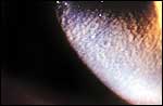
Diffuse lamellar keratitis (DLK), or sands of the Sahara syndrome
(Slide 3) ,
is a noninfectious inflammation involving the interface. DLK presents
1 to 5 days after LASIK. Patients may have decreased visual acuity
or mild irritation. Slit lamp exam reveals fine granularity limited
to the interface with no flap or stromal extension. The presence
of leukocytes and the absence of organisms can be seen on gram stain.
The cause of DLK is multifactorial. Possible causes include contaminants from instruments or sterilizers, bacterial endotoxins or exotoxins, rust or lubricant from the microkeratome, bacterial components from the eyelid margin, overlying epithelial defects, and red blood cells.
There are four grades of DLK:
- Grade 1 (Slide 4): White granular cells are present in the periphery of the interface. There is usually no decrease in best-corrected visual acuity. Recommended treatment is prednisolone acetate 1% every waking hour.
- Grade 2 (Slide 5) : White granular cells are present in the center and periphery of the interface. There is no decrease in best-corrected visual acuity. Similar to grade 1 DLK, treatment is prednisolone acetate 1% every waking hour. Oral prednisone may be considered, as well.
- Grade 3 (Slide 6) : Dense white aggregates of cells appear centrally with relative clearing peripherally. Best-corrected visual acuity is decreased more than 1 line. Recommended treatment includes prednisolone acetate 1% every waking hour, oral prednisone, and manually lifting the flap to irrigate.
- Grade 4 (Slide 7A and Slide 7B) : Permanent stromal scarring is seen with variable degrees of stromal melting. Best-corrected visual acuity is decreased, and there may be induced irregular astigmatism and hyperopia. No treatment has been proven to help. The treatment for grade 3 DLK may be considered.
Interface Debris
Inflammation secondary to interface debris should be removed
and irrigated, if it is believed to be decreasing best-corrected visual acuity.
Wrinkles and Folds
Peripheral wrinkles that are not in the visual axis and not affecting
best-corrected visual acuity may be disregarded. However, if wrinkles
in the visual axis of the flap exist and the best-corrected visual
acuity is reduced, then the flap should be repositioned. If the
wrinkles are localized to one quadrant, then only that area must
be refloated. If the wrinkles are central, then the whole flap must
be refloated. A Paton spatula can be used to wipe back all of the
epithelium that may have migrated onto the exposed stromal bed,
in order to prevent epithelial ingrowth.
Shifted and Dislodged Flaps
Shifted flaps are a rare occurrence after LASIK. These shifted
flaps are usually caused by flap dryness due to poor blinking, stromal
edema for excessive irrigation, epithelial trauma, or eye rubbing.
The chance of a displaced flap may be eliminated by requiring patients
to wear moisture chambers following surgery.
Late Postoperative Complications
Epithelial Ingrowth
Epithelial ingrowth (Slide 8) is usually noted within the first few weeks after
surgery and occurs most commonly after a flap complication or enhancement
surgery. Rates of epithelial ingrowth vary widely from 1.5% to 31.4%.
Most cases of epithelial ingrowth occur at the peripheral edge of
the flap and do not expand toward the center. This type of ingrowth
does not require treatment. However, epithelial cells can migrate
into the visual axis, decreasing best-corrected visual acuity, inducing
irregular astigmatism, causing ocular irritation, and worst of all,
predisposing a patient to stromal melts.
The hallmark of management of epithelial ingrowth is prevention: it is important to pay meticulous attention to all torn epithelium at the edges of the flap and clear any epithelium or debris from the stromal bed before repositioning the flap. Also, avoid large transition zones on small beds and shield the hinge area as necessary. Visually significant ingrowth or an area of epithelial ingrowth greater than 1 mm to 2 mm requires treatment. The flap can be lifted after clearing the edges of epithelial tags or debris with the Paton spatula, and then the stromal and undersurface of flap edges are debrided using a clean sponge for each pass.
Stromal Melts
Stromal melts are rare. These melts are caused by epithelial ingrowth,
DLK, or inflammatory interface debris. The underlying etiology of
keratolysis is not known. It is believed that flap stromal deprivation
from glucose is the cause. Therefore, in the case of epithelial ingrowth,
the ingrowth acts as a mechanical barrier in the interface between
the flap stroma and the glucose-rich aqueous humor. Inflammatory cytokines
have also been proposed.
Overcorrection and Undercorrection
Enhancements for over- or undercorrections should be performed at
least 3 months postoperatively with stable manifest refractions 1
month apart. Obtaining accurate preoperative measurements is the most
effective way to avoid unnecessary enhancements. The surgeon should
be constantly reassessing and adjusting the nomogram in accordance
to postoperative outcomes. Most, if not all, nomograms depend on the
type of laser; the amount of corneal tissue hydration; the humidity
and the temperature of the laser suite; the physical location of the
laser; and possibly the patient's age. Algorithms for LASIK differ
from PRK due to differences in ablation rates of Bowman membrane and stroma.
Regression
Regression is less common following LASIK than with PRK, possibly
due to the preservation of Bowman membrane. High myopia, hyperopia,
and high astigmatism are prone to regression following primary LASIK.
There is no evidence that prophylactic steroids prevent regression.
Haze
Haze is also less common following LASIK than PRK. Interface haze
can respond to topical steroids and may be due to toxins in the fluids
used intraoperatively.
Central Islands
Central islands appear to be due to the older software of broad beam
lasers systems and have been associated with increasing the diameter
of the optical treatment zone. Uneven stromal hydration and/or obstruction
to the laser energy plume may also play a role. Rarely with flying-spot
or scanning-slit lasers have central islands occurred. Central islands
have a tendency to resolve more readily after PRK than with LASIK.
Irregular Astigmatism
Wrinkles or folds in the flap are the most common causes of irregular
astigmatism following LASIK. Flap displacement, central islands, decentered
ablations, and epithelium or debris in the interface also can cause
irregular astigmatism. Irregular astigmatism can be a major cause
of reduced best-corrected visual acuity. Treating the underlying cause
should be the goal of retreatment to correct for the irregular astigmatism.
Small Optical Zone
Patients with large scotopic pupils and high corrections are at risk
for halos and glare at night. A patient with a thin cornea and a high
correction may not be suitable for LASIK if the calculated residual
stromal bed of 250 µm cannot be achieved. If the calculated stromal
bed is less than 250 µm, there is a risk of keratectasia. If the cornea
is thick enough, the optical zone may be enlarged. However, the surgeon
must be aware that when the optical treatment zone is enlarged, the
depth of the ablation increases accordingly.
Decentration
Decentration may present with loss of best-corrected visual acuity,
irregular astigmatism, or ghosting and night visual disturbances.
Decentration can be due to poor fixation on the part of the surgeon
or patient. Custom ablations or topography-guided laser ablations
may assist in the treatment of decentration. Generally, it may be
difficult to surgically treat decentration. Rigid gas permeable contact
lens may be of benefit.
Keratectasia
Keratectasia usually occurs when the residual stromal bed of less
than 250 µm after laser ablation has been violated. Most cases have
occurred following LASIK when the residual stromal bed was 200 µm
or in cases of forme fruste keratoconus. However, there have been
reports in which keratectasia has occurred in residual stromal beds
greater than 250 µm.
Some surgeons believe that excessive thinning of stromal bed may predispose the cornea to biomechanical weakening, causing posterior keratoconus that may not be apparent until months after LASIK. Others have proposed that IOP may be a factor in causing keratectasia. Managing keratectasia is difficult. Rigid gas permeable contact lenses are the first line of management. In severe cases involving loss of best-corrected visual acuity, penetrating keratoplasty may be the only option.
Bibliography
Castillo A, Diaz-Valle D, Gutierrez AR, Toledano N, Romero F. Peripheral melt of flap after laser in situ keratomileusis. J Refract Surg. 1998;14:61-63.
Csker S, Er H, Kahvecioglu C. Laser in situ keratomileusis to correct hyperopia from +4.25 to +8.00 diopters. J Refract Surg. 1998;14(1):26-30.
Gimbel HV, Penno EEA, eds. LASIK Complications: Prevention and Management. Intraoperative complications. 2001.
Lineberger EJ, Hardten DR, Chu RY, Lindstrom RL. Understanding the time course of DLK can eliminate visual loss. Ocular Surgery News. 1999;17(17):11-12.
Maloney RK, Sterile interface inflammation after laser in situ keratomileusis: Experience and opinions. J Refract Surg. 1998;14:661.
Smith RJ, Maloney RK. Diffuse lamellar keratitis. A new syndrome in lamellar refractive surgery. Ophthalmology. 1998;105:1721-1726.
Stulting RD, Carr JD, Thompson KP, et al. Complications laser in situ keratomileusis for the correction of myopia. Ophthalmology. 1998;106:13-20.
Wang Z, Chen J, Yang B. Posterior corneal surface topographic changes after laser in situ keratomileusis are related to residual corneal bed thickness. Ophthalmology. 1999;106:406-409.