Introduction
Juvenile open-angle glaucoma (JOAG) is a form of primary open-angle glaucoma affecting people early in life. JOAG is diagnosed in patients 3 to 20 years old. There is no known racial or sexual predilection. In the United States, the prevalence of JOAG is thought to be 1 in 50,000 people. The average age of diagnosis is 18.5 years. However, in families with a history of JOAG, children are screened early and generally diagnosed between the ages of 5 and 10 years. There are no known systemic findings.
Differential Diagnosis
Before a diagnosis of JOAG is established, other causes of glaucoma must be ruled out. The differential diagnosis in such patients is extensive and includes primary and secondary glaucomas. Late diagnosis of congenital or infantile glaucoma must be considered. Patients with congenital or infantile glaucoma often present with the classic triad of epiphora, photophobia, and blepharospasm. Upon examination they may present with buphthalmos. A thorough medical history should be taken to screen for glaucoma with known systemic associations. Entities to consider include neurofibromatosis, Sturge-Weber syndrome, Axenfeld-Rieger syndrome, Stickler syndrome, and Lowe syndrome. Secondary glaucoma also must be ruled out before a diagnosis of JOAG can be made. Considerations include glaucoma secondary to neoplasm (e.g., retinoblastoma, leukemia, juvenile xanthogranuloma) or trauma (e.g., ghost cell glaucoma, angle recession, arteriovenous fistula). Glaucoma secondary to medications (e.g., steroids), maternal infection (e.g., rubella), and intraocular infection also should be considered.
Clinical Presentation
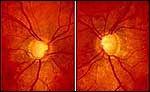
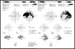
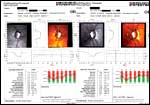
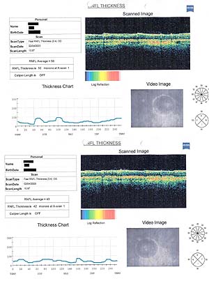
Patients with JOAG are often asymptomatic until the glaucoma is advanced. There is a high incidence of myopia among patients with JOAG; 50% to 87% of patients exhibit some degree of myopia on examination. Ophthalmologic examination reveals an elevated intraocular pressure (IOP) in both eyes. In 1995, Wiggs and colleagues found an average IOP of 38.5 mm Hg (range, 30 mm Hg to 53 mm Hg) in 23 patients with JOAG. Cupping is often evident on disc examination (Slide 1). Slit lamp examination and gonioscopy are generally unremarkable with normal angle anatomy (Slide 2). Ancillary testing reflects the patient's disease state with increased glaucomatous findings apparent with disease progression. Visual field testing, the Heidelberg Retinal Tomograph (Heidelberg Engineering Gmbh Dossenheim, Germany), and the ocular coherence tomograph may provide additional information (Slide 3, Slide 4, Slide 5). Unlike patients affected by congenital glaucoma, patients with JOAG do not exhibit buphthalmos and do not appear to have a Barkan membrane covering the anterior chamber angle on gonioscopy.

Genetic Link
A number of case series have identified families with JOAG in an apparently autosomal dominant inheritance pattern. In 1993, a locus for JOAG was mapped to the 1q21-q31 region and assigned as GLC1A. In 1997, Stone and colleagues reported the identification of trabecular meshwork-induced glucocorticoid response (TIGR) gene associated with JOAG. Concurrent to Stone's work, a group led by Kubota in Japan described a protein associated with the cytoskeleton in the retina termed myocilin (MYOC) gene. TIGR and MYOC have been determined to be identical, and in 1998 the Human Genome Organization Gene Database Nomenclature Committee assigned the gene the name myocilin (MYOC). Myocilin is normally found in the trabecular meshwork, cornea, retina, and optic nerve. Systemically, it has been identified in skeletal muscle, the heart, the stomach, bone marrow, the lung, and lymph nodes. Myocilin belongs to the family of proteins known as molecular chaperones. It is found in increased levels in eyes exposed to dexamethasone treatment, stretching, oxidative stress, and treatment with transforming growth factor ß. The normal function of myocilin protein is unknown. There have been at least 26 unique mutations in the MYOC gene sequence identified to date. The myocilin protein is divided into a myosin domain and olfactomedin domain. Most mutations are found in the olfactomedin domain of the protein. Excess myocilin protein may bind to and obstruct aqueous outflow in the eye, thereby leading to an increase in IOP. It is possible that mutant myocilin protein is unable to fulfill its physiologic function as a molecular chaperone.
Treatment
Patients with JOAG generally have severely high IOPs that are difficult to control with conventional glaucoma medication regimens. Patients with JOAG usually require surgical treatment for adequate control of their IOP. Several approaches have been tried for surgical management of JOAG. YAG goniotomy involves the placement of sharply focused YAG lesions of 4 millijoules to 6 millijoules in a linear pattern deeply into the filtration portion of the trabecular meshwork. YAG goniotomy has been proven effective for some patients with JOAG in the short-term; however, it eventually fails. Therefore, surgical goniotomy is often used as a primary means of surgical control of IOP. The goal is to maintain a stable IOP in young patients with JOAG until the patient is older, when, if necessary, more definitive filtration surgery would be more successful. Recently, the advent of selective laser trabeculoplasty (SLT) has posed a new possible short-term method of pressure control in patients with JOAG. Results have been mixed; clinicians achieved variable levels of success with SLT.
In the future, gene therapy may provide an alternative treatment modality. For example, delivering a recombinant MYOC gene into the trabecular meshwork may provide an alternative to conventional pharmacotherapy.
Conclusion
JOAG is a distinct entity, seen within a unique population, and posing a genuine treatment challenge. It is important to be aware of, and exclude, other diagnoses before settling on a diagnosis of JOAG. The care of these patients is often a lifelong commitment.
Bibliography
Copin B, Brezin AP, Valtot F, et al. Apolipoprotein E-promoter single-nucleotide polymorphisms affect the phenotype of primary open angle glaucoma and demonstrate interaction with the myocilin gene. Am J Hum Gen. 2002;70:1575-1581.
Johnson DH. Myocilin and glaucoma: A TIGR by the tail? Arch Ophthalmol. 2000;118(7):974-978.
Jonas JB, Buddle WM. Optic nerve head appearance in juvenile-onset chronic high pressure glaucoma and normal pressure glaucoma. Ophthalmology. 2000;107(4):704-711.
Lotufo D, Ritch R, Szmyd L JR, Burris JE. Juvenile glaucoma, race, and refraction. JAMA. 1989;261(2):249-252.
Melamed S, Latina MA, Epstein DL. Neodymium: YAG laser trabeculopuncture in juvenile open-angle glaucoma. Ophthalmology. 1987;94(2):163-170.
Merritt JC, Reid LA, Smith R, Harris DF. Diurnal intraocular pressure in juvenile open angle glaucoma. Ann Ophthalmol. 1979;11(2):253-260.
Rezaie T, Child A, Hitchings R, et al. Adult-onset primary open angle glaucoma caused by mutations in optineurin. Science. 2002;295:1077-1079.
Tawara A, Inomata H. Developmental immaturity of the trabecular meshwork in juvenile glaucoma. Am J Ophthalmol. 1984;98(1):82-97.
Wiggs JL. Juvenile open angle glaucoma. In: Schuman JS, Epstein DL, Allingham RR, eds. Chandler and Grant's Glaucoma. 4th ed. Baltimore: Williams & Wilkins; 1997:444-449.
Wiggs JL, Del Bono EA, Schuman JS, Hutchinson BT, Walton DS. Clinical features of five pedigrees genetically linked to the juvenile glaucoma locus on chromosome 1q21-q31. Ophthalmology. 1995;102:1782-1789.