Introduction
Patients with orbital or central nervous system disease often present with symptoms of loss of vision or visual field, double vision, eye pain, and proptosis. These symptoms many times signal potentially life- or sight-threatening disease. Many of these patients will require imaging studies as part of their evaluation.
There are two types of imaging: structural and physiologic.
Structural Imaging
Structural imaging provides information about the anatomy and pathology of orbital and skull bones, as well as the orbits, and brain structures including the blood supply.
Technology
- Computed tomography (CT)
- Magnetic resonance (MR)
- Angiography
- CT
- MR
- Conventional
CT Scanning
Physics:
- CT scanning uses a computer driven, rotating x-ray device to image in a single plane making a radiographic "slice" through the tissue of concern.
- Standard ionized radiation x-ray techniques are attenuated or weakened when they are transmitted through tissue. The intensity of the exiting radiation is then measured with special devices and imaged on film.
- Attenuation coefficient.
Windows:
Defines the relationship of the attenuation coefficient (H units) to the entire gray scale.
This allows us to use the so-called window width. This width can be set to emphasize specific
characteristics of the scanned substance. Both tissue windows (H value level of 0-40) and bone
windows (H value level of 40-300) are then possible by varying the window level. Reformatting
is done on computer - only one imaging sequence is required. High attenuation (density) is
bright like calcium; low attenuation is dark like fat.
Computer analysis:
CT scans can be oriented to slice (single thin images) or volume (overlapping thicker sections)
with different thickness (1 mm to 3 mm for orbits) depending on required resolution.
Planes and angulation techniques:
Axial: Orbit scans require negative angulation and head scans require positive angulation so
these two structures require separate sequences (Slide 1).
Coronal: Direct vs. reconstruction.
Direct: Patients must be prone/supine depending on what change in angulation may be necessary to avoid artifacts from dental fillings. Direct is always the preferred coronal image type (Slide 2).
Reconstruction: Reconstruction is used when positioning for direct is not allowed. It requires ultra thin axial slices exposing patients to increased radiation.
Contrast enhancement: Iodinated contrast agents (ionic and non-ionic) are used intravenously and do not cross an intact blood-brain barrier (BBB). Orbital disorders may not require contrast as the BBB is seldom disturbed. However, contrast is crucial for evaluation of intracranial lesions especially para sellar pathology.
Ionic agents are more toxic than non-ionic agents (12.6% vs. 3%), with many adverse effects described including anaphylactic shock, nephrotoxicity, and neurotoxicity. High-risk patients may benefit from prophylactic pretreatment that usually includes oral corticosteroids and antihistamines. Patients with allergy to shellfish may also be at risk. Patients with renal disease should have renal status (BUN, creatinine) and hydration should be checked prior to exposure to dye.1
Clinical use2,3:
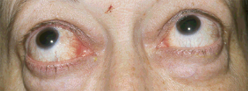
For imaging of orbital disorders, noncontrast CT is preferred (e.g.,
thyroid orbitopathy Slide 3, Slide 4,
and Slide 5 or trauma).
Brain CT is useful for acute bleeding.
Magnetic Resonance
Physics2:
Radiowaves are presented within a strong magnetic field with an appropriate receiver coil to
image tissue being examined. The particular characteristics of tissue types create different
signal intensities that are captured as an image digitally or onto film. Readings are performed
at different times which is known as pulse sequences.
Pulse sequences2:
T1 and T2 weighting: changes the intensity of the tissue signal produced by changes in the
magnetic field. The reading is taken at different times.
- T1 weighted images portray water (CSF) as low intensity (black), fat and blood signal is high intensity (bright). Great for anatomic detail (Slides 6 and Slide 7).
- T2 weighted image offers two types:
| Substance | T1 | T2 |
| Water | Dark | Bright |
| Fat | Bright | Bright |
| Bone | Dark | Dark |
| Deoxyhemoglobin | Dark | Dark |
| Methemoglobin (intracellular) | Bright | Dark |
| Gadolinium | Bright | Intermediate |
Fat saturation:
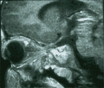
Contrast enhanced magnetic resonance images (MRIs) of orbit require specialized pulse
sequences that decrease (suppress) the high signal intensity of fat. Sequences used include
short inversion time, inversion recovery, and spectral presaturation inversion recovery
sequences. This sequence is required in orbital (allows delineation of sheath from optic nerve)
as well as base of skull imaging where fat signal may hinder visualization of
pathology2,4,5
(Slide 10, Slide 11, and
Slide 12).
Fluid attenuated inversion recovery:
Fluid attenuated inversion recovery (FLAIR), also called "fluid suppressed" or "dark fluid,"
suppresses bright CSF signal intensity in T2 so abnormalities in white matter are more evident.
This technique is especially useful for demyelinating disease.2
Diffusion weighted imaging2:
Diffusion weighted imaging (DWI) is sensitive to the microscopic random motion of the water
molecules. Ischemic areas appear as bright areas very early after TIA or definite stroke when
no abnormalities are seen on T2 or FLAIR. These ischemic lesions usually enlarge on serial DWI
over several days. DWI is helpful to identify new stroke on a patient with diffuse white matter
disease and to differentiate brain abscess (bright) from tumor (dark due to increased diffusion).
Perfusion weighted imaging:
- In perfusion weighted imaging (PWI), hemodynamically weighted MR sequences based on passage of MR contrast through the brain tissue.
- High doses of gadolinium administered by a power injector are needed.
- Signal intensity declines as contrast material passes through an infarcted area and returns to normal as it exits this area.
- PWI is useful in delineating areas of cortex at risk of infarction, and in Alzheimer and Creutzfeld-Jacobs diseases associated with decreased perfusion within the cortex.
Diffusion perfusion mismatch:
- Diffusion perfusion mismatch (DPM) is the difference in size between lesions captured by DWI and PWI.
- DPM represents ischemic penumbra, that is, the region of incomplete ischemia that lies next to the core of the infarction. This ischemic penumbra is the area that is viable if appropriate intervention is instituted promptly. The viability of this region can extend up to 48 hours after the onset of the stroke.
T2 weighted gradient2,6:
- T2 weighted gradient, or recalled echo sequences (gradient echo), is exceptionally sensitive to the presence of chronic blood products, necrosis, and calcification because of its high magnetic susceptibility.
- T2 weighted gradient is particularly useful in the diagnosis of cerebral amyloidosis and small brain vascular malformations that may present with multiple past asymptomatic hemorrhages.
Volumetric T2 weighted gradient2 offers high-resolution images in any plane.
Magnetic transfer contrast sequence2,7:
- Magnetic transfer contrast sequences improve detection of lesions that are enhanced with para magnetic contrast from adjacent background tissue.
- Magnetic transfer contrast sequence is particularly useful for the detection of cerebral metastasis.
Fast spin echo sequences result in marked reductions of MR scanning times.2
Contrast enhancement:
Paramagnetic contrast agents with gadolinium (Gd) (four types of Gd chelates are currently
available) are used in MR scanning to enhance lesions that disrupt BBB, dural/meningeal blood
supply and cranial nerve abnormality. Several studies have concluded that the routine use of
Gd is safe, even for children, and that the benefits of increased diagnostic sensitivity and
accuracy far outweigh any uncommon side effect.1,8
Two other MRI contrast agents, mangafodipir trisodium and ferumoxide, are not yet approved by the U.S. Food and Drug Administration (FDA).1
Pacemakers and aneurysm clips (metallic) are an absolute contraindication for MR use.9,10
Clinical use:
Contrast-enhanced, fat suppressed MRIs of orbit best show optic nerve pathology (demyelinating,
radiation-induced).2 Omitting IV contrast in MRI may preclude visualization of lesions that
have high perfusion or increased capillary permeability and extra axial masses which may be
iso-intense to neighboring brain (Slides 13, and
Slide 14).
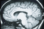
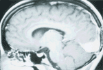
MR is the study of choice for sellar and chiasmal abnormalities,2 white matter disease, especially multiple sclerosis, as well as posterior fossa pathology. Craniopharyngiomas are recognizable by their suprasellar location, hyperintense components (fat) on T1 and contrast enhancement.
Cerebral infarction can be detected as early as 6 hours postinfarction after contrast enhancement.
Angiography
Computed tomographic angiography11:
Computed tomographic angiography (CTA) is a relatively new and minimally invasive CT technique
consisting of an intravenous bolus injection of contrast solution followed by high speed
helical or spiral CT scanning and computer-assisted generation of 3-D images of medium and
large size arteries for detecting aneurysms and stenoses. It presumably has the ability of
detecting aneurysms as small as 1.7 mm and can be used in patients with previous aneurysm clips
or who have claustrophobia or pacemakers. Patients, however, are exposed to radiation and
iodinated contrast agents and collateral flow cannot be assessed.
Magnetic resonance angiography12:
Magnetic resonance angiography (MRA) is used to study the vasculature of the head, neck and
aortic arch. It highlights flowing blood without the need for contrast material and is used in
detecting aneurysm more than 3 mm in size, carotid or vertebral basilar dissection and carotid
stenosis. This study tends to overestimate the severity of stenosis.
Techniques and sequences:
- 2 D phase contrast.
- 3 D time of flight (TOF) - best technique based on flow-related enhancement; 2 D TOF may be more helpful in areas of low flow.
- Multiple overlapping thin slab acquisitions (MOTSA) helps to overcome problems related to acoustic shadowing which can completely obscure the vascular anatomy at the site of a calcified plaque.
- Other pitfalls include susceptibility artifact (complete signal loss in regions of disturbed flow), obscured vascular lesions.
- MRA should remain a screening tool for aneurysms, arteriovenous malformations and arterial stenosis. MRA fails to demonstrate up to 20% of cerebral aneurysms less than 5 mm. Because of its limitations in resolution and sensitivity, current conventional contrast angiography remains the definitive technique to image vascular disease in cases of acute hemorrhage.2
- Another technique, magnetic resonance venography (MRV) is useful to detect venous occlusive disease.2
Conventional contrast angiography:
- Current generation angiographic studies are performed using digital subtraction imaging systems that offer high-resolution imaging and the ability to alter image contrast and vascular density retrospectively, without the need for repeated angiographic injections or conventional filming.2
- These improvements result in faster angiographic studies, lower contrast doses, and the ability to process imaging data in real time during catheter guidance and placement within the execution of interventional procedures.2
- Conventional contrast angiography remains the procedure of choice for detecting aneurysms, especially to delineate collateral circulation, carotid cavernous fistula, operable high grade carotid stenosis, and carotid stenting problems.
- Iodinated contrast material used for catheter cerebral angiography can cause up to 0.2% of significant complications (e.g., bronchospasm, laryngospasm, status asthmaticus, subglottic edema, angioedema, anaphylactic shock, cardiovascular collapse). These effects were significantly increased with the use of high osmolality contrast agents (HOCAs) than with low osmolality, mostly non-ionic contrast agents (LOCAs).1,8
- Conventional contrast angiography has a morbidity caused by vasospasm or embolic stroke of up to 5% with a mortality rate of up to 0.1%.1
- Previous reaction to contrast medium was the most important risk factor for adverse
effects to occur.1
Physiologic Imaging
Physiologic imaging provides information about the function of brain tissue, including blood flow, blood volume, and brain metabolism.
Technology
- Positron emission technology (PET)
- Single photon emission computed tomography (SPECT)
- Functional MRI
- MR spectroscopy
PET13,14,15:
- PET is an analytical nuclear medicine imaging technology that uses positron-labeled molecules.
- As the tracer compound accumulates in brain tissue, positrons are emitted that eventually convert to gamma ray energy that can be imaged onto film. PET requires a cyclotron so it is relatively expensive. FDG is a radiopharmaceutical approved by the FDA that can be injected intravenously and works by tracing the metabolism of glucose in tissue.
- The most common clinical use for PET is the study of brain neoplasms, degenerative CNS disorders, and seizures.
SPECT14:
- SPECT uses radiolabeled tracers that emit a single photon, and the level of activity is quantified as a reflection of regional cerebral blood flow. SPECT has much lower resolution and is less expensive than PET.
- Current uses for SPECT include studies of ischemic brain, cortical visual loss, and migraine.
Functional magnetic resonance imaging:
- Functional magnetic resonance imaging (FMRI) offers detailed matching of anatomic with physiologic information and avoids administration of contrast (non-invasive) without using ionizing radiation.16,17
- FMRI can show mapping of activation of the occipital cortex with repetitive visual stimulation, which is its most current use.12,16,17
- This technique helps visualizing changes in chemical composition of brain areas or changes in the flow of fluids that occur over seconds to minutes. Therefore, it is a method that assists in the objective evaluation of visual function.2
- FMRI has high spatial and temporal resolution.
- FMRI is usually performed in conjunction with a task that is dependent on a local region of the brain - the so-called blood oxygenation level dependent (BOLD) imaging.
- Patient is imaged during a control state and an activation state, and the variable of interest is the state of hemoglobin oxygenation.16,17
- Functional images are acquired in T2-weighted images format to enhance the BOLD contrast.
- Limitations of FMRI include variability within the same subject and poor reproducibility of signal change.
- FMRI requires the subject's cooperation with attention and minimal motion to avoid artifacts.
Magnetic resonance spectroscopy:
Magnetic resonance spectroscopy (MRS) evaluates the metabolic activity and concentration of
certain metabolites in tissue.
Most clinical, high field (.15 Tesla) MRI machines can be upgraded to perform MRS at a fairly low cost.
MRS is used to detect levels of N-acetyl aspartate, which is considered to be a marker of the neuron.
MRS is useful for early detection of acute stroke, neoplasms, demyelinating disease, and
neurodegenerative diseases.
Preliminary studies suggest that MRS may complement MRI for early detection of occipital
lobe abnormality.2
Considerations for selecting the most appropriate imaging modality
- Cost of diagnostic procedures based on national averages
- Facility fee (hospital, outpatient center)
- Professional fee (radiologist or non-radiologist)
- Obtain a second copy or reading of the imaging study
- Contrast use (ionic vs. non-ionic)
- Clinical guidelines for ordering the appropriate imaging study
- Obtain the patient history and perform a clinical exam to localize the lesion.
- If unsure of what test to order, call your neuroradiologist and discuss the case to decide on proper imaging technique.
- When necessary, review the images with a neuroradiologist after the procedure is performed.
- Medicolegally, the requesting health care provider is as responsible as the radiologist that read the films if the wrong diagnosis is made.
- Evaluating Neuro-Imaging
The following steps can be used to evaluate results of neuro-imaging:
- Confirm patient name and date of study
- Identify imaging methods (CT, MRI, MRA)
- Identify plane of image
- Axial - follows horizontal plane (top/bottom)
- Sagittal - follows vertical plane (front/back and right/left)
- Coronal - follows vertical plane (side/side)
- Identify sequences for MRI (T1, T2)
- Identify images with or without contrast
- Identify use of fat saturation in orbital sequences
- Confirm right and left side and eye
- Mentally identify the anatomy
- Perform a systematic review of anatomic structures
- Compare contrast and non-contrast images
- Look for artifacts, head position and motion
- Compare the study to previous imaging
- When is CT scan adequate?
- ORBIT: calcification, hemorrhage, trauma (fracture), and thyroid orbitopathy
- BRAIN: acute bleeding and intracranial calcification (noncontrast CT)
- Study the Correct Area
- Order orbit study for:
- Proptosis
- Orbital/eye pain
- Optic neuropathy (PPLOV)
- Order brain study for:
- Binocular visual field defect
- Optic neuropathy
- Other neurologic symptoms and signs (i.e., nystagmus)
- Order orbit study for:
- Use the Correct Options:
- MRI vs. CT
- Contrast
- Fat saturation
- MRV/MRA
- Angiography
- Indications for CT Scan
- Bone abnormality
- Localized orbital process (although MRI may eventually be required)
- Pacemaker dependent patient
- Ferro-magnetic foreign body around or in orbit
- Potentially psychotic reaction to claustrophobia
- Trauma
- Identification of blood within the first few hours
- Disadvantages of CT Scan
- Ionizing radiation exposure
- No sagittal imaging
- Contrast reaction
- Dental and bony artifacts
- Indications for MRI
- Diffuse orbital process
- Virtually any intracranial process when MRI is not contraindicated
- Better delineation of a process noted on CT
- Contraindications for MRI
- Pacemakers
- Metallic aneurysm clips or other prosthetic metallic devices
- Claustrophobia
- Indications for MRA
- Screening for aneurysms and other vascular pathology
- Carotid and vertebral artery pathology including dissection.
- Indications for Conventional Angiography
- Confirm vascular abnormality prior to surgery
- When MRA is negative and the clinical suspicion for a vascular lesion remains high
- Common Imaging Errors
- Failure to prescribe a dedicated study
- Inappropriate use of a dedicated study
- Omission of intravenous contrast
- Omission of specialized sequences
- Fat suppression
- Gradient echo (blood products)
- Interpretive Errors
- Failure to detect the lesion because of misleading clinical information
- Rejection of a clinical diagnosis because an expected imaging abnormality was absent
- Assumption that a striking imaging abnormality accounted for the clinical presentation.
- Failure to consider the limited clinical specificity of imaging abnormalities
Lexicon
- Echo time (TE) - The time selected to wait after the initiation of TR to receive the radiofrequency "echo" from the patient.
- Fast spin-echo (FSE) - A spin-echo sequence that results in decreased scanning time by acquiring multiple phase-encoding steps simultaneously.
- Fat saturation (fat-sat) - A sequence that decreases the signal contribution from fat in an image. This is accomplished with a special excitation pulse that is centered on the Larmor frequency of fat.
- Gradient-recalled echo (GRE, Grass) - A technique that reduces the imaging time by eliminating the 180° refocusing pulse required in spin-echo imaging. Each acquisition yields a single slice.
- Inversion recovery (STIR) - A sequence that uses the difference in relaxation times between fat and water to null the signal contribution of fat on an image.
- Larmor frequency - The characteristic frequency in which a molecule's protons (e.g., fat and water) precess in the external magnetic field (B0).
- Repetition time (TR) - The time that elapses between two consecutive excitation pulses (phase encoding steps).
- Spatial saturation (SAT) - A radiofrequency pulse applied to anatomy outside the FOV (field of view). This reduces any artifact within the FOV that would otherwise occur from motion outside the FOV.
- Spin echo - A sequence that adds a 180° pulse at one half of the TE to refocus the precessing protons at time TE. This helps maximize the signal and minimize artifacts that result from magnetic field inhomogeneities.
- Time of flight (TOF) - A term used to describe the increase in intravascular signal seen with blood flow that is perpendicular to the plane of the slice. This effect results from blood moving into the imaging plane after receiving its excitation outside the imaging plane.
- T1 (spin-lattice relaxation) - A time that corresponds to the vector realignment (along the z-axis) of the excited protons to the applied magnetic field, B0. T1 effects predominate when a short TR and a short TE are prescribed (T1 weighting).
- T2 (spin-spin relaxation) - A time that corresponds to the dispersion of the vector alignment (into the X-Y plane) of excited protons because of differences in precession rates. T2 effects predominate when a long TR and a long TE are prescribed (T2 weighting).
References
- Lee AG, Hayman LA, Ross AW. Neuroimaging contrast agents in ophthalmology.
Surv Ophthalmol. 2000;45:237-253.
- Davis PC, Newman NJ. Perspective: Advances in neuroimaging of
the visual pathways. Am J Ophthalmol. 1996;121:690-705.
- Ozgen A, Alp MN, Ariyurek M, Tutuncu NB, Can I, Gunalp I. Quantitative
CT of the orbit in Graves' disease. Br J Radiology. 1999;72(860):757-762.
- Jackson A, Sheppard S, Laitt RD, et al. Optic neuritis: MR imaging with combined
fat-and water-suppression techniques. Radiology. 1998;206(1):57-63.
- Mafee MF, Goodwin J, Dorodi S. Optic nerve sheath meningiomas. Role of MR
imaging. Radiologic Clinics of North America.
1999;37(1):37-58.
- Wolintz RJ, Trobe JD, Cornblath WT, et al. Common errors in the use of magnetic
resonance imaging for neuro-ophthalmic diagnosis. Surv Ophthalmol. 2000;45:107-114.
- Runge VM. Safety of approved MR contrast media for intravenous injection.
J Mag Reson Imaging. 2000;12:205-213.
- Boorstein JM, Moonis G, Boorstein SM, et al. Optic neuritis: Imaging with magnetization
transfer. American Journal of Roentgenology.
1997;169(6):1709-1712.
- Center for devices and radiological health magnetic resonance working group. A primer
on medical device interactions with magnetic resonance imaging systems. Washington, D.C.,
www.fad.gov 1997; 1-15.
- Price RR. The AAPM/RSNA physics tutorial for residents: MR imaging safety
considerations. RadioGraphics. 1999;19:1641-1651.
- Hashimoto H, Iida J-I, Hironaka Y, et al. Use of spiral computerized tomography
angiography in patients with subarachnoid hemorrhage in whom subtraction angiography
did not reveal cerebral aneurysms 2000. J Neurosurg.
92:278-283.
- Levin LA, Lessell S. Magnetic resonance angiography. International
Ophthalmology Clinics. 1994;34(3):293-303.
- Phelps ME. PET, the merging of biology and imaging into molecular
imaging. J Nucl Med. 2000;41:661-681.
- Thrall B. Single-photon emission computed tomography and positron emission tomography.
In: Nuclear Medicine: The Requisites. St. Louis,
Mo: Mosby; 2000:294-322.
- Coleman RE. Clinical PET: Role in diagnosis and management. J Nucl Med. 2000;41:36N-42N.