Introduction
The color of any visual stimulus may be defined as the result of a certain spectral analysis performed by the brain of the light reflected or emitted. In approximately 92% of individuals, such spectral analysis yields approximately the same color perception. This population group possesses what is called normal color vision.
The color vision of the remaining 8% of the human population differs significantly from what is considered normal. Among this 8%, approximately 2% are chromatic defective, and the remaining 6% are chromatic anomalous. In general, both anomalous and normal individuals can distinguish many of the colors that a chromatic defective individual perceives as equal. On the other hand, whereas a defective individual accepts as equal two colors that are metameric matches for a normal individual, anomalous individuals will consider them different. These two facts seem to indicate that anomalous and normal color vision are indeed different, whereas chromatic deficiencies are just a reduced form of normal vision. In fact, defective color vision can be explained if we admit that one (dichromats), two (monochromats), or three (achromats) types of cones in the retina are either missing or not functional. Anomalous individuals would possess three different kinds of photopigments and are therefore trichromats, but one or several of these pigments differ from those found in normal individuals.1-4
Table 1 summarizes the anomalies and deficiencies that arise from the different combinations of missing and variant cone photopigments. The chromatic deficiencies of dichromats are considered severe, whereas anomalous trichromats suffer mild chromatic deficiencies because their color vision does not prevent them from leading a perfectly normal life.2 It is possible to find chromatic anomalies and deficiencies that cannot be labeled as protan (lack of L-cone pigment), deutan (lack of M-cone pigment), or tritan (lack of S-cone pigment) defects.6-10 However, their incidence is quite small. Although the chromatic anomalies and deficiencies presented by both eyes of a given individual are usually the same, the literature reports instances of unilateral anomalies or deficiencies, that is, individuals with different anomalies or deficiencies in each eye11,12 or only one eye with variant chromatic vision.13-17
Most color vision defects (after ruling out side effects of different pathologies of ocular origin) are congenital and permanent. Red-green defects (protan and deutan) present the highest incidence in the general population (1% protanopic individuals, 1% deuteranopic, 4.8% deuteranomalous, and 1.2% protanomalous). Tritan individuals amount to 0.002% of the dichromatic and anomalous population. There is an autosomal dominant pattern of inheritance of tritan defects, unlike protan and deutan defects. There is a higher incidence of males with chromatic anomalies and deficiencies than females except in tritan disorders, where there is an equal incidence in males and females. Although females are carriers of chromatic anomalies and deficiencies, less than 0.4% of the female population suffer from them.4
Color vision defects caused by pathology are called acquired and usually are accompanied by visual acuity or visual field losses. Blue color vision often is affected at some stage of these pathologies.18,19 Acquired color vision defects may be associated with disorders of the prereceptoral ocular media, fundus detachment, progressive cone dystrophies, or degeneration affecting all cone classes with or without involvement of the rods, macular dystrophies and degeneration, vascular and hematologic diseases, glaucoma, hereditary dominant optic atrophy, optic nerve diseases, diseases of the central nervous systems (e.g., multiple sclerosis) or other organs (e.g., diabetes mellitus) and, finally, toxic agents such as lead, tobacco, or alcohol that affect the retina or the optic tracts. The main differences between congenital and acquired anomalies and deficiencies are summarized in Table 2.
There are many tests that can detect and classify the anomaly or deficiency suffered by a given individual. However, few can be used with children. For example, if the task consists of naming a familiar object, young children with normal color vision may fail the test just because they cannot connect picture and word.20 The child also may be incapable of understanding the task he or she must perform.21,22 In addition, the short attention span of children also is a serious problem and rules out the use of precise tests of longer duration. Some subjective psychophysical methods can yield reliable results in children older than 3 years, but tests for evaluating the color vision of adults cannot be successfully used until age 10 or 11 years.23,24
Despite these difficulties, early detection of chromatic anomalies and deficiencies is crucial to prevent problems in a child’s development and academic achievement. In many cases, color vision problems are symptoms of several diseases, related or unrelated to vision, such as dyslexia, lack of vitamin A, glaucoma, diabetes, etc.14,25 Aside from the relationship between color vision problems and these pathologies, the majority of children’s books and games largely use color for information coding. Therefore, children with color vision deficiencies may exhibit learning problems leading to poor academic achievement and suffer from an inferiority complex.26-37
Tests specifically designed for the analysis of color vision in children are available. The main difference between these tests and those for adults is that the task is easy to understand and does not take long to perform, thereby avoiding the two major problems encountered with young children. Some tests do not require any response from the child, but only the careful observation of the examiner.
When a color vision problem is detected in a child, it cannot be corrected; however, in specific cases, optical aids such as filters may be used to improve the child’s vision.3,38-44
This review analyzes the difficulties that arise when evaluating color vision in children, particularly in very young children. The specific problems posed by the tests when used in clinical practice are described, and some methods used to minimize the effect of color vision problems also are discussed.
Anomaly and Deficiency Detection
Specialized techniques are used to detect and evaluate congenital chromatic anomalies and deficiencies of color vision. These techniques include:
- spectral sensitivity, obtained from absolute thresholds, by heterochromatic matching (heterochromatic simultaneous matching, flicker photometry, or minimum distinguishable border photometry) or by increment and flicker detection on an achromatic background,
- chromatic discrimination (wavelength and colorimetric purity discrimination), and
- neutral points.
In addition to these techniques, certain colorimetric equations, by means of devices called anomaloscopes, often are used. The Rayleigh equation serves to detect protan and deutan anomalies and defects (Nagel-I and Pickford-Nicholson anomaloscopes). Several equations are available to detect tritan anomalies and deficiencies, e.g., the Engelking-Trendelenburg (Nagel-II and Pickford-Nicholson anomaloscopes) and the Pickford-Lakowski equations (Pickford-Nicholson anomaloscope). Table 3 shows the properties of dichromatic and anomalous color vision that serve as a basis for these tests.
These psychophysical measurements allow accurate detection and classification of the different chromatic anomalies and deficiencies, but require relatively sophisticated experimental devices and procedures. Therefore, they are not often used in routine vision examination. The complexity of the tasks and their long duration make the use of these tests with children difficult. For this reason, color vision in children usually is evaluated by means of printed tests. In general, these printed tests consist of several plates that check only a few of the main characteristics of dichromatic and anomalous vision. Most printed tests are based only on confusion lines and neutral points; therefore, they are far more inaccurate than the complete set of techniques mentioned above. It is not unusual for two different tests to yield contradictory results with the same observer. Therefore, several tests should be performed before making a diagnosis.
Printed tests present two main problems: pigment changes due to temperature variations and the influence of the illuminant.
In printed tests, the pigments used are sensitive to temperature variations. Chemical temperature-induced changes in the pigments alter the chromaticity of the plates, and consequently the test reliability decreases considerably, to the point of classifying anomalous trichromatic individuals as normal.
Printed tests also must be viewed only under the specific illuminant for which they have been designed. Otherwise, the chromaticity of the stimuli will be different and the results will be distorted. For instance, if we perform a test using tungstene lamps without filters, which yield a more yellowish light than the standard C illuminant recommended by the International Commission on Illumination in all printed tests, deuteranopic individuals will score less errors than they should,45-51 and deuteranomalous individuals might pass the test as normal individuals. Moreover, protanopes and extreme protanomalous individuals might demonstrate a deutan profile.51 Fluorescent lamps also are not suitable because the literature on their use in different color tests is inconclusive.49,50,52,53 Most tests should be performed in a completely dark room, using a standard C source and normal illumination to avoid glare or gloss. To avoid the problem of the illuminant, self-illuminated tests and lenses that transform common incandescent light into C or other valid illuminants can be used.45,51,54-56
Despite these two drawbacks, printed tests usually are the first choice when evaluating color vision in children because they can be administered rapidly.
Test Classification By Task
When choosing a color vision test for children, the task to be performed must be adapted to the child’s development stage. Therefore, test choice will depend on both the child’s age and the degree of accuracy needed. Although some tests designed for adults, such as the Ishihara or the Farnsworth-Munsell tests, may be adapted for children, this is the exception and not the rule.
Identification Tasks
Ishihara Test for Illiterate Individuals.
This test is best for detecting red-green anomalies and deficiencies.4,45,57-59 It consists of 13 plates with geometrical figures — squares,
circles, and lines. This test reaches 100% efficiency in children aged 7 years or older57; preliminary studies show it also is 100%
effective for 5- to 6-year-old children. With 3- and 4-year-old children, efficiency drops to 40% and 90%, respectively. The test is not reliable for
children younger than 2 years.60 The main drawbacks of the Ishihara test are that it does not grade the defect4 or contain plates to
detect tritan defects.
American Optical CO.
The American Optical CO (Hardy, Rand, Ritler) test, usually known as AO-HRR, consists of plates with geometric figures — triangles, circles,
and crosses — designed to screen tritan, protan, deutan, and tetartan defects (tetartan individuals might possess a photopigment with an absorption
peak in the yellow region of the spectrum61). Although the test grades the defect accurately, it is not a precise test for detecting
red-green anomalies because the color differences between stimuli in the four protan/deutan screening plates are so small that false positives
are frequent.58 This test can be used with children older than 5 years57; however, the literature provides evidence
of its adequacy for children in the 3- to 16-year-old age range.62
Matsubara-Kojima Plates.
This test consists of plates with 10 pictograms (e.g., a flower, a dog, and a butterfly) for red-green defect screening.63 This test can be
used in the 4- to 6-year-old57 or 3- to 7-year-old age range.64 The ambiguity of the results and the high probability of false
positives do not make this test the first choice for screening.64
Lanthony Tritan Album.
This test consists of five plates to screen and grade both acquired and congenital tritan defects. It cannot be used to evaluate protan and deutan defects.
For screening and grading, the low reliability of the test does not make its use in children advisable.4 The minimum age for this test is
4 years because its reliability for 3 year olds is only 80%, and in children younger than 2 years, the test is not reliable at all.60
APT-5.
This test has the advantage of being self-illuminated.65 Its use is recommended for children older than 5 years,66
although it also is efficient for 3 year olds.65 It is an easy, reliable test with few false positives. In addition to screening, it grades the defect.
Creamer Color Chart.
This test consists of a single plate for screening red-green defects. It is used as a preliminary test. The plate
contains two geometric figures (a circle and a star). If the individual shows difficulties in perceiving the red star, further testing with a finer test is
recommended. The test can be administered to children older than 3 years.67
Color Vision Test Made Easy.
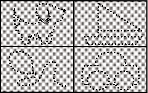
This test consists of 14 pseudoisochromatic plates with objects and drawings easily identified by children
(e.g., circles, stars, squares, ships, balloons, and dogs) (Slide 1A and Slide 1B). The efficiency of this test is comparable to that of the Ishihara test, and there are no
false positives. However, it detects only protan and deutan defects.68 It is quick and easy, and its use is recommended in children older than 3 years.

Tracing Tasks
Dvorine Color Blindness Book.
Twenty-three plates constitute this test, among which eight are used with children. These eight plates contain paths the patient must trace with a pointer.
The test is designed for screening and grading red-green defects, although grading reliability is controversial.4,58,69-71
It cannot detect tritan defects, and it is not a widely commercialized test. The test usually is not recommended for children because some find the
task difficult to understand.
Tokyo Medical College Color Vision Test for Children.
This test consists of 13 plates with pictograms. Five screen red-green defects and anomalies, and two screen tritan defects and anomalies.
Grading of the defect is accurate in all cases.4,72-74 The patient must trace the contour of each pictogram with a pointer.
The test is not advised for children ,6 years.75-79 Good visual acuity of at least 10/20 is required to do the test; therefore, it cannot
be used in children with significant amblyopia.
Farnsworth F-2 Plate.
This test consists of a single plate with two squares, one green and one blue, on a purple background. The child must trace the contours of the squares
with a pointer. Individuals with tritan defects are unable to clearly see the green square, whereas individuals with protan and deutan defects will have
difficulty seeing the blue square.3,4,23,57,80 Anomalies cannot be graded with this test. In many cases, this test does not distinguish
between protan and deutan defects, and numerous false positives occur.81 Although it is an easy, fast test, it is not advisable if precise
detection is needed.
Hahn New Pseudoisochromatic Color Vision Test.
This test consists of 15 plates with numerals and five tracing plates11,12,19-21
for screening illiterate individuals. It screens for protan, deutan, and tritan deficiencies and anomalies, and is useful in classifying and grading acquired
color vision deficiencies.82,83 It has been shown that the classification performance is particularly poor and the overriding tendency is
for protans to see neither numeral.84 The Pediatric Hahn Isochromatic Test, Type C, is a version of this test designed to be used in
primary schoolchildren and consists of 11 plates for screening, type classification, and grade estimation of color deficiencies. This test may be used
for children older than 6 years83 and seems to perform slightly better than the Ishihara test.
Pairing Tasks
Guy’s Color Vision for Children.
This test consists of two pairs of six pseudoisochromatic plates containing capital letters the individual must pair.81
Although it is recommended for children older than 3 years, the choice of both colors and letters is not optimal for this age.20,57
City University Test (First and Second Edition).
There are two versions of this test, the most precise being the second, in which the colors are more desaturated.85
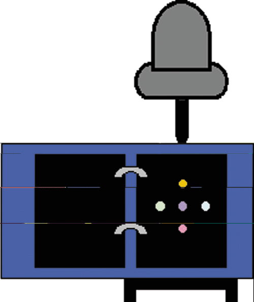
The test consists of 10 plates with a central stimuli and four peripheric stimuli (Slide 2). The observer must point out the stimulus in the periphery
that matches the central color.86 Although the test analyzes all types of chromatic deficiencies, in many cases the distinction between
protan and deutan individuals is inaccurate, and other tests must be performed for closer analysis once a chromatic defect has been noted. The
child’s capability to understand the task determines its applicability. Young children may find the task too complex.4,87,88
Pease & Allen.
This test consists of four plates with a square in one of its corners. The child must locate the position of the square in the plate. The test can be
used for children older than 3 years and allows detection of red-green and blue-yellow defects and anomalies.89 Although no false
positives appear, deuteranomalous individuals may yield false negatives.3
Verriest’s Tests.
Different tests used by Verriest and colleagues57 to test infant chromatic vision are grouped under this heading.
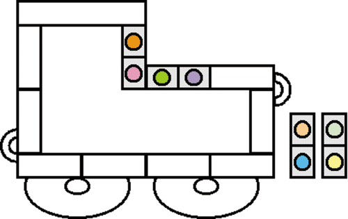
The first consists of a dominoes game of 11 pieces with a pair of colors each. For a normal trichromatic individual, the two colors of any domino would
look different but may look equal for a defective individual. The child must place the dominoes on a plate, with a drawing of a car matching the colors
of contiguous pieces (Slide 3).
The second test consists of a lottery game with six different colored cards and a cardboard that shows all the colors on top. The child must place each card under the corresponding color on the cardboard.
Both tests serve to evaluate tritan, deutan, and protan individuals and are recommended for children older than 4 years.57
Fletcher-Hamblin Simplified Color Vision Test.
This test consists of three plates. Each plate contains a mosaic of 16 squares to detect
protan, deutan, and tritan defects and anomalies. The child must choose the color that appears different from the rest. This test detects chromatic
anomalies accurately3 and is recommended for children older than 7 years.
Orientation Discrimination Tasks
Velhagen Pflügertrident Plates.
This test consists of differently oriented Snellen’s Es, and their orientation must be described by the child. The format of the test varies with the different
editions.4 It allows detection and classification but not grading of protan, deutan, and tritan defects.4 This test is
recommended for children aged 5 to 6 years. Test efficiency falls to 80% for 4 year olds and to 20% for 3 year olds. The test is invalid for children
younger than 2 years.90 Nevertheless, some authors recommend it for the 3- to 5-year-old age range.91 The difficulty
in understanding the task does not make this test a first choice for children4; however, different authors do recommend
this test.60,92
Okuma Plates.
These plates contain differently oriented Landolt’s Cs. The individual must describe the orientation of the letter.93
The test allows detection of red-green but not tritan defects and anomalies, and although the defect may be graded, this estimation is not
reliable.94 Nevertheless, this is a good routine test because it is easy and quick.95 Its use is recommended for
children older than 3 years.
Titmus Color Pediatric Test.
This test consists of a plate divided in eight regions, each containing colored circles and a Snellen’s E
at different orientations, which the individual must describe. The efficiency of this test in children is similar to that of the D-15 and
Ishihara tests.20 As with the Velhagen Pflügertrident test, which works similarly, its use is recommended for children older
than 5 years.
Hue Discrimination Tasks
Farnsworth-Munsell 100 Hue Test.
This test for adults can be easily applied to children; however, the results cannot be considered reliable for children younger than 9 years due to difficulty
of the task — putting colors in order by hue.20,23,96-98 This test allows good grading of protanope, deuteranope, and tritanope individuals.
Nevertheless, the chromatic defects and anomalies are not always correctly detected and anomalous individuals may pass as normal. The test
cannot distinguish between dichromats and anomalous trichromats. Its use is recommended as an exploratory test when a chromatic defect is
suspected.99
D-15 Panel.
Compared with the Farnsworth-Munsell test, the D-15 panel offers the advantage of being quicker. It detects and grades all types of chromatic
anomalies and dichromacies. It also diagnoses a slight tritanopic defect in children, which is frequently explained by the gradual development of blue
vision during childhood.100-102 It has been suggested the test may fail to detect chromatic anomalies correctly, so that anomalous
individuals may pass as normal,4 and conversely, normal individuals may be classified as anomalous.83
Despite its relative short duration, the task is too difficult for the test to be reliable with children younger than 10 years.
Tracking Tasks
Optokinetic.
This is a mechanical test in a revolving cylinder painted in red and green stripes, moving left or right. Both colors look identical for defective individuals,
who therefore do not follow the movement of the stripes. Ocular movements may be detected both by direct observation and photoelectrical methods.
The test can be used to detect protan and deutan defects and is recommended for children older than 3 years; however, as no verbal response is needed,
the test may be attempted at earlier ages depending on the child’s personality.103,104
Detection Without Cooperation
Visual Evoked Potential.
Protan and deutan defects can be detected by recording the neural response of the visual system by means of electrodes. The main advantage of this
test is that the individual’s age is irrelevant because no verbal response is needed. Results are reliable. Nevertheless, this test requires sophisticated
devices and specialized staff, which negates its use in everyday practice.105,106
Table 4 summarizes all of the tests described. They are classified according to advisability of their use with children.
Recommendations
Hereditary color vision defects cannot be corrected. Although several therapies have been proposed (e.g., electrical eye stimulation, iodine injections, or large doses of vitamins107-109), the reality is that there are no treatments or surgical procedures to correct a lack of color vision in some cases. However, optical aids may be used to improve the quality of the individual’s chromatic vision.3,38-44,110-113
Filters are the main optical aids available. Their use does not aim to improve sensitivity in the regions of the chromatic diagram where discrimination is poorer, but to enhance the contrast of the scenes in such a way that colors in the individual’s region of better discrimination are seen brighter than the rest. Thus, chromatic stimuli that looked identical to the dichromatic or anomalous individual may be distinguished by brightness.
Filters may be worn as ophthalmic glasses or contact lenses. However, the use of filters may cause loss of stereopsis, distortions of depth perception, low visual acuity, or brightness losses38,39,114-123 and may be used only to aid certain visual tasks.
All optical aids must be accompanied by a clear explanation of the child’s condition to his or her teacher, pointing out the child’s limitations and advising courses of action that would make things easier for the child. Color vision should be checked before beginning to use color-coded teaching material. If a color vision defect exists, it will help to photocopy the colored pages of textbooks or label pictures with words or symbols. It also is useful to label coloring utensils (crayons, pens, colored pencils, etc) with the name of the colors. White chalk should be used for writing on the blackboard because the contrast is greater than with colored chalks. In some school tasks, students with color vision defects will still need additional help.
Conclusion
The natural question arising after reviewing the different color vision tests available for children is which test is more adequate for daily practice. We believe we cannot talk of good or bad tests in general terms, but in the measure that they answer our particular needs. The choice of a color test must always address two main factors: the kind of information needed and the patient’s characteristics.
For instance, different tests are needed when evaluating one patient in the relative quiet of an examining room and when testing an entire school population for color vision problems. In the second case, a quick screening test would be sufficient in identifying those individuals with possible color problems who could be evaluated later.
Time limitation is not the only factor that guides the choice of a test. Discrimination losses in the blue region, which are rare in congenital color vision defects, are common in acquired color vision defects and are frequently linked with serious pathologies such as glaucoma, optical neuritis, or diabetes. If it is suspected that a patient may suffer one of these pathologies, a test that will study color discrimination in the blue region should be chosen. However, if only congenital color vision defects are being checked, the lack of stimuli in this particular region is not serious as tritan congenital defects are rare. In other cases, accurate grading may be needed.
Once the practitioner’s objectives are clear, it is the patient’s characteristics that determine the final choice of the test. Although the tests are recommended for certain age groups, the child’s intellectual development is what really matters. For instance, a hyperactive child should not be expected to correctly perform the D-15 panel, even if his or her age is right a priori: the child would be unable to concentrate for the necessary time. The child’s capability to understand ideas and concentrate in a given task should determine which test to use. The examiner should, therefore, decide which test is best for each particular occasion.
References
- Pokorny J, Smith VC. Congenital and Acquired Colour Vision Defects. London, England: Grune-Stratton; 1979.
- Artigas JM, Capilla P, Felipe A, Pujol J. Óptica Fisiológica. Psicofísica de la Visión. New York, NY: McGraw-Hill InterAmericana; 1995.
- Fletcher R, Voke J. Defective colour vision. In: Fundamentals, Diagnosis and Management. Bristol, United Kingdom: Adam Hilger Ltd; 1985:134-209,417-450.
- Foster DH. Inherited and Acquired Colour Vision Deficiencies. Vol 7. London, England: Macmillan Press; 1991.
- Estévez O. Anomalías Cromáticas, Ciencia del Color. València, Spain: Universitat de València; 1983.
- Alpern M, Pugh EN Jr. Variation in the action spectrum of erythrolabe among deuteranopes. J Physiol (Lond). 1977;266:613-646.
- Alpern M, Wake T. Cone pigments in human deutan colour vision defects. J Physiol (Lond). 1977;266:595-612.
- Alpern M, Moeller J. The red and green cone visual pigments of deuteranomalous trichromacy. J Physiol (Lond). 1977;266:647-675.
- Bowmarker JK, Dartnall HJ, Mollon JD. The violet-sensitive receptors of primate retinae. J Physiol (Lond). 1979;292:31P.
- Bowmarker JK, Dartnall HJ, Mollon JD. Microspectrophotometric demonstration of four classes of photoreceptor in an old world primate, Macaca fascicularis. J Physiol (Lond). 1980;298:131-143.
- Nimeroff I. Deuteranopic convergence point. J Opt Soc Am. 1970;60:966-969.
- MacLeod DI, Lennie P. Red-green blindness confined to one eye. Vision Res. 1976;16:691-702.
- Graham CH, Hsia Y. The spectral luminosity curve for a dichromatic eye and a normal eye in the same person. Proc Natl Acad Sci USA. 1958;44:46-49.
- Graham CH, Hsia Y. Some visual functions of a unilaterally dichromatic subject. In: Visual Problems of Colour. Vol 1. London, England: HMSO; 1958:279-295.
- Graham CH, Hsia Y. Color defect and color theory: studies on normal and color-blind persons including a unilaterally dichromatic subject. Science. 1958;127:675-682.
- Bender BG, Ruddock KH, de Vries de Mol EC, Went LN. The colour vision characteristics of an observer with unilateral defective colour vision: results and analysis. Vision Res. 1972;12;2035-2057.
- Walls GL. Notes on four tritanopes. Vision Res. 1964;4:3-16.
- Kaiser PK, Boynton RM. Human Color Vision. Washington, DC: Optical Society of America; 1996.
- Díez MA, Capilla P, Luque MJ. Detección de patologías mediante tests psicofísicos, II: patologías que afectan a los caminos visuales paralelos. Ver y Oír. 1999;133:155-167.
- Alexander KR. The Titmus Pediatric Color Perception Test as a color vision screener: a comparative study. American Journal of Physiological Optics. 1975;52:338-342.
- Vics II. A clinical evaluation of color testing and color perception. American Journal of Optometry. 1966;43:582-592.
- Wildman PR. Teacher awareness of color deficiency and predictive results. American Journal of Optometry. 1966;43:305-312.
- Taylor WO. Diagnosis of colour-vision defects in very young children. Lancet. 1970;1:781-782.
- Verriest G, Hermans G. Les aptitudes visuelles professionnelles. Bull Soc Belge Ophthalmol. 1975;169:1-552.
- Díez MA, Capilla P, Luque MJ, Detección de patologías mediante tests psicofísicos, I: características de los caminos visuales paralelos. Ver y Oír. 1999;132:49-57.
- Sloan LL. Testing for deficient color perception in children. Int Ophthalmol Clin. 1963;3:697-705.
- Gallagher JR, Gallagher CD. Color vision screening of preschool and first grade children. Arch Ophthalmol. 1964;72:200-211.
- Delthil S, Sourdille J, Perdriel G. Appréciation des fonctions visuelles chez l’enfant de 2 à 6 ans. Bulletin de la Societe d’Ophtalmologie de France. 1968;68:1-167.
- Bacon L. Colour vision defect—an educational handicap. Medical Officer. 1971;125:199-209.
- Schneider H. Farbenfehlsichtigkeit, Hindernis bei Mengenlehre. Deutsch Arzteblatt. 1972;17:1051.
- Espinda SD. Colour vision deficiency in third and six grade boys in association to academic achievement and descriptive behavioral patterns. Disability Abstract International. 1971;32:786.
- Espinda S. Color vision deficiency: a learning disability? J Learn Disabil. 1973;6:163-166.
- Snyder C. The psychological implications of being color blind. Journal of Special Education. 1973;7:51-54.
- Inamura T. Concerning the social adaptability of persons with congenital defective color vision [abstract]. Folia Ophthalmologica Japonica. 1977;28:890-892.
- Ohba N, Fujino T, Tanino T, Yano M, Totsuka S, Inaba Z. Anomalous vision in daily activities of congenital color vision defectives. Folia Ophthalmologica Japonica. 1977;28:876-878.
- Richards OW. Color vision marked improvement: age 10 to 14. The Optical Journal and Review of Optometry. 1975;112:7-12.
- Grosvenor T. Are visual anomalies related to reading ability? J Am Optom Assoc. 1977;48:510-517.
- Richer S, Adams AJ. An experimental test of filter-aided dichromatic color discrimination. American Journal of Optometry and Physiological Optics. 1984;61:256-264.
- Richer S, Adams AJ. Development of quantitative tools for filter-aided dichromats. American Journal of Optometry and Physiological Optics. 1984;61:246-255.
- Jennings JE. Color-Vision and Color-Blindness. 2nd ed. Philadelphia, Pa: Davis; 1905.
- Stonebridge EH. Practical help for color defectives. Ophthalmological Optics. 1968;68:62.
- McKay Taylor CO. The Optician. 1978;175:9.
- Birch-Hirschfeld. Neodym Glass. Berlin, Germany: Karger; 1932.
- Phillips RA, Kondig W. Recognition of traffic signals viewed through colored filters. J Opt Soc Am. 1975;65:1106-1113.
- National Research Council. Procedures for Testing Color Vision: Report of Working Party 41 of the National Research Council. Washington, DC: National Academy Press; 1981.
- Reed JD. The effect of illumination in changing the stimuli in pseudo-isochromatic plates. J Opt Soc Am. 1944;34:350.
- Hardy LH, Rand G, Rittler MC. Effect of quality of illumination on results of the Ishihara test. J Opt Soc Am. 1946;36:86-94.
- Volk D, Fry GA. Effect of quality of illumination and distance of observation and performance in the Ishihara test. American Journal of Optometry. 147;24:99-122.
- Schmidt I. Effect of illumination in testing color vision with P.I.C. plates. J Opt Soc Am. 1952;42:951-955.
- Katavisto M. Pseudoisochromatic plates and artificial light. Acta Ophthalmologica-KjoBenhavn. 1961;39:337-390.
- Higgings KE, Moskowitz-Cook A, Knoblauch K. Color vision testing: an alternative source of illuminant C. Modern Problems in Ophthalmology. 1978;19:113-121.
- Sloan LL. An improved screening test for red-green color deficiency composed of available pseudoisochromatic plates. J Opt Soc Am. 1945;35:761-766.
- Farnsworth D, Reed JD, Shilling CW. The effect of certain illuminants on scores made on pseudoisochromatic tests. In: Color Vision Report, 4. New London, Conn: US Naval Medical Research Laboratory; 1948.
- Pokorny J, Smith VC. Evaluation of single-pigment shift model of anomalous trichromacy. J Opt Soc Am. 1977;67:1196-1209.
- Crone RA. Quantitative diagnosis of defective color vision. A comparative evaluation of the Ishihara test, the Farnsworth dichotomous test and the Hardy-Rand-Ritler polychromatic plates. Am J Ophthalmol. 1961;51:298-305.
- Dain SJ, Honson VJ. Selection of an optimal light source for the F-M 100 Hue test. Doc Ophthalmol. 1989;52:425-432.
- Verriest G. Colour vision tests in children, part I. Attiv Fondazione Giorgio Ronchi. 1981;36:83-90.
- Belcher S, Greenshields K, Wright WD. A colour vision survey. Br J Ophthalmol. 1958;51:355-359.
- Birch J, McKeever LM. Survey of the accuracy of new pseudoisochromatic plates. Ophthalmic Physiol Opt. 1993;13:35-40.
- Mantyjarvi M. Colour vision testing in pre-school-aged children, Ophthalmologica. 1991;202:147-151.
- Hardy LH, Rand G, Rittler MC. The H.R.R. polychromatic plates. J Opt Am Soc. 1945;44:509-523.
- Cammock T, Cairns E. Concurrent validity of a children’s version of the Stroop Color-Word Test: the Fruit Distraction Test. Percept Mot Skills. 1979;49:611-616.
- Kojima K, Matsubara H. Color Vision Test Plates for Infants. Tokyo: Handaya Co; 1957.
- Lee DY, Cotter SA, French AL. Evaluation of Kojima-Matsubara color vision tets plates: validity in young children. Optom Vis Sci. 1997;74:726-731.
- Stager DR, Everett M, Swanson WH. The importance of controlled illumination in color vision testing in a pediatric ophthalmology clinical practice. J Pediatr Ophthalmol Strabismus. 1993;30:39-42.
- Swanson WH, Everett M. Color vision screening of young children. J Pediatr Ophthalmol Strabismus. 1992;29:49-54.
- Cotter SA, Lee DY, French AL. Evaluation of a new color vision test: Color Vision Test Made Easy. Optom Vis Sci. 1999;76:631-636.
- Sloan LL, Habel A. Tests for color deficiency based on the P.I.C. principle: a comparative study. Arch Ophthalmol. 1956;55:229-239.
- Peters GA. The new Dvorine colour perception test. Optometry Weekly. 1954;11:1801-1803.
- Steen JA, Lewis MF. Color defective vision and day and night recognition of aviation color signal light flashes. Aerospace Medicine. 1972;43:34-36.
- Umazume K, Seki R, Obi S. Trial manufacture of a new color vision test plates. Acta Soc Ophthalmol Jpn. 1954;58:732-735.
- Umazume K, Seki R, Obi S. Studies in trial make of new color vision test plates. Acta Soc Ophthalmol Jpn. 1956;60:1780-1782.
- Umazume K, Seki R, Obi S, Shimizu K. Studies in trial make of new color vision test plates. Acta Soc Ophthalmol Jpn. 1955;59:765-766.
- Mukai M, Yamamoto K, Yoshinaga Y, Sato K, Ohta M. Studies in the color test plates for infants. Jap Mschr Prakt Augenheilk. 1960;54:715-719.
- Hukami K. Studies on Tokyo Medical College color vision test for school children. Folia Ophthalmologica Japonica. 1960;11:842-845.
- Hukami K. Evaluation of Kojima-Matsubara’s pseudo-isochromatic plates for children. Folia Ophthalmologica Japonica. 1961;12:392-396.
- Hukami K. Color vision test for children under 6 years of age. Folia Ophthalmologica Japonica. 1961;12:590-594.
- Hukami K, Iwata C. Color discrimination test for children aged 4 to 7 years. Folia Ophthalmologica Japonica. 1967;18:1067-1071.
- Pinckers A. The Farnsworth tritan plate. Ophthalmologica. 1972;164:137-142.
- Ohtani K, Ohta Y, Kogure S, Kato H, Shimizu K, Seki T. Farnsworth tritan plate. J Clin Ophthalmol. 1974;28:25-30.
- Gardiner P. A colour vision test for young children and the handicapped. Dev Med Child Neurol. 1973;15:437-440.
- Hahn CS. New Pseudoisochromatic Colour Vision Test. 4th ed. Seoul, Korea: Hanil Medical Instruments Manufacturing Co; 1991.
- Hahn CS. Evaluation of congenital color vision deficiencies. Korean J Ophthalmol. 1991;5:26-29.
- Dain SJ, Gray S, Tran L. Colorimetric analysis and perfomance assessment of the Hahn New Pseudoisochromatic Colour Vision Test. Color Research and Application. 1988;23:69-77.
- Chauhan BC, Block MT. City tests and information theory. Ophthalmic and Physiological Optics. 1986;6:157-163.
- Fletcher RJ. A modified D-15 test. Modern Problems of Ophthalmology. 1972;11:22-24.
- Taylor SP. The effect of restricted viewing time on the performance of colour defectives using the City University Colour Vision Test. Ophthalmic Physiol Opt. 1984;4:49-52.
- Birch J. Clinical use of the City University Test (2nd edition). Ophthalmic Physiol Opt. 1997;17:466-472.
- Pease PL, Allen J. A new test for screening color vision: concurrent validity and utility. American Journal of Optometry and Physiological Optics. 1988;65:729-738.
- Mantyjarvi M. Velhagen Pfügertrident pseudoisochromatic plates in screening congenital red-green vision defects. Graefes Arch Clin Exp Ophthalmol. 1991;229:145-146.
- Kliavina AE, Marre M, Marre B. The study of color vision in children. Oftalmologicheskii Zhurnal. 1990;8:458.
- Marre M, Marre E, Lange C, Roitzsch R, Buchmann JH, Sender R. Early diagnosis of congenital disorders of color vision with the Velhagen "Pflügerhaken Color Charts for evaluating color perception" in 3375 preschool children. Klin Monatsbl Augenheilkd. 1990;196:21-25.
- Okuma T, Masuda H, Kawada C, Shinjo U. Ishihara-Okuma’s new test plates for colour defectives. Acta Soc Ophthalmol Jpn. 1973;77:1359-1365.
- Lanthony P. The Okuma’s test: an evaluation. Ophthalmologica. 1990;200:84-88.
- Littlewood R, Hyde F. Screening for congenital colour vision defects. A comparison between the Okuma and Ishihara plates. Aust N Z J Ophthalmol. 1993;21:31-35.
- Guérin-Riccard Y. Contribution à L’étude et à L’intérêt du Diagnostic Précoce, dès l’âge Scolaire, des Anomalies du Sens Coloré en Matière D’orientation Profesionnelle [thesis]. Paris, France; 1962.
- Taylor WO. An advisory clinic for colour defective children. Trans Ophthalmol Soc UK. 1966;86:591-607.
- Birch J, Chisholm IA, Kinnear P, et al. Clinical testing methods. In: Congenital Acquired Color Vision Defects. New York, NY: Grune & Stratton; 1979:83-135.
- Birch J. Use of the Farnsworth-Munsell 100-Hue test in the examination of congenital color vision defects. Ophthalmic and Physiological Optics. 1989;9:156-162.
- Sassoon HF. Blue vision in children. What is the clinical significance of diminished acuity? Clin Pediatr. 1973;12:351-354.
- O’Connell EM, Davis M, Sassoon HF. Blue perception and learning disabilities in students. Journal of Optometry Vision Theortical. 1974;5:50-57.
- Sassoon HF, Davis M, O’Connell EM. Vision tests as predictors of learning disabilities. J Am Optom Assoc. 1977;48:49-55.
- Maurer D, Lewis TL, Cavanagh P, Anstis S. A new test of luminous efficiency for babies. Invest Ophthalmol Vis Sci. 1989;30:297-303.
- Cavanagh P, Anstis S, Mather G. Screening for color blindness using optokinetic nystagmus. Invest Ophthalmol Vis Sci. 1984;25:463-466.
- White CT, Katoka RW, Martin JI. Colour-evoked potentials: development of a methodology for the analysis of the processes involved in colour vision. In: Desmedt J, ed. Visual Evoked Potentials in Man: New Developments. Oxford, United Kingdom: Claredon; 1977:250-272.
- Ver Hoeve JN, France TD, Bousch GA. A sweep VEP test for color vision deficits in infants and young children. J Pediatr Ophthalmol Strabismus. 1996;33:298-302.
- Kemp CM, Jacobson SG, Faulkner DJ, Walt RW. Visual function and rhodopsin levels in humans with vitamin A deficiency. Exp Eye Res. 1988;46:185-197.
- Reddy V, Vijayalaxmi. Colour vision in vitamin A deficiency. Br Med J. 1977;1(6053):81.
- Rieger G. The effect of combined iodine treatment in Ball Hall on the color perception of patients. Klin Monatsbl Augenheilkd. 1988;193:416-419.
- La Bissoniere P, ed. Int Contact Lens Clin. 1974;1:48-55.
- Schmidt I. Comments on the X-Chrom lens. J Am Optom Assoc. 1972;43:199-201.
- Zeltzer HI. The X-Chrom contact lens and color deficiency. Opt J Rev Optom. 1973;110:15-19.
- Kemmetmüller H, Keck G, Cabaj A. L’effet des lentilles de contact teintées sur le sens chromatique. Contactologica. 1980;2:273-280.
- Welsh KW, Vaughan JA, Rasmussen PG. Aeromedical implications of the X-Chrom lens for improving color vision deficiencies. Aviat Space Environ Med. 1979;50:249-255.
- Hartenbaum NP, Stack CM. Color vision deficiency and the X-Chrom lens. Occup Health Saf. 1997;66:36-40,42.
- Schlanger JL. The JLS lens: an aid for patients with color vision problems. Am J Optom Physiol Opt. 1985;62:149-151.
- Kassar BS, Dresner SC, May JG, Marx MS, Safir A. Evaluation of the X-Chrom lens and color deficiency. CLAO J. 1984;10:100-103.
- Matsumoto ER, Johnson CA, Post RB. Effect of X-Chrom lens wear on chromatic discrimination and stereopsis in color deficient observers. Am J Optom Physiol Opt. 1983;60:297-302.
- Paulson HM. The X-Chrom lens for correction of color deficiency. Mil Med. 1980;145:557-560.
- Knoblauch K, McMahon MJ. Discrimination of binocular color mixtures in dichromacy: evaluation of the Maxwell-Cornsweet conjecture. J Opt Soc Am. 1995;12:2219-2229.
- Adams AJ, Tague MK. Performance of air traffic control tasks by protanopic color defectives. Am J Optom Physiol Opt. 1985;62:744-750.
- Polizzotto L. Effects of using an orange filter on the color perception of dichromats. Am J Optom Physiol Opt. 1984;61:532-537.
- Richards OW. Limited improvement of color deficient vision with colored filters. J Am Optom Assoc. 1983;54:537-539.