Introduction
Cystoid macular edema (CME) is an important cause of visual loss in a large number of surgical and nonsurgical conditions. The diagnosis of CME is usually straightforward and based on clinical and angiographic criteria. However, because the precise pathogenesis of CME remains elusive, its treatment can be challenging.
Etiology
Postsurgical CME was originally described by Irvine and later by Gass in patients who had undergone cataract extraction. It usually occurs 4 to 12 weeks postoperatively in cataract surgery patients (Slides 1 and 2). In the past, approximately 5% to 15% of intracapsular surgical patients experienced visual loss due to CME. However, modern techniques of extracapsular cataract extraction have decreased the rates to 1% to 3%. The rate is higher in patients who have had complicated cataract surgery with vitreous loss, retained lens cortex, vitreous strands to the wound, poorly positioned intraocular lenses (IOLs), or iris incarceration. The incidence of clinical CME following Nd:YAG capsulotomy is approximately 1%. CME tends to occur several months after the procedure.
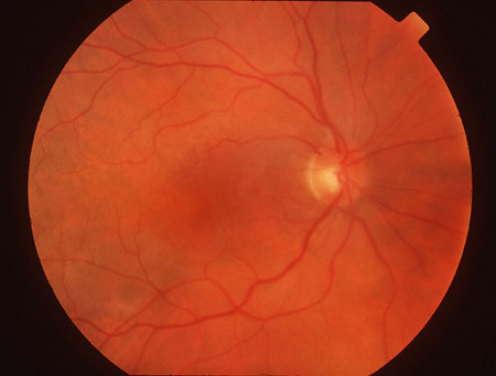
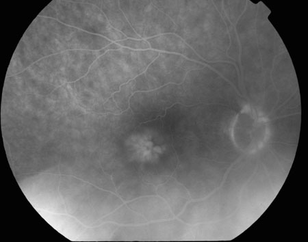
Other causes of postsurgical CME include:
- Penetrating keratoplasty
- Glaucoma filtering procedures
- Scleral buckling procedures
- Prophylactic treatment of retinal tears
Although the etiology of postsurgical CME is unknown, vitreomacular traction, inflammation, breakdown of the blood-ocular and blood-aqueous barriers, and photoxicity may be important factors.
CME is also associated with ocular diseases, such as uveitis. The association of CME with uveitides, like pars planitis, ocular sarcoidosis, and birdshot retinochoroidopathy (Slides 3 and 4) underscores the role of inflammation in the pathogenesis of CME. The development of CME can also complicate central and branch vein occlusions. The etiology of the macular edema in these cases is presumed to be secondary to alterations in the blood-retinal barrier distal to the occlusion site.
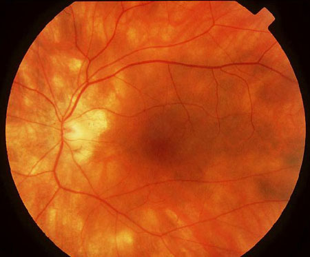
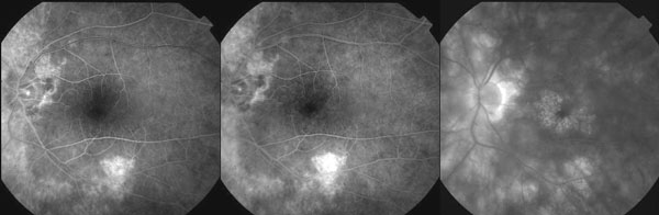
Other ocular diseases associated with CME include:
- Diabetic retinopathy
- Hypertensive retinopathy
- Epiretinal membranes
- Intraocular tumors
- Macroaneurysms
- Retinitis pigmentosa
- Choroidal neovascularization
Clinical Findings
The diagnosis of CME can often be made clinically with slit lamp biomicroscopy and a fundus contact lens. The anterior segment may reveal low grade inflammation, and in postsurgical cases, there may be complications such as vitreous or iris incarceration, IOL pupillary capture, corneal edema, or posterior capsular rupture.
The macula has a poor foveal reflex and appears thickened with multiple translucent intraretinal cystoid spaces best viewed using a thin, angled slit beam. In more subtle cases, a central foveal yellow spot can often be appreciated. Other findings include hemorrhage within the cystic spaces, serous macular detachment and optic disc edema. Chronic cases may demonstrate mottling of the retinal pigment epithelium.
Two conditions that can limit the potential for visual recovery in CME patients are epiretinal membranes (ERM) and foveal cysts. An ERM can be identified in 10% to 20% of cases of CME. A grayish, translucent membrane, straightening of retinal vessels and retinal striae are all clues to the presence of an ERM. A foveal cyst forms from the coalescence of intraretinal cysts. Foveal cysts can result in permanent visual loss if they unroof and form lamellar holes.
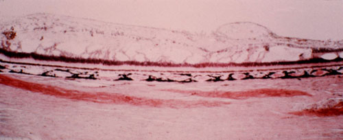
The clinical findings of CME are often mirrored histopathologically (Slide 5). Prominent cystoid spaces are present primarily in the outer plexiform layer of the fovea; cystoid spaces may also be seen in the inner nuclear, ganglion, and nerve fiber layers. These changes may stem from the accumulation of serous exudate in the extracellular space. Other histopathological findings include subretinal fluid, shortening of the inner and outer segment of the photoreceptors, and atrophy or proliferation of the retinal pigment epithelium.
Differential Diagnosis
- Macular hole
- Diabetic macular edema
- Photic maculopathy
- Retinal detachment
- Endophthalmitis
- Hypotonous retinopathy
- Adult vitelliform foveomacular dystrophy
- Nicotinic acid maculopathy
- X-linked retinoschisis
Diagnostic Features
Fluorescein angiography is the most important diagnostic test to detect the presence of CME: in the early phases, dilation and leakage of the perfoveal retinal capillaries can be appreciated; in the later phases, pooling of dye in a petalloid pattern in the perifoveal region occurs. In more severe cases, a honeycomb pattern of dye pooling can be seen outside the perifoveal region. Leakage of capillaries in the optic nerve head resulting in late staining of the optic nerve is usually present as well. Conditions such as nicotinic acid maculopathy or X-linked juvenile retinoschisis that resemble CME clinically can be distinguished from CME by their failure to demonstrate leakage on fluorescein angiography.
The amount of dye leakage present on the angiogram correlates poorly with visual acuity, hence the distinction between clinical CME that is associated with visual loss and angiographic CME that may or may not be associated with visual loss.
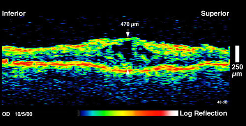
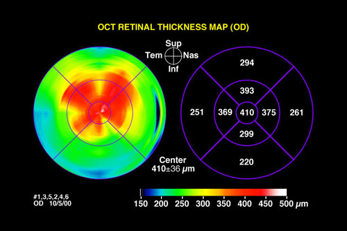
Retinal thickness is a better correlate of visual acuity. Optical coherence tomography (OCT) is an excellent method of quantifying retinal thickness as well as identifying the presence of cystoid changes in the retina 6A (Slides and 6B).
Treatment of Postsurgical CME
While postsurgical CME usually resolves spontaneously in 3 to 12 months, it can cause permanent visual loss in approximately 1% of patients undergoing extracapsular cataract extraction.
Medical treatment has centered upon decreasing ocular inflammation. Topical, peribulbar and oral corticosteroids, which block the release of arachadonic acid, have been studied for the treatment of CME. While visual improvement has been documented in many cases, no randomized, double-masked studies have been conducted to confirm these findings. In addition to decreasing inflammation, corticosteroids may also decrease CME by elevating intraocular pressure. CME may recur when the corticosteroids are discontinued. Corticosteroids can also impede wound healing, exacerbate hepatic keratitis, and promote steroid-induced glaucoma.
Oral and topical cyclooxygenase inhibitors (COIs), which block the synthesis and release of prostaglandins, offer the potential to treat ocular inflammation without risk of glaucoma. Studies of COIs for the prevention of CME in surgical patients have failed to demonstrate any impact on visual outcome. When COIs were administered to patients with established clinical CME, some improvements in visual acuity were found, but "on-off" effects upon starting or discontinuing the COIs were also noted. One randomized, prospective, double-masked, placebo-controlled trial showed that 1 drop of topical 0.5% ketorolac administered 4 times a day was effective in improving visual acuity in clinical CME patients. Limitations of the study included a short, 3-month follow-up period, and failure to compare ketorolac with corticosteroids.
The medical treatment of postsurgical CME has also included carbonic anhydrase inhibitors (CAIs), such as oral acetazolamide, on the theory that they may improve the ability of the retinal pigment epithelial pump to remove retinal edema. However, only a small number of patients benefited in these nonrandomized studies and "on-off" effects were also noted. The possible benefits of CAIs must also be weighed against the risks of bone marrow depression, aplastic anemia, and Stevens-Johnson syndrome.
Surgical treatments for pseudophakic CME include Nd:YAG vitreolysis, vitrectomy, and grid laser photocoagulation. Some studies have suggested that Nd:YAG vitreolysis may provide some visual benefit for patients with CME and vitreous incarceration in the cataract wound. However, the studies were uncontrolled and involved the concomitant use of topical steroids. In addition, since many of the patients had CME for only short periods of time, it is possible that they may have improved spontaneously without intervention.
Vitrectomy offers the possibility of excising incarcerated vitreous, relieving any vitreomacular traction, and removing inflammatory mediators in the vitreous. A multicenter, randomized, prospective clinical trial demonstrated that vitrectomy conferred a small benefit in aphakic patients with CME. Vitrectomy for pseudophakic patients with vitreous incarceration or iris capture may also provide some visual benefits. The benefits of vitrectomy in psuedophakic CME patients without any anterior segment complications are unclear.
Grid laser photocoagulation for chronic CME resulted in some visual improvement in several small, uncontrolled studies. However, this treatment may pose unnesessary risk of accelerated retinal pigment epithelial atrophy and choroidal neovascularization, especially in elderly patients.
Treatment of Nonsurgically Induced CME
The management of nonsurgically induced CME is based upon the underlying etiology. In uveitis, the treatment of CME focuses on reducing the inflammation. Corticosteroids, nonsteroidal anti-inflammatory drugs, CAIs, and immunosuppressive therapy have all been used with varying degrees of success. Vitrectomy in cases of uveitis with chronic CME unresponsive to corticosteroids has also shown some short-term visual improvement.
The Central Vein Occlusion Study demonstrated that macular grid photocoagulation for CME had no significant impact on final visual acuity. CME following branch retinal vein occlusion (BRVO), however, can be treated with macular grid photocoagulation according to the Branch Vein Occlusion Study criteria. Specifically, BRVO patients must have:
- Macular edema of more than 3 months duration
- Visual acuity of 20/40 or worse
- Intact foveal capillary zone
The average final visual acuity of treated patients was 20/40; 65% of treated patients gained 2 or more lines of vision.
CME secondary to retinitis pigmentosa has been treated with CAIs with moderate success; CAIs have also been used to treat CME associated with ERMs. More studies are required to define the optimal treatment for CME in these and other less common conditions.
Bibliography
Cox SN, Hay E, Bird AC. Treatment of chronic macular edema with azetazolamide. Arch Ophthalmol. 1988;106:1190-1195.
Fishman GA, Glenn AM, Gilbert LD. Rebound of macular edema with continued use of methazolamide in patients with retinitis pigmentosa. Arch Ophthalmol. 1993;111:1640-1646.
Flach AJ, Dolan BJ, Irvine AR. Effectiveness of ketorolac tromethamine 0.5% ophthalmic solution for chronic aphakic and pseudophakic cystoid macular edema. Am J Ophthalmol. 1987;103:479-486.
Flach AJ, Jampol LM, Weinberg D, et al. Improvement in visual acuity in chronic aphakic and pseudophakic cystoid macular edema after treatment with topical 0.5% ketorolac tromethamine. Am J Ophthalmol. 1991;112:514-519.
Gass JDM. Lamellar hole: A complication of cystoid macular edema after cataract extraction: A clinicopathological case report. Trans Am Ophthalmol Society. 1975;73:271.
Gass JD, Norton EW. Cystoid macular edema and papilledema following cataract extraction: A fluorescein and fundoscopic study. Arch Ophthalmol. 1966;76:646-661.
Harbour JW, Smiddy WE, Rubsamen PE, et al. Pars plana vitrectomy for chronic pseudophakic cystoid macular edema. Am J Ophthalmol. 1995;120:302-307.
Irvine AR. A newly defined vitreous syndrome following cataract surgery: Interpreted according to recent concepts of the structure of the vitreous. Am J Ophthalmol. 1953;36:599-619.
Nussenblatt RB, Kaufman SC, Palestine AG, et al. Macular thickening and visual acuity: Measurement in patients with cystoid macular edema. Ophthalmology. 1991;94:1134-1139.
Steinert RF, Wasson PJ. Neodymium:YAG laser anterior vitreolysis for Irvine-Gass cystoid macular edema. J Catract Refract Surg. 1989;15:304.