Introduction
Central serous chorioretinopathy (CSC) is a common idiopathic disorder characterized by a serous detachment of the neurosensory retina. CSC typically occurs in adults ranging from 20 to 50 years old. Patients may present over the age of 50, but these cases must be differentiated from choroidal neovascularization (CNV) secondary to age-related macular degeneration (AMD). Men are affected 10 times more often than women in the 20- to 50-year-old age group. However, the male/female ratio decreases to 2:1 in patients older than the age of 50, as women are more commonly affected in this age group. Affected patients are often reported to have type A personalities with a history of stressful events prior to presentation. Other conditions associated with CSC include endogenous hypercortisolism, pregnancy, and systemic corticosteroid treatment. CSC occurs most commonly in Caucasians, Hispanics, and Asians, but is relatively uncommon in African Americans. The pathogenesis of CSC remains unknown. Many theories exist regarding the formation of the serous retinal detachment including a focus of hypersecreting retinal pigment epithelium (RPE) cells, and a breakdown in the blood-retina barrier. Prünte and Flammer in 1996 hypothesized that capillary and venous congestion in the choroid with focal ischemia of choroidal lobules was the cause of CSC. In their study, leakage from the RPE or a retinal pigment epithelial detachment occurred only in areas of capillary or venous congestion delineated by indocyanine green angiography (ICG). Prünte and Flammer felt that the choroidal congestion caused a hyperpermeable state with leakage of fluid and plasma proteins from the choroidal capillaries. Fluid would then pool underneath the RPE cell layer causing a serous retinal pigment epithelial detachment. With stretching of the RPE cells, a mechanical breakdown of the blood-retinal barrier occurs with leakage of fluid through the RPE cell junctions into the subretinal space. A model of CSC has been reproduced in animals given intravenous injections of epinephrine. In addition, there is a relationship between corticosteroid use and the development of CSC. Therefore, a hormonal imbalance or dysregulation of the sympathetic nervous system may lead to choroidal or RPE abnormalities producing CSC.Clinical Findings
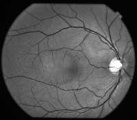
Patients present with a monocular decline in visual acuity typically in a range of 20/20 to 20/200, with an average acuity of 20/30 or better. Visual acuity may be improved with a slight hyperopic correction. Symptoms include metamorphopsia, micropsia, central scotoma, diminished color vision, and a delay in retinal recovery time after exposure to bright lights. Some patients complain of migraine-like headaches just prior to ocular symptoms. The anterior segment, vitreous, optic nerve and retinal vasculature are normal on examination. The classic presentation of CSC is a round, well-delineated serous retinal detachment in the macula. Pigment epithelial detachments (PED) are often observed, as they may be an integral part of the pathogenesis of the disorder. The PED is a discrete round elevation that may be present under the superior portion of the detached macula, or just outside the superior boarder of the serous macular detachment (Slide 1A. and Slide 1B.) This serous retinal detachment is usually inferior to the PED from the gravitational effects on the subretinal fluid. Pigment epithelial detachments may also be present as solitary lesions in the macula (Slide 2A. and Slide 2B.) RPE atrophy and yellow-white deposits may represent evidence of prior episodes of CSC in either the affected or fellow eye. RPE atrophy may also represent shifting of subretinal fluid. Yannuzzi described subretinal tracts of RPE atrophy occurring outside the macula in the inferior peripheral retina secondary to prior retinal detachments. Subretinal precipitates or turbid serous macular detachments represent fibrin or protein deposition. An uncommon finding is multiple bullous retinal detachments.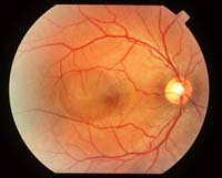
Differential Diagnosis
- Age-related macular degeneration
- Optic nerve pit
- Macular hole
- Harada's disease
- Metastatic cancer
- Presumed ocular histoplasmosis syndrome
- Choroidal metastasis
- Hypertension
- Retinoschisis
- Lymphoma
- Optic neuritis
Diagnostic Features
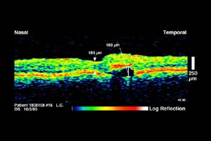
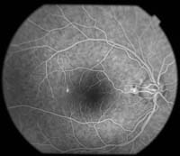
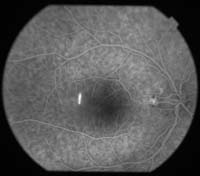
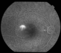
The diagnosis of central serous chorioretinopathy is made through obtaining patient history and clinical examination. Often the typical presentation of a young man with a serous macular detachment is sufficient. Biomicroscopy demonstrates a blister-like elevation of the retina. If the diagnosis is uncertain or one cannot exclude a CNV, then fluorescein or ICG angiography should be performed. Optical coherence tomography is useful in documenting subretinal fluid and pigment epithelial detachments (Slides 2A and 2B and 3). Fluorescein angiography demonstrates pinpoint hyperfluorescence in the early phase of angiography in over 95% of cases. The punctate area of hyperfluorescence represents fluorescein dye leakage through a defect in the RPE. The hyperfluorescence enlarges concentrically and stains the subretinal fluid in the later phases of the angiogram. Up to 20% of angiograms may demonstrate the classic smokestack pattern of leakage (Slides 4A, 4B, 4C). In this angiographic pattern, there is an initial punctate area of hyperfluorescence that rises in a linear fashion secondary to osmotic pressure gradients in the subretinal fluid. In most cases, there is one area of leakage that tends to occur in a circular zone 1 mm from the center of the fovea. Approximately 25% of cases may exhibit more than one area of leakage. ICG is advantageous because it delineates the choroidal circulation better than fluorescein. In most cases there is a direct correlation between the area of active leakage on fluorescein angiography and ICG. However, choroidal abnormalities identified on ICG may occur independently of areas previously identified as abnormal on fluorescein angiography. Hyperfluorescence on ICG may also present bilaterally in eyes that have inactive disease representing diffuse disturbance of the RPE and choroid. The hyperfluorescence on ICG is related either to hyperpermeability of the choroidal vessels, or to capillary and venous congestion in areas of delayed arterial filling. ICG characteristics of pigment epithelial detachments in CSC are different from AMD. In CSC, there is early staining with late hypofluorescence surrounded by a distinct ring of fluorescence at the margins. In AMD, serous PEDs remain hypofluorescent throughout the entire angiogram.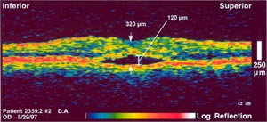
Clinical Course
In most cases of CSC, the serous retinal detachments are self-limited and resolve without intervention. Visual acuity typically remains excellent with most patients retaining a final visual acuity of 20/40 or better. Klein followed 34 untreated eyes of patients with CSC and observed resolution of the serous macular detachment in all patients with 94% achieving a final visual acuity of 20/30 or better. Improvement in visual acuity may continue for up to 1 year. Despite complete resolution, patients may complain of metamorphopsia, decreased contrast sensitivity, nyctalopia, and dyschromatopsia. Twenty percent to 50% of patients will experience a recurrence within 1 year, but recurrences can occur years later. Often, clinical examination will reveal evidence of prior asymptomatic attacks. Nearly 5% of patients will have a complicated clinical course with bullous retinal detachments, severe degeneration of the RPE, cystoid macular degeneration, and CNV.
Treatment
The 2 main available treatment options are observation and focal laser to the area of leakage on fluorescein. No known medical treatment exists, and treatment with beta-blockers has not proven beneficial. Resolution of the serous retinal detachment with observation occurs over a 3- to 4-month period. When focal photocoagulation is applied to the area of leakage on fluorescein, the serous detachment has been shown to resolve over a 2- to 3-week period. Final visual acuity tends to be the same whether treatment is by observation or laser. The risks of laser treatment include accidental photocoagulation of the fovea, CNV, and scotomata. Since most cases are self-limited with patients' recovering visual acuity at 20/30 or better, the initial treatment of CSC should be observation. Laser treatment is recommended for patients who require binocular vision and rapid resolution of their visual impairment for occupational reasons. Laser should also be considered in patients with nonresolving subretinal fluid for more than 4 to 6 months, or poor visual outcome in the opposite eye from prior serous retinal detachments. Laser should be avoided to the foveal avascular zone.
Bibliography
Bibliography Hall LS, Guyer DG, Yanuzzi LA. Central serous chorioretinopathy. In: Guyer DG, et al (eds.). Retina, Vitreous, Macula. Philadelphia: WB Saunders; 1999:206-216.
Klein ML, Van Buskirk EM, Friedman E, et al. Experience with nontreatment of central serous choroidopathy. Arch Ophthalmol. 1974;91:247-250.
Prunte C, Flammer J. Choroidal capillary and venous congestion in central serous chorioretinopathy. Am J Ophthalmol. 1996;121:26-34.
Watzke RC, Burton TC, Woolson RF. Direct and indirect laser photocoagulation of central serous choroidopathy. Am J Ophthalmol. 1979;99:914-918.
Wirostko WJ, Pulido JS. Central serous chorioretinopathy. In: Yanoff M, Duker JS. Ophthalmology. Mosby; 1998.
Yannuzzi LA, Shakin J, Fisher Y, et al. Peripheral retinal detachment and retinal pigment epithelial atrophic tracts secondary to central serous pigment epitheliopathy. Ophthalmology. 1984,91:1554-1572