Age-related macular degeneration (AMD) is the most common cause
of central vision loss in persons older than 50 years of age in developed
countries. The incidence of AMD increases after age 50 with approximately one
third of people over the age of 90 affected. There are two clinical forms of
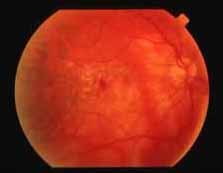
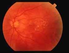
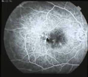
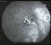
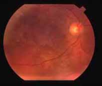
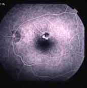
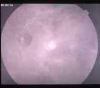
AMD: neovascular AMD
(Slides 1A,
1B,
2A and
2B) and
nonneovascular AMD. Although the nonneovascular form of AMD accounts for 90% of
cases, the overwhelming majority of eyes suffer severe vision loss as a result
of the neovascular form of AMD.
The pathogenesis of AMD remains poorly understood. Genetic
influences in AMD are difficult to study as most patients with this disease are
of advanced age. Some families demonstrate an autosomal dominant pattern and a
high degree of concordance is observed in monozygotic and fraternal twins.
Nutrition has also been suggested to play a role in the pathogenesis of AMD but
most studies have been inconclusive. Dietary supplementation with zinc,
antioxidants, and carotenoids (lutein and zeaxanthin) may help to protect
against severe vision loss in AMD but further investigation is merited.
Elevated cholesterol levels and increased dietary fat intake have been
suggested to increase the risk of AMD. Among other environmental factors,
systemic arterial hypertension and cigarette smoking are associated with an
increased risk of vision loss while increased ultraviolet light exposure
remains controversial as a risk factor for AMD.
Patients may be asymptomatic or may present with blurred vision
and/or metamorphopsia. Difficulty with dark adaptation and decreased reading
ability are also noted frequently. Patients with neovascular AMD typically
present with abrupt vision loss while patients with nonneovascular AMD present
with insidious loss of vision.
Drusen are a chief finding in the clinical presentation of both
forms of AMD. Drusen represent focal, yellow excrescences at the level of the
retinal pigment epithelium (RPE). Drusen may be divided into three types: hard
drusen, soft drusen, and basal laminar drusen.
Hard drusen are typically small (less than 63 µm in
diameter) and as they are not age-related and are ubiquitous, they are not
believed to be related to AMD. Basal laminar drusen are seen as uniformly
sized, round, raised yellow subretinal lesions that appear semitranslucent on
retro-illumination. The presence of basal laminar drusen confers an increased
risk of neovascular AMD. Soft drusen are larger, typically greater than 63
µm in diameter and have poorly demarcated borders (
Slide 3). These lesions are age related and reflect
diffuse disease of Bruch membrane. Therefore, their presence is a risk factor
for the development of neovascular AMD.
Other clinical findings such as focal RPE, hyperpigmentation,
punctate areas of RPE, hypopigmentation, and geographic atrophy (
Slide 4) are characteristics of nonneovascular AMD that
can lead to vision loss as a result of associated photoreceptor loss.
Neovascular AMD is characterized by the development of
choroidal neovascularization (CNV). CNV can result from cracks
in Bruch membrane, along with other ultra-structural abnormalities, that
allow ingrowth of new vessels from the choriocapillaris. The ingrowth of these
new vessels can lead to the following clinical findings:
- RPE detachment (hemorrhagic, serous,
fibrovascular)
- Subretinal fluid and/or blood
- RPE tear
- Intraretinal fluid and/or blood
- Subretinal fibrosis
- Disciform scar
- Intraretinal or subretinal lipid
It is the above features that are responsible for the majority
of vision loss in patients with AMD. Neovascular AMD most often occurs along
with features of nonneovascular AMD but may rarely occur without the atrophic
changes of nonneovascular AMD.
- Pattern dystrophy in the RPE
- Stargardt’s disease
- Best’s disease
- Central serous chorioretinopathy
- Bilateral juxtafoveal telangiectasis
- Toxic maculopathy (e.g., chloroquine)
- CNV from other causes (e.g., myopia, angioid streaks)
Although the clinical examination can make the diagnosis,
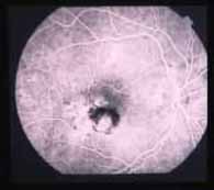
fluorescein angiography is useful to determine the presence and extent of CNV
if it is clinically suspected. Two patterns of CNV are recognized: classic and
occult CNV. Classic CNV is characterized by well-demarcated, bright areas of
hyperfluorescence that can be noted in the early phase of the angiogram (
Slide 5). Progressive dye leakage occurs in the later
frames.
Occult CNV is recognized to occur as one of two patterns:
fibrovascular RPE detachment and late leakage of undetermined source. In a
fibrovascular RPE detachment, areas of irregular elevation of the RPE can be
seen on stereoscopic angiography and stippled hyperfluorescence is noted in the
mid-phases of the angiogram. In late leakage of undetermined source, areas of
fluorescein leakage with a pattern of speckled hyperfluorescence are noted in
the late phases of the angiogram (
Slide 6). The source
of the leakage cannot be determined by the early phases of the angiogram.
RPE detachments due to hemorrhage or fluid beneath the RPE also
have characteristic features on fluorescein angiography. Hemorrhagic RPE
detachments demonstrate blocked fluorescence throughout the angiogram study.
RPE detachments due to fluid (serous RPE detachment) demonstrate well-defined
dome-shaped areas of homogenous bright fluorescence in the early phases with
persistence of hyperfluorescence into the late phases of the angiogram.
Other ancillary tests may be useful in the diagnosis and
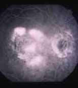
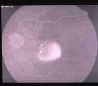
management of AMD. Indocyanine green (ICG) angiography has been used, with
variable success, to help better delineate lesion boundaries that are not
well-defined on fluorescein angiography (
Slides 7A
and
7B). The ICG appearance of CNV may be classified
as a hot spot (focal areas of hyperfluorescence less than one disk diameter in
area), plaquoid hyperfluorescence (either well defined or poorly defined), or
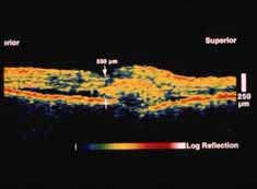
as a combination. Optical coherence tomography (OCT) may also be effective as
an ancillary test. OCT can distinguish between the various types of RPE
detachments, quantify the extent of retinal thickening, and image choroidal
neovascular membranes in the subretinal space (
Slide
8).
Currently, there is no proven treatment or prophylaxis for
nonneovascular AMD. Nutritional supplementation is being investigated as a
means of prophylaxis. Laser treatment of drusen in eyes with nonneovascular AMD
is being investigated as a means of preventing the development of CNV in eyes
at risk. This treatment is still unproven and early studies have shown that the
risk of CNV may actually increase with laser therapy to drusen.
The Macular Photocoagulation Study (MPS) was a multicenter,
randomized clinical trial that demonstrated a positive treatment effect with
laser photocoagulation in eyes with CNV secondary to AMD. The MPS divided
lesions into extrafoveal, juxtafoveal, and subfoveal lesions depending on
lesion proximity to the center of the fovea. Based on MPS results,
well-demarcated extrafoveal CNV should be strongly considered for treatment
with laser photocoagulation. Well-demarcated juxtafoveal CNV should be
considered for treatment with the understanding that there is a high recurrence
rate for such lesions. Well-demarcated new and recurrent subfoveal CNV may be
considered in selected patients. In these cases, both the patient and the
physician should be willing to accept an immediate loss of vision following
treatment with the understanding that long-term results are better with
treatment.
The MPS recommendations include only eyes with well-demarcated
lesions. However, the majority (85%) of eyes affected by neovascular AMD
present with occult lesions that do not meet MPS eligibility criteria.
Furthermore, high recurrence rates and the immediate reduction of vision
experienced by some patients make laser photocoagulation a less than perfect
treatment. Therefore, a number of clinical trials are being conducted to
improve treatment outcomes and broaden the treatment criteria for patients with
neovascular AMD.
Surgical therapies such as submacular surgery to directly
remove CNV and macular translocation are under investigation but currently
remain unproven. Angiogenesis inhibitors such as thalidomide are being
investigated as adjunctive treatment to laser photocoagulation. Photodynamic
therapy (PDT), transpupillary thermotherapy, and external beam radiation
therapy are other investigational treatment modalities for the treatment of CNV
associated with AMD.
PDT is the most promising therapy, whereby photosensitive dye
is given intravenously prior to treatment of the CNV with a low-level light
source. Theoretically, subfoveal lesions can be treated with minimal damage to
the overlying retina. Phase I/II studies have been promising and long-term
follow-up from phase III trials is anticipated.
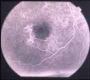
Slides
9 and
10 demonstrate the dramatic decrease in
leakage following PDT. In
Slide 9, diffuse leakage is
noted in the right eye of a patient with AMD. One month following PDT, there is
a reduction in the amount of leakage shown in
Slide
10.