Introduction
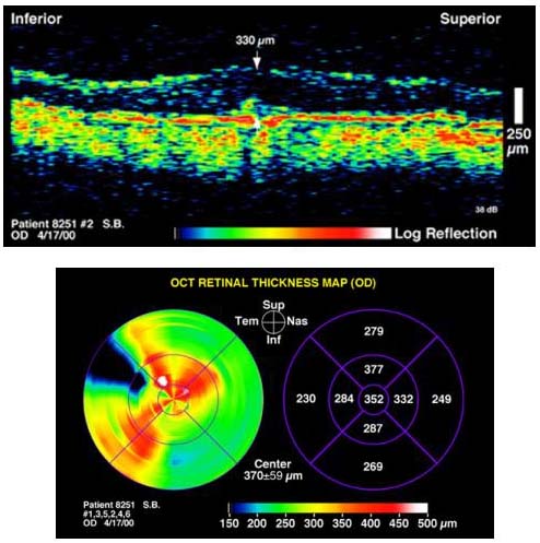
This tutorial reviews the natural history and treatment outcomes of subfoveal choroidal neovascularization (CNV) in ocular histoplasmosis syndrome, idiopathic cases, pathologic myopia, and angioid streaks. In these conditions, subfoveal CNV generally impacts patients younger than 50 years and is not amenable to thermal laser photocoagulation. Aside from observation, the two primary treatment options are photodynamic therapy and submacular surgery. Submacular surgery is an option in these patients because they typically have well-defined CNV located in the subretinal space anterior to the retinal pigment epithelium (RPE). These anatomical characteristics may allow the surgeon to remove the membrane with minimal damage to the RPE and choriocapillaris. Case selection is facilitated by imaging modalities, such as optical coherence tomography, that can accurately confirm the location of choroidal neovascular complexes in the subretinal space. To date, studies of submacular surgery in CNV not related to age-related macular degeneration have been small or retrospective and the results must be weighed accordingly. Until more definitive conclusions are available from the Submacular Surgery Trial, a thorough understanding of these studies and their limitations (Tables 1, 2, 3 and 4) will enable the clinician to more effectively manage young patients with subfoveal CNV.
Ocular Histoplasmosis Syndrome
Ocular histoplasmosis syndrome is a common cause of CNV in young adults. The classic clinical triad for ocular histoplasmosis syndrome includes CNV, peripapillary atrophy, and peripheral "punched-out" chorioretinal lesions or "histo spots." Ocular histoplasmosis syndrome is also characterized by the absence of vitreal inflammation. The fungus Histoplasma capsulatum has been implicated as the etiologic agent for ocular histoplasmosis syndrome based on positive skin testing and activation of the chorioretinal lesions upon exposure to the histoplasmin antigen. The most important evidence, however, is epidemiological:
- The highest prevalence of ocular histoplasmosis syndrome occurs in areas where H. capsulatum is endemic, such as the Ohio and Mississippi River valleys.
- Ocular histoplasmosis syndrome is rare outside endemic areas
- Most ocular histoplasmosis syndrome patients live in or have previously lived in one of these endemic areas
- More than 90% of patients with ocular histoplasmosis syndrome have positive skin tests for H. capsulatum.
Although the organism has been identified in eyes from patients with disseminated histoplasmosis, it has never been cultured from or histopathologically observed in a patient with ocular histoplasmosis syndrome. The presence of HLA markers B7, DR2, and DQ1 may genetically predispose patients to ocular histoplasmosis syndrome. Myopic patients with ocular histoplasmosis syndrome may also be at higher risk for developing CNV.
CNV from ocular histoplasmosis syndrome usually impacts adults in the third and fourth decades of life. Lewis and coworkers1,2 found the average age of patients with symptomatic ocular histoplasmosis syndrome was 40 years in first eyes and 44 years in second eyes. In one endemic area, the prevalence of ocular histoplasmosis syndrome was 4.4 % and the prevalence of macular disciform scarring was 0.1%. Although ocular histoplasmosis syndrome is much less prevalent in African Americans, they are still at risk for significant visual loss secondary to macular disciform scarring.
Natural History
There is a significant risk for visual loss in patients with ocular histoplasmosis syndrome with subfoveal CNV. Sixty-seven percent to 86% of these eyes will have a final visual acuity of 20/200 or worse. Spontaneous involution of CNV with a final visual acuity of 20/40 or better has been reported in up to 14% of eyes with ocular histoplasmosis syndrome and subfoveal CNV. More favorable visual outcomes have been found in young patients, and patients with better initial visual acuities and smaller neovascular complexes with a surrounding ring of pigmentation. This pigmentation is often evident as a dark rim of hypofluorescence on fluorescein and indocyanine green angiography. The risk for developing macular CNV in fellow eyes is 20% to 25%. The Macular Photocoagulation Study Group found a 2% incidence per year of CNV in fellow eyes of patients with ocular histoplasmosis syndrome with juxtafoveal or extrafoveal CNV after 5 years of follow-up; 8% of patients with bilateral CNV were legally blind. The average time interval between CNV in first and second eyes is 4 years.3 The presence of macular histo spots is the most significant risk factor for the development of CNV in fellow eyes. Recurrent CNV after laser photocoagulation for juxtafoveal or extrafoveal CNV can be problematic: in the Macular Photocoagulation Study, 40% of persistent or recurrent CNV in patients with ocular histoplasmosis syndrome was subfoveal; the median visual acuity in these patients was 20/125.
Treatment
Treatment of subfoveal CNV in presumed ocular histoplasmosis syndrome is challenging. Anecdotal reports have suggested that corticosteroids may be beneficial for treatment of CNV in ocular histoplasmosis syndrome, but no prospective controlled studies have verified these findings. A small pilot study of thermal laser photocoagulation of subfoveal CNV had equivocal results and was discontinued due to low enrollment. Due to its location anterior to the retinal pigment epithelium, subfoveal CNV in ocular histoplasmosis syndrome may be amenable to surgical removal. Thomas and coworkers reported results of surgical removal of subfoveal CNV in 67 eyes with OHS.4 Mean follow-up time was 10.5 months. Preoperative visual acuity was 20/200 or worse in 48% of eyes. Thirty-nine percent of eyes received previous laser treatment. Postoperatively, 48% of eyes had vision 20/200 or worse. The mean visual improvement was 1.4 Snellen lines. Visual acuity was 20/40 or better in 31% of eyes. Recurrent CNV occurred in 37% of eyes; in eyes followed for 12 months or more, the rate increased to 46%. Recurrence did not have a significant effect on visual outcome. Surgical complications occurred in 16% of eyes, including retinal detachment in four eyes and macular pucker in one eye. A follow-up study by the same group reported on surgical outcomes of 117 consecutive eyes with presumed ocular histoplasmosis syndrome and subfoveal CNV. After a median follow-up time of 13 months, the mean visual improvement was 3 lines of visual acuity.
Preoperative visual acuity was significantly correlated with visual outcome. Forty-four percent of eyes with a visual acuity of 20/100 or better obtained a postoperative visual acuity of 20/40 or better; in contrast, 25% of eyes with preoperative visual acuity of 20/200 or worse obtained a visual acuity of 20/40 or better. Recurrent CNV developed in 44% of eyes and, similar to their earlier study, was not significantly associated with a poorer visual outcome. Surgical complications developed in 14% of eyes.
Another study reported surgical outcomes on 63 eyes with ocular histoplasmosis syndrome and subfoveal CNV followed for a median of 24 months. Preoperatively, 81% of eyes had visual acuity of 20/200 or worse; mean visual acuity was 20/200. Fifty-one percent of eyes had received previous laser surgery. Postoperatively, 63% of eyes had visual acuity of 20/200 or worse. Mean visual acuity was 20/200; 35% of eyes improved by two or more lines of visual acuity. Factors that correlated with improved visual prognosis include younger age, the lack of previous laser photocoagulation, and in contrast to previous studies preoperative visual acuity of 20/200 or worse. There was a 38% CNV recurrence rate after surgery; 67% of cases were subfoveal, often necessitating additional surgery. In contrast to earlier studies, most eyes with recurrent CNV had a poor visual prognosis. Three percent of patients developed a retinal detachment.
While encouraging when compared to natural history studies, these surgical results must be interpreted cautiously due to the lack of controls, the short follow-up periods, and the risk of postoperative complications. More conclusive evidence on the role of surgery in the management of subfoveal CNV in ocular histoplasmosis syndrome will be forthcoming in the randomized, prospective, controlled, multicenter Submacular Surgical Trial.
Myopia
Pathological myopia occurs in 2% of myopes (Slide 1). The pathogenesis of pathologic myopia remains unclear, though genetic and environmental factors may play a role. Pathologic myopia is characterized by excessive axial length with secondary degenerative changes in the sclera, choroid, Bruch's membrane, retinal pigment epithelium, and retina. These changes are concentrated in the ora-equatorial zone and the posterior pole, and typically occur in eyes with more than 6 D of myopia and more than 25 mm of axial length. Ora-equatorial zone findings include "white without pressure," lattice degeneration, and pigmentary and paving-stone degeneration. Common posterior pole changes include optic nerve crescents, chorioretinal atrophy, lacquer cracks, and posterior staphyloma. Posterior staphyloma is an important cause of blindness in pathologic myopes; other vision-threatening complications include retinal detachment, chorioretinal atrophy, lacquer cracks, and CNV.
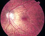
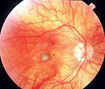
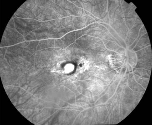
CNV occurs in 5% to 10% of patients with pathologic myopia and is subfoveal in more than 50% of cases (Slide 2, Slide 3, Slide 4, Slide 5, Slide 6). Linear breaks in Bruch's membrane secondary to stretching and degeneration of the choroid may place these eyes at risk for CNV. These breaks often appear clinically as lacquer cracks. Lacquer cracks have been associated with CNV in 82% of eyes with pathologic myopia; microscopic defects in Bruch's membrane may be linked to CNV in eyes without lacquer cracks. It is unclear, however, what causes the growth of CNV through the breaks in Bruch's membrane. Quiescent CNV in myopes is often associated with a Fuch's spot. More than 95% of CNV in pathologic myopes demonstrates mild hyperfluorescence confined to the early transit phase of the angiogram with minimal leakage beyond the borders of the lesion. This CNV generally develops into a small atrophic scar, often with a surrounding rim of pigmentation characteristic of a Fuch's spot. Since this pattern of CNV occurs in eyes with advanced myopic degeneration, its growth may be limited by underlying circulatory disturbances in the choroid and choriocapillaris due to mechanical stretching. CNV associated with more widespread leakage on angiography and the development of exudative disciform changes typically occurs in eyes with mild myopic degeneration.
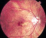
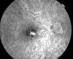
Natural History
Most natural history studies of CNV in pathologic myopia have not been limited to subfoveal CNV, and therefore results must be interpreted conservatively. One study followed 27 myopic eyes with CNV for a mean of 25.5 months; six eyes had received previous laser photocoagulation. Fifty-one percent of eyes lost 2 or more lines of visual acuity compared to baseline; 44% progressed to 20/200 or worse. Hampton and coworkers followed 42 eyes with CNV, most of them for less than 2 years.5 Fifty-eight percent had subfoveal CNV. Twenty-six percent had visual acuity of 20/200 or worse at baseline. The researchers reported that 43% of eyes lost 2 or more lines of visual acuity and that 60% were left with 20/200 or worse visual acuity. Patients older than 30 years had a poorer visual prognosis. Another study reported on 70 eyes with CNV followed for 41 months. The CNV in the macula was not subclassified. The average age of the patients was 52 years. Fifty-seven percent had vision 20/200 or worse at baseline. Forty-six percent of eyes had decreased visual acuity from baseline; 57% had visual acuity of 20/200 or worse. However, poor visual acuity in the majority of cases was due to posterior staphylomas, not CNV; most of the eyes developed stable, nonexudative atrophic scars. An additional study followed 100 eyes with primarily juxtafoveal CNV for five years. During the study period, all patients developed subfoveal CNV; their final visual acuity was 20/160. Older patients may have a poorer visual prognosis. In one study of 22 eyes with CNV and pathologic myopia, the average age of the patients was 63 years. Fifty percent had vision of 20/200 or worse at baseline. Fifty-five percent of eyes had subfoveal CNV. At 1-year follow-up, 73% had vision worse than 20/200, though 64% had the same or better vision at baseline.
One prospective natural history study of subfoveal CNV has been published from the Verteporfin in Photodynamic Therapy (VIP) Study Group.6 In this study, patients with myopia and subfoveal CNV were randomly assigned to verteporfin (n=81) or placebo (n=39). The median age was 46 years. Baseline median visual acuity was 20/64. Only 5% of eyes had baseline visual acuity worse than 20/100. At the 1-year follow-up, 26% of eyes in the placebo group had a visual acuity of 20/160 or worse; the median visual acuity was 20/80. These encouraging outcomes are due in part to the good baseline visual acuities of the VIP study eyes. They may not be comparable to the aforementioned natural history studies, which had much higher percentages of eyes with 20/200 or worse baseline visual acuity.
Treatment
Thermal laser photocoagulation of subfoveal CNV results in an immediate permanent decrease in vision with an absolute scotoma. In pathologic myopes, the progressive enlargement of the laser scar can result in additional loss of visual acuity. This knowledge, combined with the generally small atrophic scars that develop in myopes with CNV, has limited the value of laser photocoagulation in these patients.
Subretinal surgery may offer some visual benefits in pathologic myopes with subfoveal CNV, especially those younger than 40 years. In one study, surgical removal of subfoveal CNV was performed in 10 eyes with a mean follow-up of 7 months. The median patient age was 39 years. Two eyes had received previous laser photocoagulation. Three eyes (30%) had a preoperative visual acuity of 20/200 or worse. The study reported a mean decrease of 1 line of preoperative visual acuity. Visual acuity was 20/200 or worse in four (40%) of the eyes. Two (20%) eyes had recurrent CNV.
Adelberg and coworkers surgically removed subfoveal CNV in five eyes with pathologic myopia, four of which had received previous laser photocoagulation.7 The mean age of the patients was 44 years; the mean follow-up time was 11.5 months. Three (60%) eyes had preoperative vision of 20/200 or worse. Four eyes (80%) had 20/200 or worse vision postoperatively. One eye had recurrent CNV.
Another study examined surgical outcomes in 21 eyes with subfoveal CNV secondary to myopia for a median of 12 months. None had previous laser treatment. The median age of the patients was 53. Eighty-one percent had preoperative vision worse than 20/200. Postoperatively, visual acuity stabilized or improved in 86% of eyes; visual acuity was 20/200 or worse in 62% of eyes. Nineteen percent of eyes had recurrent CNV. Ten percent of eyes had retinal detachments and 19% had cataract progression.
An additional study examined surgical outcomes in 23 eyes with a mean follow-up of 24 months. Thirty-one percent had preoperative vision of 20/200 or worse. Sixty-one percent had undergone previous laser photocoagulation. Visual acuity improved or stabilized in 65% of patients, with 48% of eyes left with a postoperative visual acuity of 20/200 or worse. Postoperative complications included retinal detachment (4%), endophthalmitis (4%), vitreous hemorrhage (4%), nuclear sclerotic cataracts (22%), and recurrent CNV (57%). Recurrent CNV was associated with a poorer prognosis. Patient age younger than 40 years significantly correlated with improvement of 2 or more lines of vision after surgery.
It is difficult to assess the role of subretinal surgery in the management of patients with subfoveal CNV secondary to pathologic myopia. Most studies have been retrospective and uncontrolled and have included cases of recurrent as well as primary CNV. In addition, their results are not directly comparable to the VIP natural history data due to the poorer baseline visual acuities. Despite these drawbacks, subretinal surgery may be an option in patients younger than 40 years. Randomized, prospective, controlled trials are needed to accurately assess the value of subretinal surgery in treating subfoveal CNV secondary to pathologic myopia. This would also facilitate comparison with outcomes from observation and photodynamic therapy in the VIP trial.
Idiopathic Choroidal Neovascularization
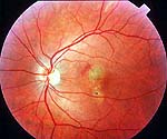
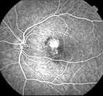
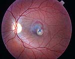
CNV in patients younger than 50 years often occurs secondary to degenerative changes such as pathologic myopia or angioid streaks, inflammatory disease such as ocular histoplasmosis or multifocal choroiditis, or trauma via choroidal rupture. Idiopathic CNV, however, occurs in this age group in otherwise healthy eyes (Slide 7, Slide 8, Slide 9, Slide 10). The prevalence of idiopathic CNV in the United States is not known. However, a study in France (where ocular histoplasmosis syndrome is not endemic) found that idiopathic CNV comprised 17% of all cases of CNV in patients younger than 50 years. The average age of the patients was 37 years; 30% had subfoveal CNV. Ten percent of patients had bilateral CNV. The presence of both B7 and DR2 HLA markers may genetically predispose an individual to idiopathic CNV. The relative risk for developing idiopathic CNV may also increase with diopters of myopia.
Natural History
Although the natural history of idiopathic subfoveal CNV has not been studied prospectively, it may have a better prognosis than CNV secondary to age-related macular degeneration, myopia, or presumed ocular histoplasmosis syndrome. A retrospective study by Ho and coworkers reported outcomes on 19 eyes with idiopathic CNV followed for an average of 88 months.8 The mean patient age was 32 years. The median initial visual acuity was 20/100; the median final visual acuity was 20/70. Thirty-one percent of patients had an initial visual acuity of 20/200 or worse; 31% of eyes had a final acuity of 20/200 or worse. Most significantly, 90% of patients improved or maintained their initial visual acuity. Age, sex, and initial visual acuity were not predictive of final visual acuity; CNV less than 1 disc area, however, was associated with a better visual outcome.
Treatment
No treatment for subfoveal idiopathic CNV has been shown to be effective in a randomized, controlled clinical trial. However, several small retrospective studies on the use of submacular surgery to remove CNV in the subretinal space have demonstrated encouraging results. In one study, eight eyes were followed for a mean of 6 months; three had received previous laser treatment. Preoperative visual acuity was 20/200 or worse in all cases. Four (50%) eyes remained 20/200 or worse postoperatively; two (25%) had 20/40 or better visual acuity. Recurrent CNV developed in four (50%) cases.
Another study followed for a mean of 18 months four eyes that underwent surgical removal of idiopathic subfoveal CNV. Two eyes had high myopia. One eye had received previous laser treatment. All had 20/200 or worse visual acuity preoperatively. One eye improved to 20/25; three eyes remained 20/200 or worse postoperatively. One eye developed a retinal break. Bottoni and coworkers removed idiopathic choroidal neovascular membranes from six eyes and followed them for a median of 18 months.9 Three (50%) eyes had preoperative visual acuity of 20/200 or worse. Postoperatively, two (33%) eyes had 20/200 or worse visual acuity and three had 20/40 or better visual acuity. Two (33%) eyes developed recurrent CNV. These studies must be interpreted with caution because of their retrospective nature and small size. The definitive role of submacular surgery in the management of idiopathic CNV will be defined by the submacular surgery trials.
Angioid Streaks
Angioid streaks are visible linear, full-thickness breaks in Bruch's membrane that extend radially from the peripapillary area (Slide 11 and Slide 12). They typically appear in the second decade of life and are usually bilateral and symmetric. The color of the streaks depends upon fundus pigmentation, ranging from dark brown in heavily pigmented patients to red in lightly pigmented patients. When the streaks atrophy, they take on a yellow appearance. A mottled peau d'orange fundus and optic disc drusen have also been associated with angioid streaks. This characteristic ophthalmoscopic appearance usually makes the diagnosis of angioid streaks straightforward; early in the disease, however, fluorescein and indocyanine green angiography may be helpful in revealing more subtle streaks and fundus mottling.
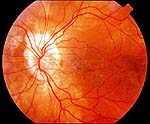
The pathogenesis of angioid streaks is unknown. Histopathologically, there is evidence of extensive calcium deposition in Bruch's membrane and breaks that correspond to the clinically observed angioid streaks. There is also atrophy of the underlying choriocapillaris and overlying RPE. The thickened and brittle Bruch's membrane may crack when subject to the routine stretching of the extraocular muscles; the characteristic radial pattern of the streaks may arise because the base of the vector forces exerted by the extraocular muscles is at the scleral foramen. These cracks in Bruch's membrane place patients at high risk for the development of CNV.
Angioid streaks have been classically linked to systemic diseases such as pseudoxanthoma elasticum (PXE), Paget's disease, and sickle cell anemia, all of which have histopathological evidence of abnormal calcium deposition in Bruch's membrane. PXE is the most common association occurring in up to 60% of patients with angioid streaks. Rarer systemic associations include Ehlers-Danlos syndrome, sickle-thalassemia, hereditary spherocytosis, and abetalipoproteinemia. Idiopathic angioid streaks occur in 35% to 50% of patients.
Visual outcomes are poor in patients with angioid streaks. In a group of 62 patients followed for a mean of 3.6 years, 50% were legally blind at their most recent follow-up visit. Aside from traumatic retinal hemorrhage, the most significant visual-threatening complication of angioid streaks is the development of CNV (Slide 13 and Slide 14). CNV can occur in 72% to 86% of eyes with angioid streaks and is bilateral in 49% to 60% of cases. The CNV is usually well defined and in the immediate proximity of a streak. The risk of CNV increases with age. Patients with PXE are at high risk for CNV-induced visual loss in the third and fourth decades of life. Clinical signs associated with CNV include the diffuse type of streaks, streaks longer than 3 disc diameters, and streaks passing through the fovea.
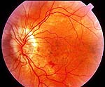
There are few natural history studies of CNV in patients with angioid streaks. Piro and coworkers retrospectively reported on 33 eyes that presented with active CNV and were followed without treatment for a range of 1 month to 14 years.10 Although the subclassification of CNV was not specified, 50% of eyes were legally blind at the time of their last examination. Laser treatment may not alter this course: in the same study, 11 patients were treated for extrafoveal lesions; eight patients (73%) were legally blind at the time of their last visit.
Although recent studies suggest that eyes with extrafoveal CNV may benefit from laser photocoagulation, especially if Macular Photocoagulation Study treatment criteria are rigidly adhered to with respect to intensity and extent of treatment, there is no effective treatment for subfoveal CNV. Several small series have reported discouraging results with submacular surgery. Thomas and coworkers reported on four eyes with subfoveal CNV secondary to angioid streaks followed for a mean of 7 months.4 Preoperative and postoperative visual acuity was 20/200 or worse in all eyes. One eye developed recurrent CNV. Adelberg and coworkers removed subfoveal choroidal neovascular membranes from five eyes with angioid streaks.7 The mean patient age was 47 years. The mean follow-up was 19 months. One eye had improvement of visual acuity from 20/300 to 20/50; the remaining eyes had postoperative vision worse than 20/200, including one eye that decreased from 20/80 to counting fingers. One eye developed a retinal detachment postoperatively. No recurrent CNV was reported.
Conclusions
Although further studies of photodynamic therapy and data from the Submacular Surgery Trial will ultimately determine the impact of these treatment modalities on the natural history of subfoveal CNV, the following trends can be gleaned from the data (see Tables 1, 2, 3 and 4):
- Patients with ocular histoplasmosis syndrome have poor visual outcomes when untreated; the most favorable results were observed after submacular surgery, especially if patients were younger than 40 years with good visual acuity and no history of previous laser photocoagulation.
- Younger patients with pathologic myopia and good initial visual acuity have a better natural history than those with ocular histoplasmosis syndrome; submacular surgery may be beneficial in patients with poorer visual acuity, especially patients older than 50 years.
- Untreated idiopathic cases have the most favorable visual outcomes compared to their counterparts with ocular histoplasmosis syndrome, myopia, or angioid streaks; it is unclear whether submacular surgery results are better than the natural history of the disease.
- Patients with angioid streaks have poor visual outcomes regardless of observation or treatment; although similar to their counterparts with idiopathic CNV, submacular surgical results are only available on a small number of patients.
References
- Lewis ML, Schiffman JC. Long-term follow-up of the second eye in ocular histoplasmosis. Int Ophthalmol Clin. 1983;23:281-285.
- Lewis ML, Van Newkirk MR, Gass JDM. Follow-up of presumed ocular histoplasmosis syndrome. Ophthalmology. 1980;87:390-399.
- Dreyer R, Green WR. The pathology of angioid streaks: a study of twenty-one cases. Trans Pa Acad Ophthalmol Otolaryngol. 1978;31:158-167.
- Thomas MA, Dickinson JD, Melberg NS, et al. Visual results after surgical removal of subfoveal choroidal neovascularization. Ophthalmology. 1994;101:1384-1396.
- Hampton GR, Kohen D, Bird AC. Visual prognosis in disciform degeneration in myopia. Ophthalmology. 1983;90:923-926.
- Verteporfin in Photodynamic Therapy Study Group. Photodynamic therapy of subfoveal choroidal neovascularization in pathologic myopia with verteporfin: One year results of a randomized clinical trial-VIP report #1. Ophthalmology. 2001;108(5):841-852.
- Adelberg DA, Del Priore LV, Kaplan HJ. Surgery for subfoveal membranes in myopia, angioid streaks and other disorders. Retina. 1995;15;198-205.
- Ho AC, Yannuzzi LA, Pisicano K, et al. The natural history of idiopathic subfoveal choroidal neovascularization. Ophthalmology. 1995;102:782-789.
- Bottoni F, Airaghi P, Perego E, et al. Surgical removal of idiopathic, myopic and age-related subfoveal neovascularization. Graefe's Arch Clin Exp Ophthalmol. 1996;234:S42-S50.
- Piro PA, Scherga D, Fine SL. Angioid streaks. Natural history and visual prognosis. In Fine SL, Owens SL, eds. Management of Retinal Vascular and Macular Disorders. Baltimore, Md: Williams and Wilkins; 1983:136-139.
- Hotchkiss ML, Fine SL. Pathologic myopia and choroidal neovascularization. Am J Ophthalmol. 1981;91:177-183.
Bibliography
Aessopos A, Voskaridou E, Kavouklis E, et al. Angioid streaks in sickle-thalassemia. Am J Ophthalmol. 1994;117:589-592.
Avila MP, Weiter JJ, Jalkh AE, et al. Natural history of choroidal neovascularization in degenerative myopia. Ophthalmology. 1984;91:1573-1581.
Baskin MA, Jampol LM, Huamonte FU, et al. Macular lesions in blacks with the presumed ocular histoplasmosis syndrome. Am J Ophthalmol. 1980;89:77-83.
Berger AS, Conway M, DelPriore LV, et al. Submacular surgery for subfoveal choroidal neovascularization membranes in patients with presumed ocular histoplasmosis. Arch Ophthalmol. 1997;115:991-996.
Braley RE, Meredith TA, Aaberg TM, et al. The prevalence of HLA-B7 in presumed ocular histoplasmosis. Am J Ophthalmol. 1978;85:859-861.
Brancato R, Pece A, Avanza P, Radrizzani. Photocoagulation scar expansion after laser therapy for choroidal neovascularization in degenerative myopia. Retina. 1990;10:239-243.
Bressler NM. Submacular surgery. New information, new questions. Arch Ophthalmol 1997;115:1071-1072.
Campochiaro PA, Morgan KM, Conway BP, Stathos J. Spontaneous involution of subfoveal neovascularization. Am J Ophthalmol. 1990;109:668-675.
Clarkson JG, Altman RD. Angioid streaks. Surv Ophthalmol. 1982;26:235-246.
Cleasby GW. Idiopathic focal subretinal neovascularization. Am J Ophthalmol. 1976;81:590-596.
Cohen SY, Laroche A, Leguen Y. Etiology of choroidal neovascularization in young patients. Ophthalmology. 1996; 103:1241-1244.
Coleman K, Ross MH, Mc Cabe M, et al. Disk drusen and angioid streaks in pseudoxanthoma elasticum. Am J Ophthalmol. 1991;112:166-170.
Craig EL, Suie T. Histoplasma capsulatum in human ocular tissue. Arch Ophthalmol. 1974;91:285-289.
Curtin BJ, Karlin DB. Axial length measurements and fundus changes of the myopic eye. Am J Ophthalmol. 1971;1:42-53.
Curtin BJ. Physiologic vs pathologic myopia: genetics vs. environment. Ophthalmology. 1979;86:681-91.
Derosa JT, Yannuzzi LA, Marmor M, et al. Risk factors for choroidal neovascularization in young patients: a case-control study. Documenta Ophtalmologica. 1996;91:207-222.
Duqesnoy RJ, Annen K, Meredith TA. Association of presumed ocular histoplasmosis with HLA-B7 and DRw2. Transplant Proc. 1979;11:1877-1878.
Elliot JH, Jackson DJ. Presumed histoplasmic maculopathy: clinical course and prognosis in nonphotocoagulated eyes. Int Ophthalmol Clin. 1975;15:29-39.
Ellis FD, Schlaegel TF. The geographic localization of presumed ocular histoplasmosis choroiditis. Am J Ophthalmol. 1975;75:953-956.
Feman SS, Podgorski SF, Penn MK. Blindness from presumed ocular histoplasmosis in Tennessee. Ophthalmology. 1982;89:1295-1298.
Fine SL. Discussion re: Visual prognosis of disciform degeneration in myopia by Hampton GR, Kohen D, Bird AC. Ophthalmology. 1983;90:926.
Fine SL, Wood WJ, Singerman LJ, et al. Laser treatment for subfoveal neovascular membranes in ocular histoplasmosis syndrome: results of a pilot randomized clinical trial. Arch Ophthalmol. 1993;111:19-20.
Ganley JP. Epidemiologic characteristics of presumed ocular histoplasmosis. Acta Ophthalmol Suppl. 1973;119(suppl):1-63.
Gass JDM, Wilkinson CP. Follow-up study of presumed ocular histoplasmosis syndrome. Trans Am Acad Ophthalmol Otolaryngol. 1972;75:994-1005.
Gass JDM, Clarkson JG. Angioid streaks and disciform macular detachment in Paget's disease (Osteitis deformans). Am J Ophthalmol. 1973;75:576-586.
Gass JDM. Biomicroscopic and histopathologic considerations regarding the feasibility of surgical excision of subfoveal neovascular membranes. Am J Ophthalmol. 1994;118:285-298.
Gelisken O, Hendrikse F, Deutman AF. A long-term follow-up study of laser coagulation of neovascular membranes in angioid streaks. Am J Ophthalmol. 1988;105:299-303.
Giovannini A, Amato GP, Mariotti C, et al. OCT imaging in choroidal neovascularization and its role in the determination of patient's eligibility for surgery. Br J Ophthalmol. 1999;83:438-442.
Godfrey WA, Cross DE, Ziemanski MC, et al. HLA-B7 in presumed ocular histoplasmosis. Transplant Proc. 1979;11;1874-1876.
Goldstein BG, Buettner H: Histoplasmic endophthalmitis. A clinicopathologic correlation. Arch Ophthalmol. 1983;101:774-777.
Grossniklaus HE, Green WR. Pathologic findings in pathologic myopia. Retina. 1992;12:127-133.
Hagedoorn A. Angioid streaks and traumatic ruptures of Bruch's membrane. Br J Ophthalmol. 1975;59: 267.
Holekamp NM, Thomas MA, Dickinson JD, et al. Surgical removal of subfoveal choroidal neovascularization in presumed ocular histoplasmosis. Ophthalmology. 1997;104:22-26.
Iida T, Hagimura N, Kishi S, et al. Indocyanine green angiographic features of idiopathic submacular choroidal neovascularization. Am J Ophthalmol. 1998;126:70-76.
Iida T, Hagimura N, Sato T, et al. Optical coherence tomographic features of idiopathic submacular choroidal neovascularization. Am J Ophthalmol. 2000;130:763-768.
Jalkh AE, Weiter JJ, Trempe CL, et al. Choroidal neovascularization in degenerative myopia: role of laser photocoagulation. Ophthalmic Surg. 1987;18:721-725.
Jampol LM, Acheson R, Eagle RC et al. Clacification of Bruch's membrane in angioid streaks with homozygous sickle cell disease. Arch Ophthalmol. 1987;105; 93-98.
Klein ML, Fine SL, Knox DL et al. Follow-up study in eyes with choroidal neovascularization caused by presumed ocular histoplasmosis. Am J Ophthalmol. 1977;83:830-835.
Kleiner RC, Enger C, Fine SL. Subfoveal neovascularization in the ocular histoplasmosis syndrome: a natural history study. Retina. 1988;8:225-229.
Klintworth GK, Hollingsworth AS, Lusman PA, Bradford WD. Granulomatous choroiditis in a case of disseminated histoplasmosis. Arch Ophthalmol. 1973;92:45-48.
Lafaut BA, Leys AM, Scassellati-Sforzolini B, et al. Comparison of fluorescein and indocyanine green angiography in angioid streaks. Graefe's Arch Clin Exp Ophthalmol. 1998;236:346-353.
Levin DB, Bell DK. Traumatic retinal hemorrhages with angioid streaks. Arch Ophthalmol. 1977;95:1072-1073.
Levy JH, Pollock HM, Curtain BJ. The Fuchs' spot: An ophthalmoscopic and fluorescein angiographic study. Ann Ophthalmol. 1977;9:1433-1443.
Lim JI, Bressler NM, Marsh MJ, Bressler SB. Laser treatment of choroidal neovascularization in patients with angioid streaks. Am J Ophthalmol. 1993;116:414-423.
Macular Photocoagulation Study Group. Persistent and recurrent neovascularization after krypton laser photocoagulation for neovascular lesions of ocular histoplasmosis. Arch Ophthalmol. 1989;107:344-352.
Macular Photocoagulation Study Group. Laser photocoagulation of subfoveal neovascular lesions in age-related macular degeneration: Results of a randomized clinical trial. Arch Ophthalmol. 1991;109:1220-1231.
Macular Photocoagulation Study Group. Five year follow-up of fellow eyes of individuals with ocular histoplasmosis and unilateral extrafoveal or juxtafoveal choroidal neovascularization. Arch Ophthalmol. 1996;114:677-688.
Mansour AM, Shields, JA, Annesley WH, et al. Macular degeneration in angioid streaks. Ophthalmologica. 1988;197:36-41.
Mansour AM. Systemic associations of angioid streaks. Int Ophthalmol Clin. 1991;31:61-68.
Mansour AM, Ansari NH, Shields JA, et al. Evolution of angioid streaks, Ophthalmologica. 1993;207:57-61.
McLane NJ, Grizzard S, Kousseff BG, et al. Angioid streaks associated with hereditary spherocytosis. Am J Ophthalmol. 1984;97:444-449.
Michaels DD. Visual Optics and Refraction. A Clinical Approach. 2nd ed. St. Louis, Mo: CV Mosby; 1980:513.
Olk RJ, Burgess DB, McCormick PA. Subfoveal and juxtafoveal subretinal neovascularization in the presumed ocular histoplasmosis syndrome. Ophthalmology. 1984;91:1592-1602.
Paton D. Angioid streaks and sickle cell anemia: A report of two cases. Arch Ophthalmol. 1959;62:852-858.
Pece A, Avanza, Introini U. Indocyanine green angiography in angioid streaks. Acta Ophthalmol Scand. 1997;75:261-265.
Quaranta M, Cohen SY, Krott R. Indocyanine green videoangiography of angioid streaks. Am J Ophthalmol. 1995;119:136-142.
Sawelson H, Goldberg RE, Annesley WH, Tomer TL. Presumed ocular histoplasmosis syndrome. The fellow eye. Arch Ophthalmol. 1976;94:221-224.
Schlaegel TF: Granulomatous uveitis: an etiologic survey of 100 cases. Trans Am Acad Ophthalmol Otolaryngol. 1958;62;813-825.
Schlaegel TF, Kenney D. Changes around the optic nervehead in presumed ocular histoplasmosis. Am J Ophthalmol. 1966;62:454-458.
Schlaegel TF. The natural history of spots in the disc-macula area. Int Ophthalmol Clin. 1975;15;19-28.
Schlaegel TF. Corticosteroids in the treatment of ocular histoplasmosis. Int Ophthalmol Clin. 1993;23:111-123.
Schneiderman TE, Kalina RE. Subretinal hemorrhage precedes development of angioid streaks. Arch Ophthalmol. 1994;1622-1623.
Secretan M, Kuhn D, Soubrane G, Coscas G. Long-term visual outcome of choroidal neovascularization in pathologic myopia: natural history and laser treatment. Eur J Ophthalmol. 1997;7:307-316.
Shields JA, Federman JL, Tomer TL, et al. Angioid streaks I. Ophthalmic variations and diagnostic problems. Br J Ophthalmol 1975;59:257-266.
Shilling JS, Blach RK. Prognosis and therapy of angioid streaks. Br J Ophthalmol. 1975;95:301-305.
Smith JL, Gass JDM, Justice J. Fluorescein fundus photography of angioid streaks. Br J Ophthalmol. 1964;48:517-521.
Smith RE, Ganley JP. An epidemiological study of presumed ocular histoplasmosis. Trans Am Ophthalmol Otolaryngol. 1971;75:994-1005.
Spitznas M, Boker T. Idiopathic posterior subretinal neovascularization (ISPN) is related to myopia. Graefe's Arch Clin Exp Ophthalmol. 1991;229:536-538.
Tabandeh H, Flynn HW, Scott IU, et al. Visual acuity outcomes of patients 50 years of age and older with high myopia and untreated choroidal neovascularization. Ophthalmology. 1999;106:2063-2067.
Uemura A, Thomas MA. Subretinal surgery for choroidal neovascularization in patients with high myopia. Arch Ophthalmol. 2000;118:344-350.
Woods AC, Whalen HE. The probable role of benign histoplasmosis in the etiology of granulomatous uveitis. Trans Am Ophthalmol Soc. 1959;57:318-343.