Introduction
Inflammatory glaucoma encompasses a number of etiologies ranging from local ocular inflammation to ocular manifestations of systemic diseases. Despite the etiology of the inflammation, glaucoma develops from a common pathophysiology. Common features of inflammatory glaucoma will be discussed, followed by specific discussion of selected inflammatory conditions.
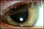
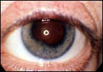
In addition to cases in which inflammation is obvious, inflammatory glaucoma should be considered in cases of uniocular open-angle glaucoma when there is no indication of trauma, exfoliation, or pigmentary dispersion syndrome. Symptoms may range from mild to severe with a course that is acute, chronic, or relapsing. Acute iritis is classically characterized by anterior segment hyperemia, miosis, and the presence of cells and flare in the anterior chamber. Iridocyclitis has the addition of keratic precipitates on the corneal endothelium and, in some cases, cells in the anterior vitreous. Early diagnosis can be difficult because patients may present with a rare white cell or trace flare, with cells or keratic precipitates appearing after several days. Other findings on clinical examination may include inflammatory cells on the trabecular meshwork, keratic precipitates on the trabecular meshwork (Slide 1), posterior synechiae (Slide 2), or inflammatory foci in the fundus. In uveitis, the intraocular pressure (IOP) may be low, normal, or high depending on the balance of the inflammatory effects of decreasing aqueous fluid inflow and increasing aqueous outflow resistance (Slide 3).
Pathophysiology of Inflammatory Glaucoma
Inflammation causes a decrease in aqueous fluid inflow by reversible and irreversible mechanisms that are at least partly mediated by cytokines. Initially, altered aqueous fluid production is reversible, but over time structural damage to the ciliary body processes occurs. In some cases, a cyclitic membrane forms over the ciliary processes, resulting in irreversible impairment of aqueous inflow.
In other cases, inflammation increases aqueous outflow resistance by a number of reversible and irreversible mechanisms. Mechanical obstruction by inflammatory debris is one contributor to increased outflow resistance. Other reversible mechanisms include keratic precipitates on the trabecular meshwork and swelling of phagocytic cells lining lamellae within the trabecular meshwork or adjacent to Schlemm's canal. With chronicity, an endothelial or fibrovascular membrane can line the inner surface of the trabecular meshwork, irreversibly increasing outflow resistance. Other permanent changes from inflammation include peripheral anterior synechiae, the obliteration of channels within the trabecular meshwork, pupillary block, and iris bombé.
The balance of these inflammatory effects determine whether the IOP is low, normal, or high. For instance, early in the inflammatory process, the decrease in aqueous production by poorly functioning ciliary body processes may outweigh the obstruction by inflammatory debris, therefore resulting in a low or normal IOP. However, with corticosteroid treatment, the aqueous production may return to normal. The damage to the outflow system becomes more critical, resulting in increased IOP.
Treatment
The treatment of inflammatory glaucoma typically requires the use of aqueous suppressants and corticosteroids. In addition to beta-blockers and carbonic anhydrase inhibitors, epinephrine-like compounds and brimonidine are useful in suppressing aqueous production. If medical management fails, seton or filtration surgery may be effective. Goniotomy or trabeculotomy can be helpful in pediatric patients with juvenile rheumatoid arthritis, sarcoidosis, or idiopathic uveitis. Perioperative use of steroids and antimetabolites are often used to help control inflammation. Laser trabeculoplasty has not been effective in treating inflammatory glaucoma.
Corticosteroid-Induced Glaucoma
Although corticosteroids are useful in controlling inflammation, the benefits from using corticosteroids must be weighed against the risk for corticosteroid-induced glaucoma. Corticosteroid-induced glaucoma typically occurs after 3 to 6 weeks of corticosteroid use but can occur as early as 1 week. Corticosteroid use stimulates a number of changes at the cellular and extracellular levels that are thought to result in increased outflow resistance. Some of these changes include increased deposition of extracellular matrix materials like glycosaminoglycans, induction of myocilin and related proteins, and decreased phagocytic and protease activity. With corticosteroid exposure, a characteristic fingerprint-like material resembling basement membrane accumulates in the extracellular space and densely packed fine fibrils abnormally accumulate beneath the inner wall of the endothelium of Schlemm's canal. Trabecular cells appear to be transformed into myofibroblast-like cells with a reorganized cytoskeleton of cross-linked actin networks.
The changes induced by corticosteroids further add to the complexity of changes observed in patients with inflammatory glaucoma and must be considered when deciding whether to treat inflammation. In severe cases, immunosuppression with methotrexate, azathioprine, cyclosporine, or other agents may be required.
Posner-Schlossman Syndrome
Posner-Schlossman syndrome, also known as glaucomatocyclitic crisis, is a unilateral nongranulomatous iritis with an acute onset and recurrent course. During attacks, IOP tend to be high, often ranging from 40 mm Hg to 70 mm Hg. The initial episode usually occurs in the third to sixth decade of life and rarely after 60 years of age. Patients may complain of a slight ocular discomfort, blurred vision, and colored halos. On examination, the eye usually appears quiet with little or no hyperemia. Keratic precipitates may not be noticed until 2 to 3 days after the onset of symptoms. The angle may appear normal or have a few tiny inflammatory precipitates on the trabecular meshwork. Usually Posner-Schlossman syndrome does not lead to synechiae. The attack often subsides in 1 to 3 weeks, with or without treatment. However, because visual field loss can occur with increased duration of the disease, treatment with aqueous suppressants and steroids is usually initiated with some patients requiring trabeculectomy with an antimetabolite.
Fuchs' Heterochromic Iridocyclitis
Fuchs' heterochromic iridocyclitis is a predominantly unilateral, idiopathic, low-grade iridocyclitis characterized by iris atrophy, posterior subcapsular cataract formation, and glaucoma. Onset is usually gradual in the third and fourth decades of life. Patients may be asymptomatic or complain of a monocular reduction in visual acuity. On examination, the eye appears white and quiet with trace flare and occasional cell. Characteristic filament or star-shaped, fine, colorless keratic precipitates can best be seen with retroillumination. Histologically, plasma cells can be seen infiltrating the iris to produce the characteristic hypochromia and loss of the pupillary collarette. Gonioscopy may reveal a few fine new blood vessels on the ciliary body band, scleral spur, and corneoscleral meshwork. Steroids are not typically used in cases of Fuchs' heterochromic iridocyclitis because no evidence suggests that controlling inflammation influences IOP in these cases. If treatment is necessitated, a trabeculectomy or a combined cataract extraction with trabeculectomy may be performed. The failure rate for surgical intervention in patients with Fuchs' heterochromic iridocyclitis is high.
Trabecular Meshwork Keratic Precipitates
Keratic precipitates on the trabecular meshwork occur rarely and appear as colorless or tapioca-colored broad, flat inflammatory precipitates projecting from the corneoscleral meshwork. Trabecular meshwork keratic precipitates appear in white and quiet eyes with few aqueous cells and few or no inflammatory precipitates on the cornea. IOP is elevated bilaterally and does not correlate with the amount of keratic precipitates present. Aqueous suppressants and hourly topical corticosteroid use are usually effective. The keratic precipitates typically resolve in 1 to 2 weeks. Unfortunately, the keratic precipitates and elevated IOP often recur after taper of the corticosteroid. These patients must continue to be followed because recurrence is common and patients are often asymptomatic. Some patients may later develop sarcoidosis, ankylosing spondylitis, rheumatoid arthritis, or chronic uveitis.
Sarcoid Uveitis
Sarcoid uveitis represents one of the ocular manifestations of systemic sarcoidosis, which is characterized by noncaseating granulomas involving pulmonary, cutaneous, and ocular tissues. Ocular manifestations of sarcoidosis include iridocyclitis, vitritis, chorioretinitis, retinal periphlebitis, and optic disc inflammation. Examination findings include inflammatory precipitates on the trabecular meshwork, large peripheral anterior synechiae, posterior synechiae, iris nodules, iridocyclitis, chorioretinitis, retinal periphlebitis described as candle-wax drippings, optic disc inflammation, and vitritis, which is sometimes described as a string of pearls. Histologically, "Schlemm's canalitis" is suggested by inflammation around Schlemm's canal and is believed to contribute to the narrowed and fibrotic Schlemm's canal observed in patients with sarcoid. Sarcoidosis typically occurs in young adults and black patients. Treatment of patients with sarcoid uveitis can be challenging, often requiring the careful balance of the beneficial effects of corticosteroids with the risk for corticosteroid-induced glaucoma. Chronic sarcoid uveitic patients with secondary glaucoma often have a poor outcome. Aqueous suppressants frequently are not sufficient to control IOP, and surgical intervention is required. The specific use of seton versus trabeculectomy is debated and requires careful consideration.
Juvenile Rheumatoid Arthritis
Juvenile rheumatoid arthritis is the most common uveitic disorder in children. Symptoms include fever, generalized lymphadenopathy, hepatosplenomegaly, pericarditis, rash, and arthritis. Uveitis is mainly seen with pauciarticular arthritis involving fewer than four joints. Although juvenile arthritis is more common in boys, uveitic disease is seen more commonly in girls. The bilateral and usually asymptomatic uveitis has small, nongranulomatous keratic precipitates located inferiorly on the cornea. Secondary glaucoma is treated with aqueous suppressants. However, care should be taken using carbonic anhydrase inhibitors in these patients because of the risk for metabolic acidosis with concurrent aspirin use. Medical management often fails, necessitating surgical intervention. Filtering surgery also fails frequently, but trabeculodialysis, goniotomy, or setons with adjunctive antimetabolites may be more successful in these patients.
HLA-B27 Associated Diseases
In addition to juvenile rheumatoid arthritis, other HLA-B27 associated diseases can present with uveitis and include ankylosing spondylitis and Reiter's syndrome. Uveitis is typically unilateral and self-limited, but recurrence is common, usually in the same eye.
Miscellaneous Etiologies
Although outside the scope of this forum, other inflammatory conditions in which glaucoma occurs include pars planitis, scleritis, episcleritis, and Behcet's disease. Infectious etiologies also producing inflammation include the herpes viruses, cytomegalovirus, Toxoplasma gondii, human immunodeficiency virus, mycobacteria, Pneumocystis carinii, Histoplasma capsulatum, Candida,congenital rubella, syphilis, leprosy, cocciodiomycosis, mumps, onchocerciasis, and toxocariasis.
Conclusion
Inflammatory glaucoma results from a number of inflammatory conditions, but all cases represent a balance between the inflammatory effects of decreased aqueous fluid inflow and increased aqueous outflow resistance. Aqueous suppressants and corticosteroids serve as the first line of therapy in these conditions; however, management of these conditions often proves challenging.
Bibliography
Allingham RR. Glaucoma due to intraocular inflammation. In: Chandler and Grant's Glaucoma. Epstein DL, Allingham RR, Schuman JS, eds. Baltimore, MD: Williams & Wilkins; 1997:375-394.
Caprioli J, Samson CM, Foster CS, et al. Uveitis and glaucoma. J Glaucoma. 2000;9:463-467.
Ceballos EM, Beck AD, Lynn MJ. Trabeculectomy with antiproliferative agents in uveitic glaucoma. J Glaucoma. 2002;11:189-196.
Ceballos EM, Parrish RK, Schiffman JC. Outcome of Baerveldt glaucoma drainage implants for the treatment of uveitic glaucoma. Ophthalmology. 2002;109:2256-2260.
Dickerson JE Jr, Steely HT Jr, English-Wright SL, Clark AF. The effect of dexamethasone on integrin and laminin expression in cultured human trabecular meshwork cells. Experimental Eye Research. 1998;7:353-358.
Freedman SF, Rodriguez-Rosa RE, Rojas MC, Enyedi LB. Goniotomy for glaucoma secondary to chronic childhood uveitis. Am J Ophthalmol. 2002;133:617-621.
Hamanaka T, Takei A, Takemura T, Oritsu M. Pathological study of cases with secondary open-angle glaucoma due to sarcoidosis. Am J Ophthalmol. 2002;134:17-26.
Jabs CA, Johns CJ. Ocular involvement in chronic sarcoidosis. Am J Ophthalmol. 1986;102:297-301.
Jampel HD, Jabs DA, Quigley HA. Trabeculectomy with 5-fluorouracil for adult inflammatory glaucoma. Am J Ophthalmol. 1990;109:168-173.
Jap A, Sivakumar M, Med M, Chee SP. Is Posner Schlossman syndrome benign? Ophthalmology. 2001;108:913-918.
Johnson D, Gottanka J, Flugel C, et al. Ultrastructural changes in the trabecular meshwork of human eyes treated with corticosteroids. Arch Ophthalmol. 1997;115:375-383.
Obenauf CD, Shaw HE, Sydnor CF, Klintworth GK. Sarcoidosis and its ophthalmic manifestations. Am J Ophthalmol. 1976;86:648-655.
Wax M, Herndon LH, Lessor R. Steroid-induced glaucoma in a young woman. J Glaucoma. 1998;7:353-358.