Supported in part by an unrestricted research grant from Research to Prevent Blindness, Inc., New York, New York
Introduction
Primary open-angle glaucoma (POAG) is a progressive optic neuropathy with characteristic optic nerve excavation and corresponding visual field defects (Slide 1).1 Gonioscopically, the angles are open and the intraocular pressure (IOP) may be elevated in 60% to 70% of patients. The disease is multifactorial and usually bilateral, although it is not necessarily symmetrical. All other causes of damage to the nerve fiber bundles should be excluded. Following is an attempt to update current thinking on the etiology of POAG.
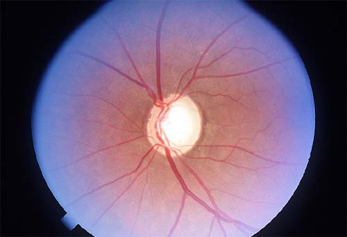
Etiology of POAG
The current treatments for glaucoma focus on lowering IOP. However, this approach may not be successful because 25% to 38% of patients may continue to lose visual field and develop blindness even when IOP has been reduced to the normal range.2-4 The research of Hattenhauer and associates has suggested that 27% of glaucoma patients become blind in at least one eye after 20 years or more with the disease.5 It is well known that the underlying pathology in POAG is the death of retinal ganglion cells (Slide 2). The cells preferentially damaged in glaucoma are the magnocellular retinal ganglion cells.6-7 Experimental animal studies of the retina and human autopsy studies of lateral geniculate nucleus in glaucoma also point to the damage of larger retinal ganglion cells that project to magnocellular layers of the lateral geniculate.8-10
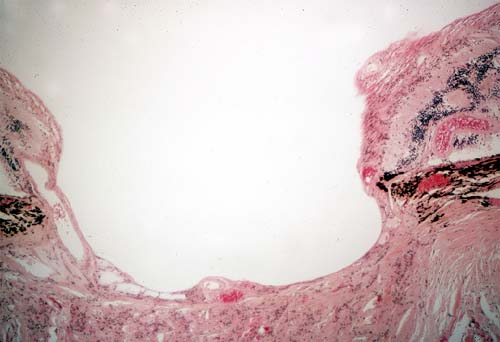
The most prevalent theories attempting to explain glaucomatous optic neuropathy are the mechanical theory and the vascular theory. In the mechanical theory, the emphasis is on the damage to the optic nerve neurons at the level of the lamina cribrosa by the elevated IOP.11 Alternately, the raised IOP may attenuate the sensitive microcirculation to the optic nerve head. On the other hand, the vascular theory suggests that eyes with inherently poor vascular supply to the optic nerve head are more predisposed to damage by elevated or normal IOP.12 But the cause-and-effect relationship between nerve damage and vascularity has not been established. Sponsel and coworkers13 found that in patients with glaucoma or ocular hypertension, the eye with the higher velocity of retinal leukocyte flow was associated with better visual function with regard to visual field and contrast sensitivity. It is controversial whether increased blood velocity translates to enhanced perfusion pressure to a particular area. Further support for the vascular theory came after the development of the laser Doppler flowmetry (LDF) technique to evaluate the circulation of the optic nerve.14 Studies have shown diminished blood flow in the optic nerves of eyes with POAG.15,16 Similarly, optic nerve flow was decreased in patients with low systemic blood pressure and increased in patients with hypertension.17 Whether LDF measures the entire blood flow to the optic nerve head is not determined, though most investigators believe LDF penetrates as far as the level of the lamina cribrosa.18 Because neither theory could explain all cases of glaucoma, the trend was to combine the two views together.
In the late 1960s and early 1970s, another theory was proposed that correlated elevated IOP with blocking axoplasmic flow at the lamina cribrosa.19,20 The resultant interruption of trophic factors to the ganglion cell body may cause the cells to initiate a suicidal response resulting in programmed cell death or apoptosis.6
The focus has shifted more to understanding the response of nerve tissue to trauma and aging. Based on the data emerging from studies of central nervous system trauma, the new concepts of excitatory neurotoxins and apoptosis were applied to understanding the damage in glaucoma. When nervous tissue is severely injured, regardless of the cause, it follows the same common final pathway before neuronal death. The injurious events may relate to ischemia/hypoxia, trauma, hypoglycemia, stroke, and various acute or chronic degenerative and hereditary neuronal diseases.21 The functional damage to the nervous tissue continues to progress even after the primary cause has been removed. These new concepts may help clinicians understand why some glaucoma patients continue to exhibit progressive neuropathy even after an offending factor such as high IOP has been controlled. Also, there is evidence that up to 50% of retinal ganglion cell axons may be lost by the time visual field loss and abnormal cupping are detected.22-23
The term secondary degeneration has been applied to progressive neuropathy that spreads to adjacent areas beyond the initially injured neuron site. The aim of therapeutic neuroprotection is to protect these initially spared neurons from secondary degeneration. The biochemical events surrounding the area of nerve injury involve the release of the excitatory amino acids glutamate and aspartate. These amino acids have the ability to excessively stimulate the nerve and cause neuronal fatigue, toxicity, and ultimately nerve death.24 The cytotoxic effects of glutamate on the inner layers of the retina are well known.25,26 Dreyer and coworkers27 discovered significantly higher levels of glutamate in the vitreous samples of glaucoma patients compared to normal individuals. Similarly, Brooks and coworkers28 found significantly high vitreal glutamate concentration in dogs with primary glaucoma compared to normal animals. Even relatively minor but chronic elevation of glutamate may be toxic to the retinal ganglion cells.
After the release of glutamate at the injury site, Na+ enters the cell. There is concomitant entry of chloride ions and water, which causes cellular swelling. These events constitute the acute phase of neuronal trauma. Depending on the severity of the insult, the cell may recover or proceed to further loss of function and death. In the second or delayed phase, there is cellular influx of Ca++ and, once the calcium homeostasis is altered, a wide variety of abnormal biochemical reactions ensue. There is release of cytotoxic enzymes such as protease, endonuclease, and lipase that destroy the cell membrane. Free radicals accumulate and further disturb the essential metabolic functions of the cells. Glutamate toxicity also releases G protein via its stimulation of metallotrophic receptors, which in turn activate phospholipase C. The end result is major disruption of normal cellular function.
Another important pathway for cellular death is apoptosis. This active process is different from necrosis and, when triggered by calcium, ion imbalance enables the cell to die without liberating its digestive enzymes. Apoptosis appears to be controlled by genes, which may be artificially altered in the future to prevent the initiation of the deadly program. Quigley and coworkers6 have shown that ganglion cell death in glaucoma shares certain similarities with classic apoptosis. Retinal cells in glaucomatous optic neuropathy display chromatin condensation and involution or shrinkage. Neufeld and coworkers29 have demonstrated increased levels of nitric oxide synthase (NOS) isoforms 1, 2, and 3 in the optic nerve head of patients with POAG. The presence of NOS-1 and NOS-2 suggests that nitric oxide may reach toxic levels in the optic nerve in glaucoma. Excitotoxicity, even when mild, can cause neuronal apoptosis.30 Excitotoxicity of retinal ganglion cells is mediated by overstimulation of a subtype of glutamate receptor, the N-methyl-D-aspartate (NMDA). Dreyer and associates31 have shown that agents that interfere with translation or transcription of these proteins are also effective in preventing NMDA-induced excitotoxicity. Overstimulation of NMDA receptors activates NOS, which mediates increased levels of nitric oxide and superoxide anion.
A new theory as to the sequence of events leading to glaucomatous nerve damage has been introduced. The first stage may be triggered in susceptible patients by factors such as elevated IOP. In the second stage, damaged ganglion cell axons are influenced by neurotropin deprivation and/or the released excitatory amino acids. With the loss of neurotrophic support of the ganglion cells, slow death is inevitable. Consensus among the proponents exists that these events are interconnected and once initiated are difficult to control with current therapies for glaucoma.
Hayreh and associates32 raised the issue of nocturnal hypotension in the development and progression of glaucomatous optic neuropathy. The physiologic drop in blood pressure at night, for example, may have adverse effects on a glaucoma patient with compromised optic nerve circulation. Hayreh's group33 also prospectively investigated the effects of topical beta-blocker drops on nocturnal blood pressure, heart rate, and visual field function. The study showed that in patients with normal-tension glaucoma who were on beta-blocker therapy, there was a significantly more marked visual field progression (P=.0003) than in those not using topical beta-blockers. These patients also exhibited a significantly greater decrease in mean diastolic blood pressure (P=.009) at night compared to patients with ischemic optic neuropathy.
The age-dependent reduction in the number of optic nerve fibers is also an important consideration.34 High-pass resolution and histologic studies have suggested the average loss of 10,000 nerve fibers every year after the age of 40 years. As the average number of nerve fibers is approximately 1.2 million, a person in his or her mid-80s may have lost approximately 40% of neurons due to age-related events alone.
References
-
The American Academy of Ophthalmology Quality of Care Committee Glaucoma Panel. Primary open angle glaucoma preferred practice pattern. American Academy of Ophthalmology. San Francisco, Calif; 1996.
-
Hart WJ, Yablonski M, Kass MA, et al. Multivariate analysis of the risk of glaucomatous visual field loss. Arch Ophthalmol. 1979;97:1455-1458.
-
Wilson R, Walker AM, Dueker DK, et al. Risk factors for rate of progression of glaucomatous visual field loss: A computer-based analysis. Arch Ophthalmol. 1982;100:737-741.
-
O'Brien C, Schwartz B, Takamoto T, et al. Intraocular pressure and the rate of visual field loss in chronic open angle glaucoma. Am J Ophthalmol. 1991;111:491-500.
-
Hattenhauer MG, Johnson DH, Ing HH, et al. The probability of blindness from open-angle glaucoma. Ophthalmology. 1998;105:2099-2104.
-
Quigley HA, Nickells RW, Kerrigan LA, et al. Retinal ganglion cell death in experimental glaucoma and after axotomy occurs by apoptosis. Invest Ophthalmol Vis Sci. 1995;36:774-786.
-
Quigley HA, Dunkelberger GR, Green WR. Chronic human glaucoma causing selectively greater loss of large optic nerve fibers. Ophthalmology. 1988;95:357-363.
-
Dandona L, Hendrickson A, Quigley HA. Selective effects of experimental glaucoma on axonal transport by retinal ganglion cells to the dorsal lateral geniculate nucleus. Invest Ophthalmol Vis Sci. 1991;32:1593.
-
Chaturvedi N, Hedley-Whyte ET, Dreyer EB. Lateral geniculate nucleus in glaucoma. Am J Ophthalmol. 1993;116:182-188.
-
Yücel YH, Zhang A, Gupta N, et al. Loss of neurons in magnocellular and parvocellular layers of lateral geniculate nucleus in glaucoma. Arch Ophthalmol. 2000;118:378-384.
-
Mueller H. Anatomische beitrage zur ophthalmologie: Ueber nervean-veranderungen an der eintrittsstelle des schnerven. Arch Ophthalmol. 1858;4:1.
-
Von Jaeger E. Ueber glaucoma und seine heilung durch iridectomie. Z Ges Aertze Wein. 1858;14:484.
-
Sponsel WE, DePaul KL, Kaufman PL. Correlation of visual function and retinal leukocyte velocity in glaucoma. Am J Ophthalmol. 1990;109:49-54.
-
Riva CE, Harino S, Petrig BL, et al. Laser Doppler flowmetry in the optic nerve. Exp Eye Res. 1992;55:499-506.
-
Grunwald JE, Piltz J, Hariprasad SM, et al. Optic nerve and choroidal circulation in glaucoma. Invest Ophthalmol Vis Sci. 1998;39:2329-2336.
-
Kaiser HJ, Flammer H, Hendrickson P. Ocular blood flow: new insights into the pathogenesis of ocular diseases. Basel: Karger; 1996.
-
Gruenwald JE, Piltz J, Hariprasad SM, et al. Optic nerve blood flow in glaucoma: Effect of systemic hypertension. Am J Ophthalmol. 1999;127:516-522.
-
Koelle JS, Riva CE, Petrig BL, et al. Depth of tissue sampling in the optic nerve head using laser Doppler flowmetry. Laser Med Sci. 1993;8:49-54.
-
Lampert PW, Vogel MH, Zimmerman LE. Pathology of the optic nerve in experimental acute glaucoma: Electron microscopic studies. Invest Ophthalmol. 1968;7:199-213.
-
Anderson DR, Hendrickson A. Effect of intraocular pressure on rapid axoplasmic transport in monkey optic nerve. Invest Ophthalmol Vis Sci. 1974;13:771-783.
-
Schumer RA, Podos SM. The nerve of glaucoma. Arch Ophthalmol. 1994;112:37-44.
-
Quigley HA, Addicks EM, Green WR, et al. Optic nerve damage in human glaucoma. II. The site of injury and susceptibility to damage. Arch Ophthalmol. 1981;99:635-649.
-
Quigley HA, Addicks EM, Green WR. Optic nerve damage in human glaucoma: III. Quantitative correlation of nerve fiber loss and visual field defect in glaucoma, ischemic neuropathy, disc edema and toxic neuropathy. Arch Ophthalmol. 1982;100:135-146.
-
Olney JW. Inciting excitotoxic cytocide among central neurons. Adv Exp Med Biol. 1986;203:631-645.
-
Otori Y, Wei JY, Barnstable CJ. Neurotoxic effects of low doses of glutamate on purified rat retinal ganglion cells. Invest Ophthalmol Vis Sci. 1998;39:972-981.
-
Lucus D, Newhouse J. The toxic effects of sodium L-glutamate on the inner layers of the retina. Arch Ophthalmol. 1957;58:193-201.
-
Dreyer EB, Zurakowski D, Schumer RA, et al. Elevated glutamate levels in the vitreous body of humans and monkeys with glaucoma. Arch Ophthalmol. 1996;114:299-305.
-
Brooks DE, Garcia GA, Dreyer EB, et al. Vitreous body glutamate concentration in dogs with glaucoma. Am J Vet Res. 1997;58:864-867.
-
Neufeld AH, Hernandez MR, Gonzales M. Nitric oxide synthase in the human glaucomatous optic nerve head. Arch Ophthalmol. 1997;115:497-503.
-
Bonfoco E, Krainc D, Ankarcrona M, et al. Apoptosis and necrosis: two distinct events induced respectively by mild and intense insults with NMDA or nitric oxide, superoxide in cortical cell cultures. Proc Natl Acad Sci USA. 1995;92:7162-7166.
-
Dreyer EB, Zhang D, Lipton SA. Transcriptional or translational inhibition blocks low dose NMDA-mediated cell death. Neuroreport. 1995;6:942-944.
-
Hayreh SS, Zimmerman MB, Podhajsky P, et al. Nocturnal arterial hypotension and its role in optic nerve and ocular ischemic disorders. Am J Ophthalmol 1994;117:603-624.
-
Hayreh SS, Podhajsky P, Zimmerman MB. Beta-blocker eyedrops and nocturnal arterial hypotension. Am J Ophthalmol. 1999;128:301-309.
-
Kerrigan-Baumrind LA, Quigley HA, Pease ME. Number of ganglion cells in glaucoma eyes compared with threshold visual field tests in the same person. Invest Ophthalmol Vis Sci. 2000;41:741-748.