Introduction
Sturge-Weber syndrome is one of several syndromes classified under the heading of phakomatosis because it involves pathology in the skin, eye, and brain. The first reported description of Sturge-Weber syndrome associated with eye involvement has been attributed to Schirmer in 1860. He reported a patient with facial nevus flammeus and ipsilateral buphthalmos. Sturge later reported on a patient who had these findings, along with neurologic findings, and proposed that there were vascular abnormalities in the brain and eye along with the skin. Weber verified neurologic involvement radiologically.
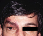
Inheritance of disease is sporadic and eye involvement is almost exclusively unilateral. It is believed that men and women are equally affected. Histopathology is significant for hamartomatous malformation of cephalic blood vessels causing hemangiomas of the skin, meninges, and eye. A layer of endothelium lines these dilated blood vessels. Typically, the lesions remain constant throughout life. Calcification of brain tissues adjacent to hemangiomas causes seizures, mental retardation, hemiparesis, and hemianopia. The facial lesion (port-wine stain) occurs along a distribution similar to the branches of the trigeminal nerve. However, the hemangioma often traverses the midline (Slide 1).
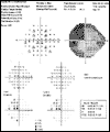
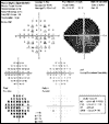
Ocular manifestations include choroidal, episcleral, and conjunctival hemangiomas, glaucoma, and heterochromia of the iris1. The fundus appearance is described as "tomato-catsup" (a reddish-orange color) when choroidal hemangiomas are present. Choroidal hemangiomas usually occur in the posterior pole, causing decreased vision by several mechanisms including hyperopia (secondary to anterior displacement of the macula), exudative retinal detachment, cystoid macular edema, and retinal pigment epithelium (RPE) fibrosis and atrophy. Homonymous quadrantanopia secondary to hemangiomatous involvement of blood vessels adjacent to the occipital lobe can also be a cause of visual change (Slide 2A and Slide 2B).
Epidemiology
Approximately 30% to 40% of patients with Sturge-Weber syndrome have glaucoma. Sixty percent of patients with glaucoma have early-onset glaucoma (arbitrarily defined as increased intraocular pressure [IOP] in patients with Sturge-Weber syndrome who are younger than 3 years old), leading to buphthalmos. The other 40% of patients develop glaucoma later in life. Some believe that there is an increased risk for glaucoma if the upper eyelid contains a hemangioma. Others believe that both eyelids must be involved to truly increase the risk of glaucoma.
Glaucoma
Theories
The exact mechanism of glaucoma is unknown, but there have been several theories:
- Hypersecretion/transudation from a choroidal hemangioma2
- Neurally regulated increase in aqueous production3
- Outflow obstruction by anterior chamber angle malformation
- Increased episcleral pressure
Discovery of trabecular meshwork abnormalities has led some investigators to believe that these abnormalities are the primary mechanism of glaucoma. In 1957, Barkan4 looked at the effects of goniotomy in three eyes of two patients. Because of his success with goniotomy, he believed that there was a membrane covering the angle, obstructing outflow. Goniotomy removed this membrane, thus restoring aqueous outflow. He hypothesized that this "Barkan" membrane was the direct cause of glaucoma in patients with Sturge-Weber syndrome. Christiansen and Records5 found an iris root and ciliary body anteriorly inserted into the trabecular meshwork, associated with an angle cleavage abnormality. Pathologic studies performed by Cibis and colleagues6 demonstrated that the trabecular meshwork in three eyes of two patients with Sturge-Weber syndrome and glaucoma appeared similar to the trabecular meshwork of older patients and patients with open-angle glaucoma, supporting the idea of impaired aqueous outflow as the primary cause of glaucoma associated with Sturge-Weber syndrome.
Phelps7 proposed an alternative mechanism. He examined 21 patients with Sturge-Weber syndrome and glaucoma and found that episcleral pressure was elevated relative to established normal values8 and the episcleral pressure in the unaffected eye. Also, blood refluxed easily into Schlemm’s canal. These findings led to his conclusion that increased episcleral venous pressure caused increased resistance to aqueous outflow. This in turn caused glaucoma in patients with Sturge-Weber syndrome (Slide 3).
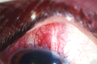
A current theory is that a combination of obstruction of outflow by anterior chamber angle malformation and increased episcleral pressure leads to glaucoma in patients with Sturge-Weber syndrome9. Some investigators believe that early-onset glaucoma is associated with angle malformation, whereas later-onset glaucoma is related to increased episcleral pressure.
Treatment
Treating glaucoma in patients with Sturge-Weber syndrome is challenging. In separate studies, Altuna and colleagues1 and Yang and colleagues10 looked at the effects of latanoprost on IOP in patients with Sturge-Weber syndrome. Altuna’s group found that at 6 months, only three of 18 patients had a 20% reduction in IOP. Yang’s group found that two of six patients had a significant reduction in IOP. However, because both of these patients had juvenile-onset glaucoma, they believed that latanoprost may be effective for this specific subset of patients who have glaucoma associated with Sturge-Weber syndrome. Van Emlen and associates11 treated seven of eight patients with both cryotherapy and medications and were able to maintain IOP less than 21 mm Hg in all patients.
Sidoti and colleagues12 investigated trabeculectomy with mitomycin C as an option for treatment. In their retrospective consecutive series review, they found that 59% of 29 patients had IOP control at 3 years (IOP between 5 mm Hg and 21 mm Hg without further interventions or complications). However, five patients developed infection (three cases of blebitis and two cases of bleb-related endophthalmitis). Thus, the group recommends only limited use of trabeculectomy with mitomycin C with caution. Ali and colleagues13 found success with trabeculectomy without mitomycin C in seven eyes of six patients.
Mandal14 retrospectively examined 10 eyes of nine patients who underwent trabeculotomy-trabeculectomy for glaucoma associated with Sturge-Weber syndrome. He found a mean IOP reduction from 28 mm Hg to 12 mm Hg without a need for medication. However, other studies by Boards and Shields15 and Agarwal16 did not replicate these findings.
Budenz and colleagues17 and Hamush and colleagues18 looked retrospectively at the value of implant devices in patients with Sturge-Weber syndrome and glaucoma. Budenz found that two-staged implantation of a Baerveldt device in 10 eyes kept IOP below 21 mm Hg. Hamush discovered that Ahmed implants kept IOP lower than 21 mm Hg in 79% of 11 eyes at 2 years, 59% at 3.5 years, and 30% at 5 years.
Iwach and colleagues19 retrospectively examined 36 eyes with Sturge-Weber syndrome and glaucoma. They studied types of interventions, reasons for surgical intervention, and time between original intervention and next intervention. The group found that 45% of patients had intervention because of optic nerve change, whereas 55% had intervention because of increasing IOP. Of all treatments performed in this study, the group found that goniotomy with medications was the most effective.
Choroidal Hemangioma
The choroidal hemangiomas associated with Sturge-Weber syndrome are typically described as being diffuse, becoming progressively less thick peripherally, and blending into the normal peripheral choroid. This is in contrast to isolated, or circumscribed, hemangiomas that tend to have defined borders20. Hemangiomas in patients with Sturge-Weber syndrome commonly occur in the presence of elevated episcleral and orbital venous pressure and angle malformation, or both, leading to an increase in IOP. The RPE overlying the hemangioma often undergoes fibrous transformation. A decrease in visual function in choroidal hemangiomas is usually secondary to these RPE abnormalities, exudative retinal detachments, or cystoid macular edema. Exudative retinal detachments are usually extensive. Treatment with laser, therefore, often requires initial drainage of the subretinal fluid and subsequent laser photocoagulation. This form of treatment has had only limited success.
Radiation therapy has been studied for the treatment of diffuse hemangiomas complicated by exudative retinal detachments. Zografos and associates21 looked at treatment of choroidal hemangiomas with cobalt-60 gamma brachytherapy. Of the 41 eyes in their study, two had diffuse-type choroidal hemangiomas and exudative retinal detachment. Both eyes were treated with 15-mm diameter cobalt-60 applicators placed at the presumed center of the tumor. The retinal detachments resolved in both eyes and did not recur. Vision improved in one eye and remained stable in the other.
External beam radiation treatment delivered in a precise lens-sparing fashion is an alternative, less toxic approach to the treatment of exudative retinal detachments associated with diffuse choroidal hemangioma. Studies looking at this modality have shown promising results. Using external beam radiation, Scott and colleagues22 treated five eyes with diffuse choroidal hemangioma and exudative retinal detachment. They found that treatment caused resolution of all detachments; however, visual acuity was not improved in any of these eyes.
Madreperla and colleagues23 studied the effect of lens-sparing radiation therapy on subretinal fluid secondary to diffuse choroidal hemangiomas. Five eyes with diffuse choroidal hemangioma treated with radiation (1,200 cGy to 4,000 cGy) had complete resolution of fluid, with either improvement or stabilization of vision. Schilling and colleagues24 observed 15 eyes in 12 patients with diffuse choroidal hemangioma and Sturge-Weber syndrome, with exudative retinal detachment involving or threatening the fovea. After being treated with 20 Gy of external beam radiation therapy (linear accelerators), all patients had resolution of their exudative retinal detachments. Approximately 50% of patients realized improved vision by two or more lines of acuity, while eight stayed the same or worsened (six eyes had severe glaucoma prior to treatment). Schilling also found that improvement in vision was related to rapidity of treatment after symptoms. Follow-up was 4.5 years (4 months to 24 years, median 4 years). Additionally, five of the tumors shrunk by 1 mm to 3 mm.
A potential complication of surgery in eyes with hemangiomas is expulsive suprachoroidal hemorrhage25. Some investigators advocate prophylactic sclerostomies prior to beginning glaucoma surgery to prevent this hemorrhage. Others, however, deem this unnecessary, as patients more typically will experience choroidal effusions, which usually resolve spontaneously. The mechanism of effusion is believed to involve a sudden decrease in pressure during or immediately after surgery. This causes a pressure gradient across the capillary membrane of the ciliary body and choroid, favoring transudation of fluid into the choroidal space.
Seizures
Seizures occur in approximately 80% of patients with Sturge-Weber syndrome26,27. These seizures are primarily partial motor seizures, although some patients exhibit secondary generalization. Patients may also exhibit atrophy of the cortical parenchyma; often, mental retardation and motor skill deficits are sequelae from seizures. Seizures are treated initially with standard antiepileptic drugs. Patients with intractable seizures are considered for surgical management, such as focal cerebral resection of affected lobes or subtotal hemispherectomy28. Some physicians29 are currently considering use of prophylactic antiepileptic medications to prevent cognitive defects associated with seizures in patients with Sturge-Weber syndrome.
References
- Altuna J, Greenfield D, et al. Latanoprost in glaucoma associated with Sturge-Weber syndrome: Benefits and side effects. J Glaucoma. 1999;8:199-203.
- Mehney G. Naevus flammeus associated with glaucoma. Arch Ophthalmol. 1937;17:1018-1023.
- Font RL and Ferry AP. The phakomatoses. IntOphthalmol Clin. 1972;12(1):11-22.
- Barkan O. Goniotomy for glaucoma associated with nevus flammeus. Am J Ophthalmol. 1957;43:545-549.
- Christiansen G, Records R. Glaucoma and expulsive hemorrhage mechanisms in the Sturge-Weber syndrome. Ophthalmology. 1979;86:1360-1366.
- Cibis G, Tripathi R, Tripathi B. Glaucoma in Sturge-Weber syndrome. Ophthalmology. 1984;91:1061-1071.
- Phelps C. The pathogenesis of glaucoma in Sturge-Weber syndrome. Ophthalmology. 1978;85:276-286.
- Phelps C, Armaly M. Measurement of episcleral venous pressure. Arch Ophthalmol. 1978;85:35-42.
- Weiss D. Dual origin of glaucoma in encephalotrigeminal haemangiomatosis. Trans Ophthalmol Soc UK. 1973;93:477-491.
- Yang C, Freedman S, et al. Use of latanoprost in the treatment of glaucoma associated with Sturge-Weber syndrome. Arch Ophthalmol. 1998;126:600-602.
- Van Emlen C, Goethals M, et al. Treatment of glaucoma in children with Sturge-Weber syndrome. J Ped Ophthalmol Strabismus. 2000;37:29-34.
- Sidoti P, Belmonte S, et al. Trabeculectomy with Mitomycin-C in the treatment of pediatric glaucomas. Ophthalmology. 2000;107:422-429.
- Ali M, Fahmy I, Spaeth G. Trabeculectomy for glaucoma associated with Sturge-Weber syndrome. Ophthalmic Surg. 1990;21(5):352-355.
- Mandal AK. Primary combined trabeculotomy-trabeculectomy for early-onset glaucoma in Sturge-Weber syndrome. Ophthalmology. 1999;106(8):1621-1627.
- Board RJ, Shields B. Combined trabeculotomy-trabeculectomy for the management of glaucoma associated with Sturge-Weber syndrome. Ophthalmic Surg. 1981;12(11):813-817.
- Agarwal HC, Sandramouli S, et al. Sturge-Weber syndrome: Management of glaucoma with combined trabeculotomy-trabeculectomy. Ophthalmic Surg. 1993;24(6):399-402.
- Budenz D, Sakamoto D, et al. Two-staged Baerveldt glaucoma implant for childhood glaucoma associated with Sturge-Weber syndrome. Ophthalmology. 2000;107:2105-2110.
- Hamush N, Coleman A, Wilson MR. Ahmed glaucoma valve implant for management of glaucoma in Sturge-Weber syndrome. Arch Ophthalmol. 1999;128:750-760.
- Iwach A, Hoskins D Jr, et al. Analysis of surgical and medical management of glaucoma in Sturge-Weber syndrome. Ophthalmology. 1990;97:904-909.
- Witschel H, Font R. Hemangioma of the choroid. A clinicopathologic study of 71 cases and a review of the literature. Surv Ophthalmol. 1976;20:415-431.
- Zografos L, Bercher L, et al. Cobalt-60 treatment of choroidal hemangiomas. Arch Ophthalmol. 1996;121:190-199.
- Scott T, Ausburger J et al. Low dose ocular irradiation for diffuse choroidal hemangiomas associated with bullous nonrhegmatogenous retinal detachment. Retina. 1991;11:389-393.
- Madreperla S, Hungerford J, et al. Choroidal hemangiomas: Visual and anatomic results of treatment by photocoagulation or radiation therapy. Ophthalmology 1997;104(11):1773-1778.
- Schilling H, Sauerwein W, et al. Long term results after low dose ocular irradiation for choroidal hemangiomas.Br J Ophthalmology. 1997;81(4):267-273.
- Bellows AR, Chylack L, et al. Choroidal effusion during glaucoma surgery in patients with prominent episcleral vessels. Arch Ophthalmol. 1979;97:493-497.
- Pascual-Castroviejo I, Diaz-Gonzalez C, et al. Sturge-Weber syndrome: Study of 40 patients. Pediatric Neurol. 1993;9(4):283-288.
- Sujansky E, Conradi S. Outcome of Sturge-Weber syndrome in 52 adults. Am J Med. Genet. 1995;57(1):35-45.
- Arzimanoglou A, Andermann F, et al. Sturge-Weber syndrome: Indications and results of surgery in 20 patients. Neurology. 2000;55(10) :1472-1479.
- Ville D, Enjolras O, et al. Prophylactic antiepileptic treatment in Sturge-Weber disease. Seizure. 2002;11(3):145-150.