Introduction
In 1979, Eagle and colleagues1 proposed the term iridocorneal endothelial (ICE) syndrome to unify a group of disorders that share a number of clinical characteristics. Essential iris atrophy, Chandler’s syndrome, and iris nevus (Cogan-Reese) syndrome demonstrate abnormalities of the cornea, anterior chamber angle, and iris. However, each is defined by the degree to which these structures are affected.
Reports from the late 19th century describe severe iris atrophy with glaucoma. Harms is generally credited with providing the first solid description of this condition in 1903,2 and numerous reports followed over the next half century. In 1956, Chandler3 described a syndrome involving progressive iris stromal atrophy, endothelial dystrophy with corneal edema, and glaucoma. He noted that corneal edema often occurred at normal or mildly elevated intraocular pressure (IOP). Specifically, all cases had an abnormality of the corneal endothelium, which Chandler described as a fine "hammered silver" appearance. In 1965, Chandler and Grant4 suggested that Chandler’s syndrome, as it had become known, and essential iris atrophy represent two basic forms of the same disease.
In 1969, Cogan and Reese5 described two patients with pigmented iris nodules and glaucoma. Suspicion of malignant melanoma prompted enucleation of these eyes, revealing that the lesions were composed of benign tissue resembling normal iris stroma. They also noted ‘ectopic Descemet’s membrane’ over the trabecular meshwork and iris surface. Initially, the iris nevus syndrome was believed to be a distinct disease; however, in 1978 Campbell6 offered a pathogenic mechanism to unify these disorders.
Today, three variants of ICE are generally recognized. In essential iris atrophy, as the name implies, progressive thinning and atrophy of the iris with hole formation is the predominant clinical feature. Chandler’s syndrome presents with signs and symptoms related to corneal edema. Iris nevus (Cogan-Reese) syndrome demonstrates significant and characteristic nodular iris changes.
Clinical Presentation
The ICE syndromes typically manifest in early to mid adulthood. Most studies suggest a female predominance, though it varies in degree. The disease is nearly always unilateral, although bilateral cases have been reported,7 and subclinical abnormalities of the endothelium have been noted in the fellow eye.8 Occurrence is sporadic. Familial cases have been rarely noted and may not be true cases of ICE. There are no known systemic associations.
Presenting manifestations often involve abnormalities of the iris and pupil, visual disturbances, and pain. Pupillary changes include distorted or ectopic pupil, a second ‘pupil,’ or pseudopolycoria, or dark spots on the iris, and are often noted by the patient or another casual observer. Occasionally, a subtle disturbance of the pupil will alert the ophthalmologist to the presence of disease. Visual disturbances may be transient, with blurring or halos around lights that are typically worse in the morning. Alternatively, a persistent reduction in vision may be secondary to persistent corneal edema or glaucomatous damage. Pain and foreign body sensation are due to corneal edema.
Examination reveals abnormalities of the cornea, iridocorneal angle, and iris. The central feature of these disorders is the presence of a distinctive abnormality of the corneal endothelium. As noted by Chandler, the endothelium takes on a fine, hammered metal appearance, which is similar to Fuch's dystrophy but less coarse. This change may affect the entire cornea or may occur in patches. The cornea may be otherwise clear or may reveal varying degrees of edema.
Peripheral anterior synechiae are another hallmark of disease. These are typically broad based and extend to or beyond Schwalbe’s line. They may begin focally but eventually tend to progress around the entire angle.
Iris abnormalities, as described above, include corectopia, ectropion uveae, atrophy, thinning, and hole formation (Slide 1, Slide 2, Slide 3, Slide 4, and Slide 5). Iris holes or tears are of two types. Stretch holes typically form in the axis of papillary distortion, possibly as a consequence of stretching by peripheral synechiae. Melt holes often form adjacent to areas of heavy anterior synechiae and are believed to be due to focal iris ischemia, as collateral iris vessels have been identified angiographically in some patients.
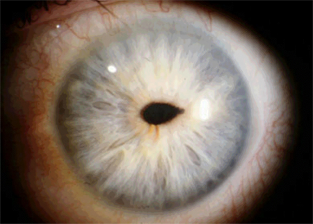
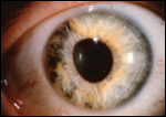
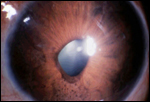
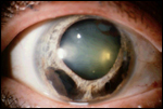
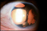
Iris nodules may also form and are the hallmark of the iris nevus syndrome. Nodules are relatively flat or pedunculated and initially may occur focally or segmentally, spreading with progression of the disease. Histopathologically, these lesions are normal iris stroma that has become surrounded or enveloped by aberrant Descemet’s membrane, leading to the clinical appearance of nodules.
Glaucoma often develops in patients with ICE syndrome, occurring in at least 50% of patients. Elevated IOP may occur with primarily open angles as a consequence of trabecular obstruction by endothelial membrane or may be secondary to synechial angle closure as the disease progresses. Glaucoma is often difficult to manage in these patients.
Pathogenesis
Numerous early theories were postulated regarding the pathogenesis of ICE syndrome. Initially, it was believed that the changes in these eyes were the result of some previous or ongoing inflammatory insult2; however, no significant inflammation has been noted. Others suggested a possible vascular or ischemic etiology; however, no supporting evidence was found.
Campbell and colleagues6 offered a mechanism of disease that explained all the clinically observed findings and unified these previously distinct entities. Published in 1978, this study was the first to present evidence that all the pathologic findings in ICE syndrome are due to an aberrant endothelial membrane. Clinical review of 82 patients demonstrated endothelial abnormalities throughout the spectrum of disease. Histopathologic review of 10 enucleated eyes revealed aberrant endothelial overgrowth of the iris in all. Six of the 10 eyes demonstrated an endothelial membrane overgrowing foci of open angle. Near complete overgrowth of the angle was noted in one eye with few synechiae but an IOP of 50 mm Hg, suggesting that, initially, this may lead to blockage of the angle without synechial angle closure. Iris nodules were identified as normal-appearing stroma surrounded by the abnormal membrane.
These findings suggested that the disease begins at the cornea, with proliferation or spread of an abnormal endothelium and deposition of aberrant Descemet’s membrane. As this membrane crosses the angle, it provides resistance to aqueous outflow and eventually leads to formation of anterior synechiae, with subsequent glaucoma. Growth of this membrane across the iris leads to synechiae and atrophy, with hole and nodule formation.
The ICE Cell
Much attention has been focused on identifying and describing the abnormality of the endothelium that initiates this sequence of events. By specular microscopy, an abnormal appearing population of endothelial cells can be identified.9,10 Normal endothelial cells appear light with dark intercellular junctions and are of fairly regular size and hexagonal shape. ICE cells are larger and more pleomorphic, appearing as dark cells with a light surround and occasional white central spot. ICE cells may coexist with relatively normal endothelial cells or may replace the entire endothelium.
Numerous studies have attempted to characterize these cells using various techniques,11-15 including light and electron microscopy, histologic staining, and immunocytochemistry. These analyses have been performed on corneal buttons and trabecular meshwork specimens obtained after keratoplasty and trabeculectomy.
Many researchers have identified similar structural features of these cells, including presence of numerous microvilli, filopodia, and surface blebs, which are findings not typical of endothelia. Some authors initially identified epithelial-like cells with intracellular keratin filaments and desmosomes, whereas others disputed these findings. The majority of data support the expression of both epithelial and endothelial markers by these cells, suggesting the possibility of an epithelial-like metaplasia of endothelial cells in response to an unknown stimulus.
A theory that received significant attention was proposed by Alvarado, who suggested that an infectious agent, possibly herpes virus, may be the metaplastic stimulus. He conducted a series of experiments attempting to identify herpes simplex viral (HSV) DNA in ICE corneas.16 Sixteen of 25 ICE specimens were positive for HSV DNA, whereas 15 of 15 controls were negative, as were 15 of 15 specimens with interstitial keratitis, bullous keratopathy, and keratoconus. Neither herpes zoster nor Epstein-Barr viral DNA was identified in any specimen tested. When the endothelium was removed from samples prior to DNA isolation, all ICE samples that previously tested positive for HSV DNA tested negative, suggesting localization of HSV DNA within the endothelium.
Glaucoma in ICE Syndrome
Glaucoma is a common characteristic of ICE syndrome, reportedly occurring in 50% to 80%9,17,18 of patients. Some studies have found increased risk of glaucoma with essential iris atrophy and iris nevus syndrome than with Chandler’s syndrome, while others have not.21 Glaucoma is most likely more common in eyes with total or diffuse endothelial involvement than with subtotal or patchy change. Sherrard19 identified increased IOP requiring treatment in 71% of patients with total ICE and 18% with subtotal disease.
Management of glaucoma in this disorder can be challenging. Medical therapy often fails to control IOP, necessitating surgical treatment. Similarly, the failure rate of filtering surgery is also high, often necessitating multiple procedures.20-25
In one study,23 22 of 25 patients with ICE and glaucoma failed medical therapy. The success rates of primary trabeculectomy were 63% at 1 year, 44% at 2 years, and 23% at 5 years. Sixty percent of first operations, 20% of second, and 17% of third were functional at 1 year.
Another study23 reported the survival of trabeculectomy with antifibrotic agent (either mitomycin-C or 5-fluorouracil) to be 73% at 1 year, 44% at 2 years, and 29% at 5 years. Tube shunt survival was 54% at 5 years.
These results indicate a high failure rate for traditional filtering surgery in ICE syndrome, with increasingly poor survival of subsequent procedures. Glaucoma drainage devices, or tube shunts, appear to have a higher success rate in the long term; however, their use is not without additional complications. Tubes have been noted to migrate anteriorly against the cornea or posteriorly toward the iris and, in some cases, may extrude from the anterior chamber, all presumably due to contractile forces generated by the expanding endothelial membrane. Some investigators have suggested cutting the tube longer than normal to allow for repositioning if necessary.
References
- Eagle RC Jr, Font RL, Yannof M. Proliferative endotheliopathy with iris abnormalities: The iridocorneal endothelial syndrome. Arch Ophthalmol. 1979;97:2104-2111.
- Harms C. Einseitige spontone luckenbildung der iris durch atrophie ohne mechanische zerrung. Klin Monatsbl Augenheilkd. 1903;41:522-528.
- Chandler PA. Atrophy of the stroma of the iris. Endothelial dystrophy, corneal edema, and glaucoma. Am J Ophthalmol. 1956;41:607-615.
- Chandler PA, Grant WM. Lectures on Glaucoma. Philadelphia, Pa: Lea & Febiger; 1965:276-285.
- Cogan DG, Reese AB. A syndrome of iris nodules, ectopic Descemet’s membrane and unilateral glaucoma. Doc Ophthalmol. 1969;26:424-433.
- Campbell DG, Shields MB, Smith TR. The corneal endothelium and the spectrum of essential iris atrophy. Am J Ophthalmol. 1978;86:317-324.
- Hemady RK, Patel A, Blum S, Nirankari VS. Bilateral iridocorneal endothelial syndrome: Case report and review of the literature. Cornea. 1994;13:368-372.
- Lucas-Glass TC, Baratz KH, Nelson LR, Hodge DO, Bourne WM. The contralateral corneal endothelium in the iridocorneal endothelial syndrome. Arch Ophthalmol. 1997;115:40-44.
- Hirst LW, Quigley HA, Stark WJ, Shields MB. Specular microscopy of iridocorneal endothelia syndrome. Am J Ophthalmol. 1980;89:11-21.
- Neubauer L, Lund OE, Leibowitz HM. Specular microscopic appearance of the corneal endothelium in iridocorneal endothelial syndrome. Arch Ophthalmol. 1983;101:916-918.
- Alvarado JA, Murphy CG, Maglio M, Hetherington J. Pathogenesis of Chandler's syndrome, essential iris atrophy and the Cogan-Reese syndrome. I. Alterations of the corneal endothelium. Invest Ophthalmol Vis Sci. 1986;27:853-872.
- Hirst LW, Bancroft J, Yamauchi K, Green WR. Immunohistochemical pathology of the corneal endothelium in iridocorneal endothelial syndrome. Invest Ophthalmol Vis Sci. 1995;36(5):820-827.
- Howell DN, Damms T, Burchette JL, Green WR. Endothelial metaplasia in the iridocorneal endothelial syndrome. Invest Ophthalmol Vis Sci. 1997;38(9):1896-1901.
- Levy SG, Kirkness CM, Ficker L, McCartney ACE. The histopathology of the iridocorneal-endothelial syndrome. Cornea. 1996;15(1):46-54.
- Levy SG, McCartney AC, Baghai MH, Barrett MC, Moss J. Pathology of the iridocorneal-endothelial syndrome. The ICE-cell. Invest Ophthalmol Vis Sci. 1995;36(13):2592-2601.
- Alvarado JA, Underwood JL, Green WR, et al. Detection of herpes simplex viral DNA in the iridocorneal endothelial syndrome. Arch Ophthalmol. 1994;112:1601-1609.
- Laganowski HC, Muir MG, Hitchings RA. Glaucoma and the iridocorneal endothelial syndrome. Arch Ophthalmol. 1992;110:346-350.
- Wilson MC, Shields MB. A comparison of the clinical variations of the iridocorneal endothelial syndrome. Arch Ophthalmol. 1989;107:1465-1468.
- Sherrard ES, Frangoulis MA, Muir MG. On the morphology of cells of posterior cornea in the iridocorneal endothelial syndrome. Cornea. 1991;10(3):233-243.
- Doe EA, Budenz DL, Gedde SJ, Imami NR. Long-term surgical outcomes of patients with glaucoma secondary to the iridocorneal endothelial syndrome. Ophthalmology. 2001;108(10):1789-1795.
- Kidd M, Hetherington J, Magee S. Surgical results in iridocorneal endothelial syndrome. Arch Ophthalmol. 1988;106:199-201.
- Kim DK, Aslanides IM, Schmidt CM, et al. Long-term outcome of aqueous shunt surgery in ten patients with iridocorneal endothelial syndrome. Ophthalmology. 1999;106:1030-1034.
- Laganowski HC, Muir MG, Hitchings RA. Glaucoma and the iridocorneal endothelial syndrome. Arch Ophthalmol. 1992;110(3):346-350.
- Lanzl IM, Wilson RP, Dudley D, Augsburger JJ, Aslanides IM, Spaeth GL. Outcome of trabeculectomy with mitomycin-C in the iridocorneal endothelial syndrome. Ophthalmology. 2000;107(2):295-297.
- Wright MM, Grajewski AL, Cristol SM, Parrish RK. 5-Fluorouracil after trabeculectomy and the iridocorneal endothelial syndrome. Ophthalmology. 1991;98(3):314-316.
Bibliography
Shields MB. Iridocorneal endothelial syndromes. In: Chandler PA, Grant WM, eds. Glaucoma. 4th ed. Baltimore, Md: Williams and Wilkins; 1997:319-326.