Rudolph S. Wagner, MD
Successful strabismus surgery depends on a thorough knowledge of the anatomy of the extraocular muscles and fascia. Average values for the anatomic insertions, tendon and muscle lengths, and the relative positions of the muscle insertions relative to the limbus, as depicted in the spiral of Tillaux, are important surgical reference points (Table 1). Experienced surgeons recognize, however, that distances are average values and measure the medial rectus muscle insertion at 3.5 mm from the limbus in a young child. Some ophthalmologists will vary the magnitude of their surgery depending on the size of the globe.
Penetration of Tenon's capsule at the point approximately 10 mm from the limbus where the muscles pass through should be avoided. Compromising this barrier to orbital fat may result in excessive inflammation, scarring, and surgical adherence syndrome, which may limit postoperative ocular motility.
Intraoperative Management
A requirement for successful strabismus surgery is the proper selection of instruments and sutures, adherence to perioperative sterile technique, and careful postoperative management.
The sterile surgical technique used in most operating rooms includes cleaning the skin in the surgical field with povidone iodide. Some surgeons instill topical antimicrobials in the eye, or 5% povidone iodide solution, or both, whereas others prefer not to apply any medications to the eye immediately before surgery. Instilling 2.5% sterile phenylephrine drops at the beginning of surgery may reduce bleeding by causing vasoconstriction and produce mydriasis. This will facilitate visualization of the retina with indirect ophthalmoscopy by the surgeon, if desired, upon completion of the procedure.
Individual surgeons develop preferences for surgical instruments, but a basic instrument set exists (Table 2). Some surgeons may prefer the surgical instrument set compiled by Helveston depending on their training and technique used. A discussion of proper selection and handling of the surgical instruments during strabismus surgery is found in Dr. Marshall Park's Atlas of Strabismus Surgery.1
In the immediate postoperative period, many surgeons instill a combination antibiotic/steroid ointment or drops and ask parents to instill these in the eye three to four times daily during the week after surgery.2 Few surgeons routinely prescribe systemic antibiotics after strabismus surgery. Because the incidence of postoperative infection after strabismus surgery is low, no data support a specific protocol regarding antibiotic use.3
Patients' eyes are not routinely patched after surgery. Bilateral patching produces unnecessary anxiety in patients in whom both eyes were operated. Concerns about exposure to pathogens upon leaving the surgical facility may prompt a surgeon to send a child home with a sterile patch on the operated eye or eyes.
Proper suture and needle selection is important. Spatula-type needles are essential for creation of the scleral tunnel for attachment of the operated muscle to the globe. A table with preferred sutures is shown (Table 3). Pre-placement of a double-armed suture (continuous suture with a needle at each end) is preferred to secure the muscle before disinsertion. A full-thickness "locking bite" is taken on opposite muscle margins (Slide1).
Passage of the needle through the sclera at the proper depth is critical. If placed too deep, then perforation into the choroids and retina may result. Too superficial placement may result in the suture is pulling through the tunnel during tying. A slipped muscle may result if superficial placement is not recognized. When the sclera is thin, as in high myopia or when satisfactory positioning of the needle due to space limitations becomes problematic, reattachment at the original insertion can be performed. This "hang back" or "hemi-hang back" technique allows passage of the needle through the thicker sclera anterior to the original muscle insertion (Slide 2).
Surgical results comparing graded recessions in which the needle is passed through the sclera posterior to the insertion vs. hanging back the muscle to a measured point posterior to the insertion are equivalent.4
The surgical decisions regarding which eye will be operated on and how much each muscle will be recessed, advanced, or resected are based on the preoperative evaluation. Intraoperative alteration of the surgical plan may need to be made in cases of reoperations or when unexpected results such as an absent muscle are encountered. The surgeon draws on his or her experience and knowledge of the aforementioned anatomic relationships in such cases.
Generally, adjustable suture surgery is reserved for adults; however, some teenagers and particularly stoic children may tolerate this procedure. Compliance is difficult to predict in most children.
The initial step in all strabismus surgery after placement of a lid retractor or speculum is fixation and proper positioning of the globe. A 4-0 silk traction suture passed through the conjunctiva and episclera near the limbus at two points 180º apart provides excellent positioning of the globe. For horizontal muscle surgery, the sutures are placed at the 6 o'clock and 12 o'clock positions.
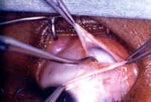
Many surgeons prefer the standard limbal incision, in which the conjunctiva is entered near the limbus and blunt dissection performed, avoiding the muscle and its insertion. This incision is required in most reoperations.5 A cul-de-sac or fornix incision is also useful, particularly for inferior oblique muscle surgery. This incision avoids the perilimbal conjunctiva and in the initial healing period results in a less visible scar (Slide 3). Surgery performed through a fornix incision provides less visualization of the operative field and requires an assistant familiar with this technique.
The standard limbal incision may be a better choice for an unassisted surgeon and when conjunctival recession is desired. Occasionally, the surgeon will encounter excessive scar tissue, which precludes either of the above incisions and requires an incision directly over the muscle. Once the conjunctival incision is made, the strabismus surgical procedure is initiated.
References
- Parks MM. Atlas of Strabismus Surgery. Philadelphia, Pa: Harper & Row; 1983.
- Kivlin JD, Wilson ME Jr. Periocular infection after strabismus surgery. Periocular Infection Study Group. J Pediatr Ophthalmol Strabismus. 1995; 32:42-49.
- Breckenridge AL, Dickman DM, Nelson LB, et al. Long-term results of hang-back medial rectus recession. J Pediatr Ophthalmol Strabismus. 2003; 40:81-84.
- VonNoorden GK. The limbal approach to surgery of the rectus muscles. Arch Ophthalmol. 1968; 80:94-97.
- Parks MM. Fornix incision for horizontal rectus muscle surgery. Am J Ophthalmol. 1968; 65:907-915.