Mark R. Levine, MD · Amarpreet Singh, MD
Introduction
Dry eye syndrome (DES) is a complex and prevalent disease that affects approximately 12% of the United States' population. The condition affects more than 3.2 million American women who are middle-aged or older.1 In contact lens wearers, computer users, patients who live or work in dry environments, and patients with autoimmune disease, the incidence is higher. To illustrate the magnitude of DES, Schiffman and colleagues2 conducted a study in which the effect of DES on a patient's life was shown to be similar to that of moderate to severe angina.
Why is DES discussed in oculoplastics? Failure to appreciate the complexity of the tear film (i.e., a decrease in aqueous tear production or meibomian gland dysfunction) may lead to an unhappy patient after ptosis or blepharoplasty surgery. A borderline condition or exacerbation of a pre-existing condition may exist postoperatively, as the lid dynamics are changed. Preoperative appreciation of the dry eye and meibomian gland dysfunction with appropriate preoperative treatment may circumvent potential postoperative complications.
In an oculoplastic practice, patients may be referred for epiphora, but present with pseudoepiphora (dry eyes). Despite having success in the majority of patients with pseudoepiphora, a small percentage of patients fall short of expected comfort despite adequate treatment.
A large number of patients with DES have meibomian gland dysfunction. In the literature, McCulley and colleagues3 found that 100% of patients with meibomianitis had signs of DES. The symptoms of DES (i.e., dryness, foreign body sensation, tearing, fluctuating visual acuity) account for the greatest percentage of presenting complaints to an ophthalmologist. A thorough understanding of how to examine, diagnose, stratify, and treat DES is essential.
Evaluation and Diagnosis
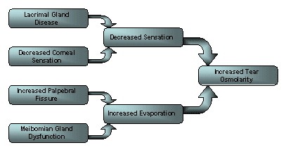
Central to virtually all dry eye disorders is a loss of water from the aqueous tear film in a quantity sufficient to increase tear film osmolarity. This loss of water and increase in osmolarity may result from a condition that decreases tear production or increases tear evaporation (Slide 1). Because numerous causes exist and the treatment options vary, it is important to determine the etiology and tailor the regimen to the cause.
Evaporative dry eye may be caused by an increase in the palpebral fissure height (i.e., increasing the surface area for evaporation), giving credence to the role of lateral tarsorraphy in the treatment. Evaporative dry eye may also be caused by meibomian gland dysfunction (MGD). The lipid layer of the tear retards aqueous evaporation (Slide 2).
Tear film is composed of an outer lipid layer, a middle aqueous layer, and an inner mucin layer. The constituents of the layers are produced by the meibomian glands, the lacrimal (and accessory lacrimal) glands, and the goblet cells. A dysfunction in any of the three may lead to a complication with tear film stability.
The neural reflex arc is often an overlooked aspect of ocular function and is crucial in maintaining a healthy ocular environment. Surgeons once believed that tear production was composed of the basal secretions of Wolfring and Krause (sympathetically innervated) and the reflex secretions of the lacrimal gland. When basic secretion is decreased and there is corneal exposure and irritation, the reflex secretors come into play.
The functions of the eyelids and ocular adenexa are integrated by two reflexes, both mediated on the afferent side by the first division of the trigeminal nerve (V1). The efferent pathways are mediated by the facial nerve (CN 7). The arm that is involved in the composition of the tear film is mediated by the parasympathetics of CN 7, whereas the arm that involves the hydrodynamic elements is run by the motor division. These are integrated to promote a healthy ocular surface (Slide 3).
When evaluating a patient with suspected DES, a number of points in the history and physical examination will help in establishing the diagnosis.
History
Early morning irritation, pain, and crusting that improve as the day progresses is a complaint more common in patients with blepharitis and meibomian gland disease than patients with DES. Foreign body sensation, pain, and irritation that becomes progressively worse throughout the day or with increased visual activity (i.e., reading, watching television, or working on a computer) are indicative of DES.
Patients may also complain of decreased visual acuity due to the instability of the tear film. A function of the tear film-air interface and cornea is its contribution to refraction. This contributes approximately 40 D of the total 60 D refractive ability of the eye (the remaining 20 D being in the crystalline lens). Because the tear-air interface is of paramount importance, a disruption in the tear film causes significant symptoms of blurred vision.
A physician must review a patient's medication list. A number of medications exacerbate DES including antidepressants, decongestants, antihistamines, blood pressure medications, diuretics, ulcer medications, tranquilizers, beta blockers, and oral contraceptives.
A physician should review a patient's medical and dietary history. Does a patient have acne rosacea and eat cheese, nuts, chocolate, or drink coffee, tea, or wine? This is indicative of meibomian gland dysfunction.
Physical examination
Ocular rosacea is often an overlooked cause of MGD but represents an important facet in management. Look for signs of ocular or facial rosacea including rhinophyma, malar flushing, and facial telangiectasias.
Inspection of tear meniscus and tear break up time
Begin by examining the tear meniscus (should be 1.0 mm and convex). Next, instill fluorescein in the inferior cul-de-sac and evaluate the stability of the tear film. This must be performed before the instillation of drops and before manipulation of the lids.
Moisten a fluorescein strip with sterile saline and touch it to the inferior tarsal conjunctiva. Place the cobalt blue filter on and, after several blinks, instruct a patient to keep his or her eyes open. Look for dry spots that appear in less than 10 seconds. This is an indication of an unstable tear film. Another test that correlates well with an abnormal tear break-up time is the inability to hold eyes open for 10 seconds without burning or stinging.
Schirmer testing
Schirmer testing is a test of aqueous tear production that is performed by placing a thin strip of filter paper in the inferior cul-de-sac (Slide 4).
Place the strip laterally to minimize the effect of corneal irritation. A patient is also asked to keep the eyes closed to minimize the effect of blinking. This test may be performed in several ways to measure different aspects of aqueous tear production.
One preference is to measure basic tear secretion after anesthetic instillation and drying out of the fornix with a cotton-tipped applicator. This allows the effect of reflex tearing to be excluded in the measurement of tear production. The strips are left in for 5 minutes and, although normal measurements may be variable, a result of <10 mm at 5 minutes is highly suggestive of aqueous tear deficiency (ATD).
A practical way to achieve a similar (although not identical) result is to perform the Schirmer test for 1 minute and multiply the result by 3. This allows for identification of patients who have moderate to severe dry eye as well as patients who are normal. However, repeated measurements may be required for patients who fall in between. The other tests (Schirmer I and II) are more specific for the evaluation of reflex tearing.
Fluorescein or rose bengal staining
The pattern of staining is more important than the presence or absence of stain. Inferior staining with fluorescein suggests probable lid etiology such as lagophthalmos, meibomianitis, or blepharitis. Using rose bengal staining (or the equally effective and less irritating lissamine green) can detect early changes in the progression of DES.
Rose bengal or lissamine green stains the conjunctiva once goblet cell loss becomes significant. The pattern of staining is more useful than the presence or absence of stain. The nasal conjunctiva stains more than the temporal conjunctiva, and the resilient cornea stains less than the conjunctiva and later in the disease process.
Gross examination of the lids
Look for eyelid malposition and blink rate (i.e., Parkinson's disease). It is important to look for punctal ectropion and punctal stenosis which may be primary or secondary to chronic meibomianitis, blepharitis, or rosacea. Pay attention to whether a patient has gross epiphora. If present, this usually indicates a structural complication (i.e., lid position or lacrimal system). Remember to irrigate the lacrimal system in a patient with gross epiphora.
Slit lamp examination of the lid margin
Inspect the lids for the presence of blepharitis with scaling or lash inflammation. Bacterial lipases can directly cause
destabilization of the tear film in addition to a toxic effect on the surface epithelium. Chronic blepharitis has associated meibomian abnormalities. The bacteria produce toxins that inflame the lid and cause keratinization of the meibomian glands and subsequent stenosis. This may result in an unstable tear film that often is associated with evaporative dry eye.4
When looking at the meibomian glands for signs of meibomian gland dysfunction, the meibomian gland secretions should be clear or slightly yellow and express easily. The natural progression of MGD is:
- Lid margin vascular engorgement and telangiectasis of the lid margin overlie the meibomian glands (Slide 5).
- Stenosis of gland orifices. When pressure is applied to the lid margin, oil secretion can be expressed through the stenosed orifices. These secretions are often turbid and/or have an appearance that is similar to toothpaste (Slide 6).
- Chronic meibomianitis may lead to the thickening and blunting of the lid margin that causes gland orifice obliteration (Slide 7).
Treatment
Treatment of DES and MGD is multifactorial. Neither a single correct treatment regimen nor a hierarchy of treatment protocols exists. The treatment must be aimed at the underlying cause.
Lid hygiene
Hot compresses applied to the eyelid margins can help to liquefy the thick meibomian gland secretions and facilitate their expression. Mild, nonirritating cleaning solutions, such as diluted baby shampoo or commercially prepared eyelid scrubs, can also be applied to the eyelids to remove clogging debris. Additionally, light pressure applied to the eyelids can aid in gland expression.
Punctal occlusion
Punctal occlusion should never be used when a patient has delayed clearance of the tears or in patients with MGD. If the outflow is blocked in a patient with an increase in inflammatory mediators (i.e., MGD), then the condition will worsen, as the inflammatory components are unable to be cleared. Punctal occlusion is indicated when the diagnosis of ATD has been made. Correct diagnosis is essential before undergoing punctual occlusion.
Artificial tears
Drops with preservatives, with a disappearing preservative and with no preservatives, are available. Because of the frequency of application, non-preserved artificial tears are recommended. Tears should be applied liberally throughout the day and, if necessary, a lubricating ointment may be used at night. This ointment may contain an antibiotic preparation.
Topical steroids
Topical steroids are useful as a temporizing measure when a patient is symptomatic and the eye or eyelids show moderate to severe signs of inflammation. In severe cases in which the ocular surface is filled with inflammatory mediators, patients will likely see results from low-dose steroids, though in most cases this is still an off-label use.
Topical corticosteroids have a beneficial effect on the subjective and objective clinical parameters of patients with moderate to severe dry eye. These effects were associated with a reduction of inflammation markers of conjunctival epithelial cells.5 Topical corticosteroids should not be used for longer than 2 weeks to minimize adverse effects (i.e., steroid-induced glaucoma).
Oral antibiotics (tetracyclines)
Tetracyclines represent the most common and most effective treatment regimen for rosacea. These drugs are believed to be effective not primarily as antibiotics but rather through a secondary effect that they exert on the meibomian glands. Tetracyclines decrease bacterial lipase, thereby altering the fatty acid composition of the meibomian gland secretions and improving their solubility. These medications are also effective in protecting the cornea from inflammatory responses by inhibiting collagenase.
Among this class of medications, tetracycline and doxycycline are the most commonly used. They are similar in their mechanism of action, adverse effect profile, and efficacy. However, differences exist. Tetracycline, having a shorter half-life, is prescribed four times per day, as opposed to doxycycline, which is given once or twice per day. The adverse effects profile is slightly more favorable for doxycycline. Common adverse reactions are gastrointestinal upset, photosensitivity, and permanent discoloration of the teeth in newborns. Frucht-Pery and colleagues6 reported a more rapid therapeutic response to tetracycline; however, no difference was found at 6 months.
Topical immunosuppressants
One immunomodulator specifically approved for dry eye is Restasis (cyclosporine A, Allergan). The mechanism of action is not clearly understood, though it is believed to be related to inhibition of T cell production in the lacrimal gland. This emulsion has been shown to improve tear production, which leads to improvement in tear film stability. Tear film stability resolves dry eye symptoms and returns the ocular surface to a more healthy state.
The Food and Drug Administration initially rejected Restasis because it failed to demonstrate a therapeutic effect. It is now known that it takes 3 to 4 months to achieve a clinically significant effect and approximately 6 months before full therapeutic potential is achieved. This is due to the life cycle of T cells (approximately 120 days).
Researchers have found that treatment of DES with topical CsA for 6 months resulted in an increase in goblet cell numbers in patients with and without Sjögren's syndrome, and a decrease in epithelial turnover in patients with non-Sjögren's keratoconjunctivitis sicca. Reducing ocular surface inflammation may have an effect on the proliferative activity of the epithelium.7 One study found a decrease in interleukin-6 levels in the conjunctival epithelium of moderate to severe dry eye patients treated with 0.05% CsA for 6 months.8
Omega-3 fatty acids
It is thought that a dysfunction in the meibomian glands leads to a defect in the lipid layer of the tear film. This, in turn, allows the aqueous layer to evaporate easily, allowing for corneal surface exposure and symptoms.
Researchers report patients with DES being successfully treated with oral flaxseed oil (1000 mg twice a day).9 The major components of flaxseed oil are omega-3 (57%), omega-6 (15%), and omega-9 (18%) fatty acids. These components act as precursors to different prostaglandins, which affect inflammation. According to Jeffrey P. Gilbard, MD,10 omega-6 fatty acids are ubiquitous in milk, ice cream, pizza, steak, and fried foods.
These foods are commonplace in the American diet. In contrast, Americans are omega-3 starved (found in salmon, herring, sardines, etc). While omega-6 fatty acids are pro-inflammatory, omega-3 fatty acids counter balance them (they are used in the production of prostaglandins and cytokines that are anti-inflammatory). Omega-6 fatty acids are converted in our blood stream into pro-inflammatory arachadonic acid and its inflammatory by-products.
In addition to the effects on the eyelids, studies suggest that flaxseed oil and other omega-3 fatty acids may be helpful in treating a variety of conditions. The evidence is strongest for heart disease and complications that contribute to heart disease,11 but the range of possible uses for flaxseed oil include arthritis, hypercholesterolemia, and breast cancer prophylaxis.12-14
By using flaxseed oil (1000 mg capsules twice a day), not only are the inflammatory mediators lessened (similar to using doxycycline for its anti-inflammatory effects), the secretions of the meibomian glands are also improved. This is continued indefinitely in addition to erythromycin ointment at night (to decrease any bacteria that elaborate inflammatory toxins). The erythromycin ointment may be stopped after several months.
Topical antibiotics
Topical antibiotics are used in conjunction with oral flaxseed oil to reduce staphylococcal load at the eyelid margin. This is an effective adjunct when treating blepharitis.
The Oculoplastic Approach
Before ptosis or blepharoplasty surgery and after a patient has been found to have ATD, he or she should be counseled about the risk of postoperative dry eye and prophylaxis should be instituted. This should include frequent application of artificial tears and lubricating ointment at bedtime with or without the use of punctal plugs. This will help to prepare the cornea adequately after more exposure from eyelid elevation. In a patient with moderate to severe ATD, Restasis is started.
If a mild case of MGD presents, add flaxseed oil (1000 mg orally, twice a day). If there is significant MGD, then a regimen of flaxseed oil 1000 mg orally, twice a day, erythromycin ointment at bedtime, warm compresses twice a day, and artificial tears (to dilute the bacterial inflammatory byproducts) is instituted. If the MGD is severe or unresponsive to the above, then it may be prudent to place the patient on oral tetracyclines as well. It may take several months to change the dryness of the tear lipid interface; however, this added precaution may make a difference in achieving an uncomplicated postoperative course.
References
- Schaumberg DA, Sullivan DA, Buring JE, Dana MR. Prevalence of dry eye syndrome among US women. Am J Ophthalmol. 2003; 136:318-326.
- Schiffman RM, Walt JG, Jacobsen G, Doyle JJ, Lebovics G, Sumner W. Utility assessment among patients with dry eye disease. Ophthalmology. 2003; 110:1412-1419.
- McCulley JP, Sciallis GF. Meibomian keratoconjunctivitis. Am J Ophthalmol. 1977; 84:788-793.
- McCulley JP, Shine WE. Eyelid disorders: The meibomian gland, blepharitis, and contact lenses. Eye Contact Lens. 2003; 29(Suppl): S93-S95.
- Avunduk AM, Avunduk MC, Varnell ED, Kaufman HE. The comparison of efficacies of topical corticosteroids and nonsteroidal anti-inflammatory drops on dry eye patients: A clinical and immunocytochemical study. Am J Ophthalmol. 2003; 136:593-602.
- Frucht-Pery J, Sagi E, Hemo I, Ever-Hadani P. Efficacy of doxycycline and tetracycline in ocular rosacea. Am J Ophthalmol. 1993; 116:88-92.
- Kunert KS, Tisdale AS, Gipson IK. Goblet cell numbers and epithelial proliferation in the conjunctiva of patients with dry eye syndrome treated with cyclosporine. Arch Ophthalmol. 2002; 120:330-337.
- Turner K, Pflugfelder SC, Ji Z, et al. Interleukin-6 levels in the conjunctival epithelium of patients with dry eye disease treated with cyclosporine ophthalmic emulsion. Cornea 2000; 19:492-496.
- Boerner C. Dry eye successfully treated with oral flaxseed oil. Ocular Surgery News. 2000; Oct 15:147.
- Gilbard J. Enriched flaxseed oil no fish tale for dry eye, meibomitis. Ophthalmology Times. 2003; 28:16-17.
- Harper CR, Jacobson TA. The fats of life: The role of omega-3 fatty acids in the prevention of coronary heart disease. Arch Intern Med. 2001; 161:2185-2192.
- Kremer JM. N-3 fatty acid supplements in rheumatoid arthritis. Am J Clin Nutr. 2000; (Suppl):349S-351S.
- Prasad K. Dietary flaxseed in prevention of hypercholesterolemic atherosclerosis. Atherosclerosis. 1997; 132:69-76.
- Stoll BA. Breast cancer and the Western diet: Role of fatty acids and antioxidant vitamins. Eur J Cancer. 1998; 34:1852-1856.