Introduction
In the Eyelid Retraction tutorial, a differential diagnosis was enumerated. In clinical practice, the most common entities related to upper eyelid retraction are:
- thyroid-related lid retraction
- overcorrection from ptosis surgery
- facial nerve palsy
- traumatic disruption of the levator muscle with scarring.
In the lower eyelid, the most common entities are:
- thyroid-related lid retraction
- lower lid external blepharoplasty
- external subciliary approach to blow-out fractures
- traumatic lid disruption.
Clinical Findings
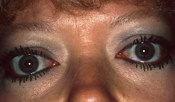
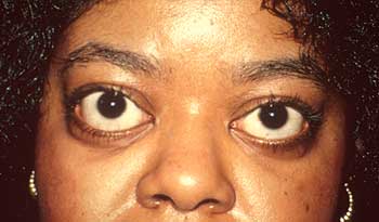
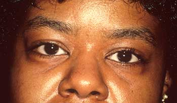
The upper eyelid margin is normally situated 1.5 mm to 2 mm below the superior limbus. The lower eyelid margin is situated at the inferior limbus. Lid retraction may be mild, moderate, or severe. The mechanism for lid retraction and thyroid-related ophthalmopathy includes
- direct inflammatory involvement of the levator/Mueller's muscle complex
- hyperactivity of the sympathetic nervous system in hyperthyroidism with activation of Mueller's muscle
- secondary overaction of the levator-superior rectus complex in response to involvement and tethering of the inferior rectus muscle.
In the lower eyelid, fibrosis of the inferior rectus muscle/inferior tarsal muscle, exerting a retracting action to the lower lid via its capsulopalpebral head, is the probable mechanism.1 In the cases of lower eyelid trauma and postsurgical trauma, intralamellar scarring or anterior lamellar shortening may vertically shorten the eyelid.
Diagnostic Testing
Prior to correction of lid retraction, the immediate treatment goal is to protect the cornea with preservative-free tear supplements, lubricating ointment or gels, punctal plugs, moist chamber shields and taping the eyelid during sleep.
If the anterior lamella is shortened following external lower lid blepharoplasty, the scar may be released with placement of a full thickness skin graft or scar release with a suborbicularis oculi fat (SOOF) procedure fixation and lateral tarsal strip. If postsurgical scarring involving the suborbicularis fascia and orbital septum between the anterior and posterior lamella is involved, release of scarring in that plane with lateral tarsal strip and Frost sutures for several days is the appropriate treatment.
The surgical approaches for treatment for dysthyroid eyelid retraction are classically divided into five groups, each of which is performed through an external or conjunctival approach:
- excision of Mueller's muscle2-3;
- levator aponeurosis weakening or recession2-3;
- excision of Mueller's muscle combined with levator aponeurosis weakening or recession
- marginal myotomy of the levator aponeurosis4;
- use of spacers such as tarsus, sclera, hard palate, ear cartilage, or AlloDerm (LifeCell, Branchburg, NJ).5
For a 1-mm to 2-mm eyelid retraction, many clinicians suggest performing a graded Mueller's muscle resection. For larger amounts of lid retraction (3 mm to 4 mm), complete extirpation of Mueller's muscle, combined with stripping or recession of the levator aponeurosis or marginal myotomy of the levator aponeurosis and Mueller's muscle, has been recommended.2-3 Materials such as tarsus, sclera, ear cartilage, and hard palate have been used as spacers interposed between the edge of the levator aponeurosis and the superior tarsal border to recess or weaken the levator muscle. However, using spacers, especially sclera, can result in prolonged eyelid edema, variable surgical outcomes (depending upon unpredictable absorption), and fixation of the overlying skin to the spacer. In most cases of significant eyelid retraction (3 mm or more) in which these techniques have been performed, patients have a high eyelid crease and a degree of superior tarsal sulcus hollowing, especially if some fat was also removed.
Surgical Procedure
When performing a graded recession of the levator/Mueller's muscle complex, I prefer an external approach to the upper eyelid without opening the orbital septum. Keeping the orbital septum intact masks the effect of the eyelid crease as fat comes forward in the pre-aponeurotic space.6 The procedure is performed under attended local anesthesia (Slide 1). The eyelid crease is marked with methylene blue with little or no skin removed (Slide 2). Xylocaine 1% (lidocaine HCl, AstraZeneca) with epinephrine and Marcaine 0.75% (bupivicaine, Cooke-Waite) are injected into the eyelid and subconjunctivally. An incision is made in the eyelid crease through the orbicularis muscle down to the superior tarsal border. Double-battery cautery is used to divide the levator aponeurotic fibers and, at the same time, recess the levator/Mueller's muscle complex in a graded fashion (greater temporally than nasally) off the conjunctiva (Slide 3). Some surgeons prefer to excise part or all of Mueller's muscle; however, I do not believe this is necessary for a good result. The cautery dissection prevents bleeding from Mueller's muscle. It is important to free up and cut cicatricial bands from Mueller's muscle to the conjunctiva, especially temporally around the lacrimal gland (Slide 4). The lateral horn of the levator is then divided to bring the lateral canthal angle down. The patient is placed in an upright position to determine the lid height and contour (Slide 5). The eyelid is placed 1 mm higher than the anticipated result to allow for a 1-mm postoperative fall from the orbicularis anesthetic block and epinephrine effect on Mueller's muscle. The Levator/Mueller's muscle complex is sutured directly to the underlying conjunctiva with 6-0 silk sutures or to the superior tarsus with a hang-back suture. The eyelid crease is closed (Slide 6, Slide 7, Slide 8, Slide 9, Slide 10, Slide 11, Slide 12 and Slide 13).
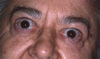
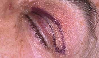
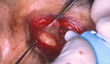
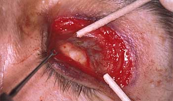
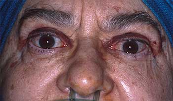
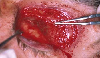
The lower eyelid retractors may be approached through an infraciliary incision or conjunctival approach. This may be combined with implantation of an ear cartilage graft, tarsal conjunctival grafts, hard palate grafts, or AlloDerm grafts between the tarsus and capsulopalpebral fascia with superior placement of the lateral canthal tendon. The purpose of the anterior superior placement of the lateral canthal tendon is to counteract the lower lid retraction and proptosis. Excessive horizontal tightening in the form of a lateral tarsal strip can cause further eyelid retraction by pulling the lower eyelid beneath the equator of the globe. Finally, a small lateral tarsorrhaphy may augment the correction, especially if a patient has significant proptosis.7
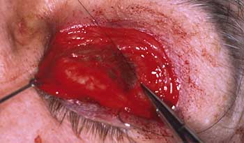
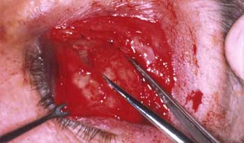
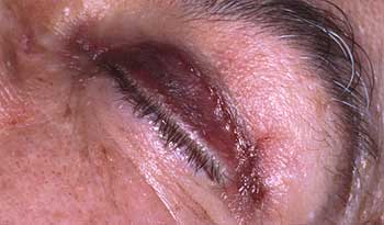
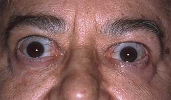
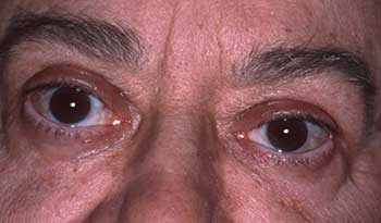
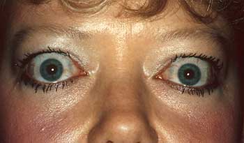
In the past, I used a subciliary incision to create a skin-muscle flap. In this procedure, the capsule of palpebral fascial complex is recessed to bring the lower lid to the inferior limbus. If the eye is mildly to moderately proptotic and tearing, superior placement of lateral canthal tendon is performed. An ear cartilage graft is harvested and placed in a cartilage crusher to contour the graft. The graft is sutured to the inferior tarsal border and to the capsulopalpebral fascial complex to position the lid height at the limbus. The vertical height of the graft is inconsequential. The skin-muscle flap is placed in position and the incision closed. Over the years, this approach has been proven successful; however, I have encountered problems with contouring the ear cartilage graft despite its being crushed to change its memory. The ear cartilage has offered stiffness and survivability as it is sandwiched between skin, orbicularis and conjunctiva. However, the contouring at times has led to a less-than-smooth lower eyelid.
Despite AlloDerm's lack of rigidity when it is hydrated, the use of AlloDerm interposed from a conjunctival approach to the inferior tarsal border to the recessed lower eyelid retractor complex, along with anterior superior placement of the lateral canthal tendon and postoperative Frost sutures, has been effective. In the procedure, the anteroposterior lamella is injected with Xylocaine 1% with epinephrine and Marcaine 0.75%. A #15 blade is used to make a lateral canthal incision. A horizontal canthotomy and inferior cantholysis are performed. A double-pronged skin hook is used to pull down the lower eyelid and a double-battery cautery is used to divide conjunctiva, inferior tarsal muscle, and capsulopalpebral fascia from its insertion to the inferior tarsal border. All bands are cut to allow the complex to retract inferiorly and to allow the lid margin to come up superiorly without resistance. The inferior tarsal muscle need not be excised as it is effectively weakened in the recession of the complex. An AlloDerm graft of substantial vertical height is placed in the lower lid and sutured to the inferior border of the tarsus, the conjunctiva, and the retractors of the lower lid. The goal is to bring the lower lid into a position just above the inferior limbus to allow for slight AlloDerm graft shrinkage and loss of vertical height. The lateral canthal tendon is then placed anterosuperiorly and sutured in the periosteum with a 5-0 nylon suture. Most important, a temporolateral and medial tarsorrhaphy with 6-0 silk sutures is placed through skin, orbicularis, and the gray line of the upper lid to the gray line, skin, and orbicularis of the lower lid and tied over cotton bumpers. This technique keeps the AlloDerm on stretch and the lid margin pulled superiorly. It also keeps the lower eyelid retractor complex inferior so that it cannot advance forward. This approach has been more successful than the previously described procedure involving ear cartilage (Slide 14 and Slide 15).
Discussion
Completely disinserting both the levator aponeurosis from the mid tarsus and Mueller's muscle from the superior tarsal border, as well as freeing the Mueller's muscle meticulously and completely from the underlying conjunctiva, is crucial for the success of this procedure. My technique differs from the ones described by Anderson in that the Mueller's muscle is not excised.2 However, all the fibrotic bands that have formed between Mueller's muscle and the conjunctiva from the inflammatory response from Graves' disease must be severed. Dissecting tissue with a hot cautery minimizes bleeding, facilitates dissection, and prevents significant postoperative inflammation. In addition, not incising the orbital septum allows the eyelid crease to be preserved, especially in cases of mild retraction. This feature represents a significant improvement over procedures in which the orbital septum is opened to approach the levator aponeurosis (with or without preaponeurotic fat removal). The disruption of the orbital septum preaponeurotic fat and levator aponeurosis anatomy results in a higher lid crease and superior tarsal sulcus hollowing.
The same philosophy applies to lower eyelid recession of the underlying capsulopalpebral Mueller's muscle complex without excising the Mueller's muscle. Obviously, the eyelid surgery follows orbital decompression first and strabismus surgery second, as the eyelid position is affected by both of the above.
References
- Tse, DT. Management of thyroid-related eyelid retraction. In: Tse DT, ed. Color Atlas of Ophthalmic Surgery. Philadelphia: JB Lippincott. 1992:387-412.
- Harvey JT, Anderson RL. The aponeurotic approach to eyelid retraction. Ophthalmology. 1981;88:513.
- Putterman AM. Surgical treatment of thyroid-related upper eyelid retraction. Graded Mueller's muscle excision and levator recession. Ophthalmology. 1981;88:507.
- Grove AS. Upper eyelid retraction and Graves' disease. Ophthalmology. 1981;88:499.
- Lisman RD, Milite JP. Management of eyelid malposition in thyroid ophthalmopathy. In: Nesi FA, Lisman RD, Levine MR, eds. Smith's Ophthalmic Plastic and Reconstructive Surgery, 2nd ed. St. Louis, MO: CV Mosby; 1998;113-116.
- Levine MR, Chu A. Surgical treatment of thyroid lid retraction: A new variation. Ophthalmic Surgery. 1991;22:90-94.
- Levine MR, Tomsak RL, El-Toukhy E. Ophthalmology Clinics of North America. Orbital Disease. 1996; 9:645-658.
Posted April 2004