Introduction
Dry eye is a common condition seen in patients with autoimmune disease, women who are postmenopausal, and the elderly.1,2 In a population-based prevalence study of people 65 years of age or older, 14.6% of 2,482 patients reported symptoms suggestive of dry eye.2,3 Extrapolating these numbers to the United States population suggests that at least 4.3 million Americans have dry eye. In another population-based study, 39,876 women in the United States reported a prevalence of dry eye disease to be 7.8% or approximately 3.2 million aged 50 years of age or older.4 Surprisingly, despite these numbers, dry eye remains remarkably underdiagnosed.3-5
Dry eye is often underdiagnosed because of its classification and lack of one specific diagnostic test.6 Two distinct categories of this condition are dry eye related to insufficient production of tears and dry eye related to increased tear evaporation. Tests for one category may be positive yet negative for another category, but both may lead to dry eye disease. To further add to the confusion, a crossover of conditions between the two groups often occurs. For example, meibomian gland dysfunction is the leading cause of evaporative dry eye disease and may also occur in a significant number of patients with aqueous deficiency.
Diagnosis
Diagnosing dry eye begins with patient history. Numerous diagnostic tests are available. Tear film osmolarity, tear lysozyme and tear lactoferrin concentrations, and conjunctival impression cytology are rarely used outside of a research setting. Generally, a clinician relies on the patient history in conjunction with Schirmer testing (with or without anesthesia), supravital conjunctival staining, tear film break up time, tear fluorescein clearance, tear meniscus height, and the presence or absence of tear film debris. Delayed tear fluorescein clearance is reported to show better correlation with the severity of ocular irritation symptoms and keratoconjunctivitis sicca than the Schirmer 1 test.6,7
The tear break-up time is the best screening test for dry eye disease. If this test is abnormal (i.e., under 5 seconds) some form of ocular surface disease may be present — most commonly, dry eye disease. The clinician can help confirm the diagnosis of dry eye with the use of supravital staining. Lissamine green is a new supravital stain that combines the diagnostic advantages of fluorescein and rose bengal. Lissamine green stains healthy epithelial cells when the cells are not protected by a mucin layer in a manner similar to rose bengal. Lissamine, like fluorescein, also stains dead or degenerated cells.8-10 Lissamine green does not cause the ocular discomfort and corneal toxicity associated with the use of rose bengal and is helpful in diagnosing dry eye (Slide 1).8,11,12
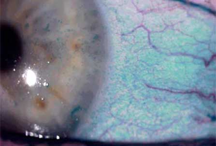
The primary advantage of lissamine green in diagnosing patients with dry eye is that it avoids the pain on installation that is associated with rose bengal in patients with keratoconjunctivitis sicca. The primary disadvantage of lissamine green is that it is less sensitive than rose bengal, more transient, and more difficult to see on slit-lamp examination. In the evaluation of candidates for refractive surgery, supravital staining can reduce the risk of complications and wound healing complications related to poor outcomes by identifying high-risk patients before surgery.13
Classification
The National Eye Institute has proposed a classification system for dry eye based on the differentiation between deficient aqueous production and evaporative tear loss. Multiple subgroups in each of the two categories exist and significant crossover can occur. However, the classification system provides a starting point for a rational treatment plan for dry eye syndrome (Slide 2). Traditional therapy has included the use of artificial tears to supplement a patient's own tear production. For patients with mild dry eye, preserved artificial tears have been effective. For patients with severe dry eye, nonpreserved unit dose packs have gained widespread acceptance. Recently, transiently preserved artificial tears in which the preservative is dissipated upon exposure to air have been developed. For patients in whom artificial tears are not sufficient, punctal occlusion, either temporary or permanent, with plugs or cautery may be effective for preserving a patient's natural tears and prolonging the effect of artificial tears that have been instilled.14
Pathogenesis
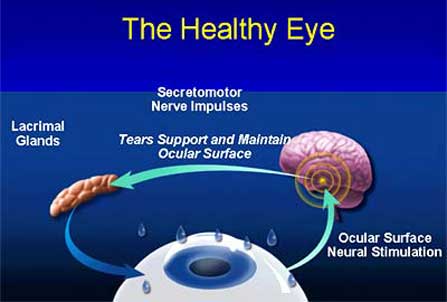
A major advance in dry eye over the past decade is the recognition that the ocular surface and lacrimal glands function as an integrated unit (Slide 3). Communication between these tissues occurs through a sensory/autonomic neural reflex loop. The sensory nerves enervate the ocular surface and nasal mucosa synapse with efferent autonomic nerves in the brain stem that stimulate secretion of tear fluid and proteins by the lacrimal glands. Ocular surface sensitivity decreases as aqueous tear production and clearance of tears from the ocular surface decrease. In turn, this decrease in surface sensation exacerbates dry eye due to a decrease in sensory-stimulated reflex tearing and decreased ability of the lacrimal glands to respond to ocular surface injury. A vicious self-perpetuating cycle of continuing inflammation is created, mediated by tears produced by lacrimal gland tissue heavily infiltrated with T-cell lymphocytes. The T-cell lymphocytes are constantly secreting inflammatory mediators and cytokines that bathe the ocular surface.15,16
The importance of inflammation in the pathogenesis of dry eye disease has also been elucidated over the past decade (Slide 4). Decreased tear production and tear clearance lead to chronic inflammation on the ocular surface. This inflammatory response consists of inflammatory cell infiltration of the ocular surface, activation of the ocular surface epithelium with increased expression of adhesion molecules and inflammatory cytokines, increased concentrations of inflammatory cytokines in the tear fluid, and increased activity of matrix degrading enzymes, such as matrix metalloproteinase-9 in the tear fluid.17 A significant positive correlation has been observed between the levels of inflammatory cytokines in the conjunctival epithelium and the severity of ocular irritation symptoms and corneal fluorescein staining. The inflammatory cytokines and other inflammatory mediators also correlate positively to the severity of conjunctival squamous metaplasia in patients with Sjögren’s syndrome keratoconjunctivitis.15,16
Inflammation has been linked not only to aqueous deficiency dry eye disease but also to that of evaporative loss. The most common cause of evaporative loss dry eye disease is posterior blepharitis or meibomian gland dysfunction. The signs and symptoms of evaporative loss dry eye disease are exacerbated by abnormalities in the lipid layer of the tear film, which is produced by the meibomian glands. Obstruction of the meibomian ducts causes accumulation of meibomian gland secretions, known as meibum. Accumulation of meibum within the meibomian gland can lead to inflammation of the gland and bacterial colonization.1,3,18-20 The colonizing bacteria have lipases that break the nonpolar wax and sterol esters into triglycerides and free fatty acids (polar lipids), thus altering the normal composition of the meibum.21 The polar lipids diffuse easily through the aqueous layer and contaminate the mucin layer, making it hydrophobic.22 This causes the tear film to become unstable, and the surface of the eye becomes unwettable (Slide 7).
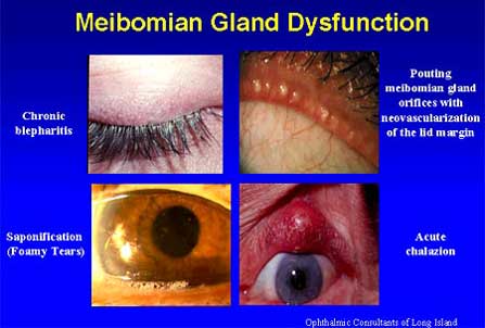
The abnormal meibum has a melting point above the ocular surface temperature, in contrast to normal meibum, which has a melting point equal to or lower than the ocular surface temperature.20,23 The abnormal meibum solidifies and obstructs the ducts, leading to further inflammation and perpetuating the vicious cycle.
Topical cyclosporine A is a highly specific immunomodulator that affects primarily T-lymphocytes, may decrease the inflammation of the meibomian glands, and reduces plugging and dysfunction.
New Pharmacologic Class
Restasis (cyclosporine ophthalmic emulsion, Allergan) is a 0.05% emulsion of cyclosporine A, the active ingredient. The vehicle is glycerin, castor oil, polysorbate 80, carbomer 1342, purified water, and sodium hydroxide. This vehicle is marketed separately as the artificial tear Refresh Endura (Allergan). Restasis appears as a white to slightly translucent homogeneous emulsion. Cyclosporine A is a cyclic polypeptide produced as a metabolite by the fungus Tolypocladium inflatum gams. Cyclosporine A has the following characteristics:
- Well-known for anti-inflammatory properties and as an immunomodulatory agent.
- Most commonly used systemically to prevent rejection of transplanted tissues.
- Used systemically to treat psoriasis and rheumatoid arthritis.
- Mechanism of action of cyclosporine on inflammatory disease processes arises from its ability to inhibit T cell-mediated inflammation.
- Prevents activation of T cells by cytokines and other agents of inflammation.
- Normalizes effects of chronic dry eye disease processes of T cells and lacrimal gland acinar cells.
- Activated T cells are responsible for the production of inflammatory cytokines and other substances. These substances lead to tissue damage, activation of more T cells, and the production of more inflammatory substances.
Phase 3 Studies
A large, multicenter, double-masked, parallel, randomized 6-month trial evaluating three treatment groups involving 877 patients was the basis for approval of Restasis. Patient demographics were 82% women, 84% caucasian, and 31% of patients had Sjögren’s syndrome. The age range was 22 to 90 years of age with a mean of 60 years of age. There were no statistically significant differences between treatment groups in any demographic variable and no statistically significant differences in previous therapy or types of concomitant medications used during study for other conditions. At baseline, patients were using artificial tears as frequently as required. In the group receiving 0.05% cyclosporine, the average baseline use was 6.25 times daily. In the group receiving 0.1% cyclosporine, the average baseline use was 5.56 times daily. These patients had been previously treated unsuccessfully with multiple products and practices.
The study evaluated several primary and secondary variables. The four primary efficacy variables included two objective variables — corneal staining and Schirmer test with anesthesia — and two subjective variables — reliance on artificial tears and blurred vision. The six secondary efficacy variables included photophobia, sandy/gritty feeling, burning or stinging, itching, feeling of dryness, and pain. Several tertiary laboratory variables to assess immune involvement included presence of inflammatory markers, goblet cell density, and status of pathological apoptosis. Tests were performed on conjunctival biopsy tissue obtained before and after treatment.
Key inclusion criteria:
- Symptomatic chronic dry eye disease despite conventional management.
- Schirmer test values of 5 mm of wetting or less in 5 minutes in at least one eye.
- The sum of the corneal and interpalpebral conjunctival staining of at least +5 on "0" to "15" scale in the same eye in which corneal staining of least +2.
- Baseline ocular surface disease index score of 0.1.
- Facial expression rating scale scores of at least 3, with "1" being the happiest face and "9" being the most unhappy face.
- Normal lid anatomy and blinking function and best-corrected Early Treatment Diabetic Retinopathy Study visual acuity scores at least +0.7 logMAR in both eyes.
Key exclusion criteria:
- Severe chronic dry eye disease (i.e., those with nasal-stimulated Schirmer scores less than 3 were not enrollable). These patients were judged to have dry eye disease so severe that there were no lacrimal gland acini left to respond to therapy.
- Permanent goblet cell loss or scarring.
- Active ocular infection.
- Ocular rosacea.
- Active severe blepharitis or lid margin inflammation.
- Inflammation not attributable to dry eye disease or any type of corneal sensitivity disorder.
- Lacrimal punctal occlusion within 3 months of entry into the study.
- Contact lens wear.
Rationale of this study involved the theory that insufficient ocular lubrication increases corneal abrasion of the superficial epithelium. Abraded corneal epithelium readily accepts stain. Although an indirect measure of inflammation and immunoreactivity, corneal staining is commonly used for chronic dry eye and is considered clinically important. Significant difference relative to the vehicle was seen in corneal staining between the 0.05% and the 0.1% cyclosporine groups at 4 months.
Significant difference relative to the vehicle was seen for 0.05% cyclosporine and the 0.1% cyclosporine at 6 months. Patients showed statistically and clinically significant mean reductions of more than 30% versus baseline in corneal staining at all time points.
Changes also occurred from baseline blurred vision. Blurring of vision occurs in chronic dry eye disease because desiccation of corneal epithelial cells causes them to shrink, crease, and opacify. This makes blurred vision an important efficacy measure. Patients treated with 0.05% cyclosporine and the 0.1% cyclosporine exhibited statistically and clinically significant reductions versus baseline and vehicle in complaints of blurred vision at all time points. At 6 months, 44% of patients treated with cyclosporine 0.05% and the 0.1% cyclosporine experienced less blurred vision. The mean vision improvement was 24%, indicating that normalizing ocular surface improvement was statistically significantly different from vehicles at 1 month, 3 months, 4 months, and 6 months (Slide 8).
Artificial tear reliance also changed from baseline. Patients in all study groups were allowed to instill artificial tears between study-medication doses throughout the trial. A reduction in the volume of artificial tears used can be a gauge of symptomatic relief. After 6 months, a reduction versus baseline in artificial tear use was seen in all groups. The decrease was much larger in the cyclosporine groups. The decrease was statistically significant for the group receiving 0.05% cyclosporine and the 0.1% cyclosporine at 6 months. Concomitant tear use was reduced in 33% of patients by at least five times daily. Tear use was reduced in 51% of patients by at least three times daily. Overall use of artificial tears fell by one-third at 6 months. These data indicate that patients’ eyes were feeling better (Slide 9). There was no difference in efficacy between the 0.05% and 0.1% concentrations of cyclosporine. This led the FDA to recommend the 0.05% cyclosporine medication.
Need for laboratory assessments is significant because there is currently no good, single test to assess function or that correlates well with common complaints. Clinical presentation of dry eye disease can have significant variation. Patients range from mildly to extremely symptomatic and staining can be very mild or pronounced. One patient may have severe corneal staining and a very low Schirmer score, whereas another patient with severe corneal staining may have a normal Schirmer score.
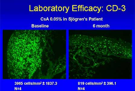
Severe dry eye may have no effect on vision in one patient, whereas another patient might complain of blurring despite only minor corneal staining. The condition may have reached clinical level, or it may be severe end-stage disease with complete lacrimal gland shut-down and severe corneal and conjunctival signs. Signs and symptoms may be the result of the primary disease or secondary to autoimmune disorder, trauma, or other factors. These factors warranted the performance of laboratory assessments. When comparing baseline (Slide 5) to 6-month values in a patient with Sjögren’s syndrome, a significant decrease is seen in the number of CD-3 positive T-cells in the conjunctiva with 0.05% cyclosporine. Similar results occurred in patients with non-Sjögren’s (Slide 6).
Consistent with a similar pathophysiology for both conditions, there were reduced inflammatory markers. HLA-DR and CD11a determined via conjunctival biopsy. Baseline values were found to be significantly elevated in patients with Sjögren’s syndrome and non-Sjögren’s syndrome in the pivotal clinical trials, as well as a separate biopsy study performed by Allergan in collaboration with the National Eye Institute. After 6 months of treatment with 0.05% cyclosporine, statistically significant decreases were seen for inflammatory markers HLA-DR and CD11a, compared with increases in the vehicle group.
Goblet cell density may be the most sensitive measure of overall ocular surface health. The number of mucin-producing goblet cells is known to be decreased in patients with moderate to severe chronic dry eye disease. As goblet cells die, density decreases results in decreased mucin production, which further destabilizes tear film. Increase in goblet cell density indicates a normalization of ocular surface. Cyclosporine treatment resulted in a significant increase in goblet cells. The increase over 6 months of treatment was 191% in the patients treated with cyclosporine, compared with only 13% in vehicle-treated group (P=.013). Vehicle provided only symptomatic relief and had no influence on goblet cell density (Slide 10).
These clinically relevant findings indicate that cyclosporine works on the underlying pathology of chronic dry eye disease. These data further suggest that use of 0.05% cyclosporine results in normalization of the ocular surface, permitting return of a stable and healthy tear film. The vehicle is a low-viscosity, oil-based emulsion that includes a water-soluble polymer, oil, and an emulsifying agent that lubricates and moisturizes, providing immediate palliative relief. The oil component remains on the tear-film surface for up to several hours and may retard aqueous evaporation. Vehicle alone, while not treating the disease, provides substantial symptomatic relief. All statistically significant results favored Restasis over its vehicle. Vehicle alone showed no significant benefit for any measured inflammatory marker.
Adverse events were monitored. Blood chemistry and hematology tests were performed to monitor changes in renal and hepatic function. No significant adverse events were reported. All adverse events were local and not related to administration of the drop medications. This is significant because these patients usually react significantly to any form of topical medication. Transient burning, usually of mild or moderate severity, was reported for 14.7% of the 293 patients treated with 0.05% cyclosporine and was the main adverse event. Other ocular adverse events reported by 3% to 6% of patients treated with 0.05% cyclosporine were stinging, discharge, and foreign-body sensation. Difference in incidence between 0.05% cyclosporine and vehicle groups was negligible.
Serious adverse events (5.8% of 293 patients in the group treated with 0.05% cyclosporine and 4.8% of the 292 patients in the vehicle group) were considered unrelated or unlikely to be related to study medications. This reasoning was based on the observation that with the exception of mild burning on contact, no discernible difference in the type or rate of adverse events in either treatment group when compared to vehicle was found.Treatment discontinuation rates due to adverse events were low and comparable — 6.5% for 0.05% cyclosporine versus 4.5% for vehicle. There were no clinically significant differences between the group treated with 0.05% cyclosporine and the vehicle-treated group in visual acuity, changes in intraocular pressure, or biomicroscopy. No cases of ocular infection in the cyclosporine-treated groups presented, although ocular infection was present in the vehicle-treated group. Additional results support the excellent safety profile of topical cyclosporine. There were no cyclosporine-related ocular infections in either the Phase 2 or Phase 3 studies. No clinically significant changes were seen in blood chemistry or hematology values (including hepatic and renal function). No patients experienced adverse effects related to blood chemistry or hematology. Also, there were no treatment-related changes in intraocular pressure, visual acuity, or biomicroscopy. No overgrowth of microbial organisms presented.
Restasis is the first treatment that targets an underlying pathological mechanism for chronic dry eye — immune-mediated inflammation. It is the only purpose-designed topical drug therapy for chronic dry eye disease. Restasis is demonstrated effective and safe in animal studies and human clinical trials. It reduces signs and symptoms of dry eye disease and has favorable pharmacokinetics while being well-tolerated with few and minor ocular adverse effects. Use is not associated with any systemic adverse effects — no cyclosporine is detectable in the blood. No microbial overgrowth or ocular infections occurred during clinical studies.
Restasis has been successful in treating patients with keratoconjunctivitis sicca and has also been used in treating patients with meibomian gland dysfunction.21 With new proven efficacy in meibomian gland dysfunction, this agent has promise for treating conditions in both arms of the dry eye disease classification, namely aqueous deficiency and evaporative tear loss.
Summary
The difficulty of diagnosing and treating dry eye disease remains problematic. However, a breakthrough in understanding of the pathogenesis of dry eye disease has led to the first drug therapy to treat the cause of dry eye and not just its symptoms or signs. Restasis offers the advantage of immunomodulation without the risk of corticosteroid adverse effects. Restasis may be the first drug to effectively treat the causative factors leading to dry eye disease.
References
- Schaumberg DA, Sullivan DA, Buring JE, Dana MR. Prevalence of dry eye syndrome among US women. Am J Ophthalmol. 2003;136:318-326.
- Fox RI, Howell FV, Bone RC, Michelson P. Primary Sjögren’s syndrome: Clinical and immunopathologic features. Seminars in Arthritis and Rheumatology. 1984;14:77-105.
- Schein OD, Munoz B, Tielsch JM, et al. Prevalence of dry eye among the elderly. Am J Ophthalmol. 1997;124:723-728.
- Schein OD, Tielsch JM, Munoz B, et al. Relation between signs and symptoms of dry eye in the elderly: A population-based perspective. Ophthalmology. 1997;104:1395-1401.
- Afonso A, Monroy D, Stern ME, et al. Correlation of tear fluorescein clearance and schirmer test scores with ocular irritation symptoms. Ophthalmology. 1999;106:803-810.
- Macri A, Pflugfelder S. Correlation of the Schirmer 1 and fluorescein clearance tests with the severity of corneal epithelial and eyelid disease. Arch Ophthalmol. 2000;118:1632-1638.
- Chodosh J, Dix RD, Howell RC, Stoop WG, Tseng SC. Staining characteristics and antiviral activities of sulforhodamine B and lissamine green. Invest Ophthalmol Vis Sci. 1994;35:1046-1058.
- Feenstra RP, Tseng SC. Comparison of fluorescein and rose bengal staining. Ophthalmology. 1992;99:605-617.
- Kim J, Foulks GN. Evaluation of the effect of lissamine green and rose bengal on human corneal epithelial cells. Cornea. 1999;183:328-332.
- Manning FJ, Wehrly SR, Foulks GN. Patient tolerance and ocular surface staining characteristics of lissamine green versus rose bengal. Ophthalmology. 1995;102:1953-1957.
- Lemp MA. Report of the National Eye Institute/Industry workshop on Clinical Trials in Dry Eyes. CLAO J. 1995;21:221-232.
- American Academy of Ophthalmology. Punctal occlusion for the dry eye. Three-year revision. Ophthalmology. 1997;4:1521-1524.
- Stern ME, Beuerman RW, Fox RI, et al. The pathology of dry eye. The interaction between the ocular surface and lacrimal glands. Cornea. 1998;17:584-589.
- Pflugfelder SC, Jones D, Ji Z, Afonso A, Monroy D. Altered cytokine balance in the tear fluid and conjunctiva of patients with Sjogren’s syndrome keratoconjunctivitis sicca. Curr Eye Res. 1999;19:201-211.
- Afonso A, Sobrin L, Monroy DC, et al. Tear fluid gelatinase B activity correlates with IL-1alpha concentration and fluorescein clearance in ocular rosacea. Invest Ophthalmol Vis Sci. 1999;40:2506-2512.
- Prabhasawat P, Tseng SC. Frequent association of delayed tear clearance in ocular irritation. Br J Ophthalmol. 1998;82:666-675.
- Marsh P, Pflugfelder SC. Topical nonpreserved methylprednisolone therapy for keratoconjunctivitis sicca in Sjogren’s syndrome. Ophthalmology. 1999;106:811-816.
- Sall K, Stevenson OD, Mundorf TK, Reis BL. Two multicenter, randomized studies of the efficacy and safety of cyclosporine ophthalmic emulsion in moderate to severe dry eye disease. CsA Phase 3 Study Group. Ophthalmology. 2000;107:631-639.
- Goto E, Tseng SC. Differentiation of lipid tear deficiency dry eye by kinetic analysis of tear interference images. Arch Ophthalmol. 2003;121:173-180.
- Phillips TE, McHugh J, Moore CP. Cyclosporine has a direct effect on the differentiation of a mucin-secreting cell line. J Cell Physiol. 2000;184:400-408.
- Perry HD, Doshi S, Donnenfeld ED, et al. Double masked randomized controlled study evaluating topical 0.05% cyclosporine A in the treatment of meibomian gland dysfunction (posterior blepharitis). Invest Ophthalmol Vis Sci. 2003;44:E. Abstract 1395.
- Jumblatt JE, Jumblatt MM. Regulation of ocular mucin secretion by P2Y2 nucleotide receptors in rabbit and human conjunctiva. Exp Eye Res. 1998;67:341-346.
- Rocha EM, Wickham LA, da Silveira LA, et al. Identification of androgen receptor protein and 5alpha-reductase mRNA in human ocular tissues. Br J Ophthalmol. 2000;84:76-84.