Vance Thompson, MD, FACS
Introduction
LASIK has become the most commonly performed surgical procedure for the correction of refractive error. Limitations to LASIK exist, especially in higher refractive errors in which patients can experience reduced accuracy and an increasing rate of complications.1,2 Patients with higher refractive errors have other options like IOL implants and refractive lensectomy.3 Ophthalmologists may be more comfortable with phakic implant technology because the preoperative, surgical, and postoperative care are similar to the care involved with cataract surgery.
The Verisyse lens (Advanced Medical Optics, Santa Ana, Calif) was recently approved by the Food and Drug Administration (FDA) for the treatment of patients with myopia from 5 D to 20 D. The Verisyse phakic lens is the same as the Artisan myopia lens (made by Ophtec, Groningen, The Netherlands, and distributed internationally). Verisyse is the marketing name used in the United States.
Verisyse Phakic Lens Specifications
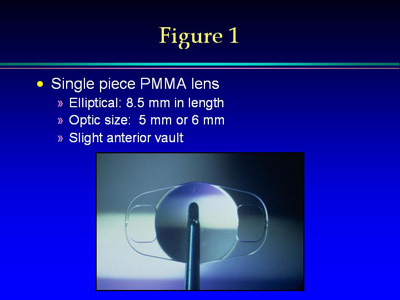
The Verisyse myopia lens is a single-piece PMMA ultraviolet light-absorbing lens designed for implantation into the anterior chamber of a phakic human eye (Slide 1).
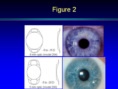
The lens is available for the correction of high myopia between 5 D and 20 D. The lens has either a 5-mm (lens model 206) or 6-mm (lens model 204) convex-concave optic that is incorporated into an 8.5-mm elliptically shaped plate lens design (Slide 2).
The lens has two enclavation (iris fixation) arms on either side for fixation to the relatively immobile mid-peripheral iris stroma. The lens has a slight anterior vault to provide adequate space for aqueous flow and avoid iris chaffing. Lens model 206 (5-mm optic) is available in 1-D increments from 8 D to 20 D. Lens model 204 (6-mm optic) is available in 0.5-D increments from 8 D to 15 D.
The hyperopic lens is being tested in FDA-monitored clinical trials and is not available for general use in the United States at the time of this writing.
Preoperative Evaluation
Inclusion criteria to be in the FDA-monitored clinical trials are patient age of 21 years or older, stable refractive error (defined as a 0.5 D change in refraction over a 6-month period), myopia of 5 D to 20 D, endothelial cell counts greater than 2,000 cells/mm², and anterior chamber depth of 3.2 mm or greater.
Preoperative evaluation for Verisyse lens implantation includes documentation of uncorrected vision, manifest refraction, best-corrected vision, and cycloplegic refraction.
Corneal topography, keratometry, corneal thickness, axial length measurement, anterior chamber depth, and endothelial cell counts should also be obtained. A slit lamp examination is performed to rule out or treat pre-existing blepharitis, dry eye, or other ocular surface conditions.
IOP and assessment for iris trans-illumination defects also should be assessed because patients with high myopia are at an increased risk for pigment dispersion syndrome and pigmentary glaucoma. A dilated evaluation is performed to rule out lens pathology such as cataract and to perform a quality retinal/vitreous examination with scleral depression if necessary because patients with myopia are at an increased risk for retinal pathology.
Surgery
The Verisyse lens implant procedure can be performed under retrobulbar, peribulbar, or topical anesthesia. Peribulbar anesthesia was the most common anesthetic choice in the U.S. FDA monitored clinical trials and is my preferred method of anesthesia.4 It is my opinion that when fixating the Verisyse implant to the iris, the iris can be sensitive and topical anesthesia may not be enough to keep a patient comfortable.
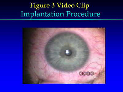
Marks are made at the limbus for creating a 6.2-mm limbal incision (for inserting a 6-mm optic; 5.2-mm limbal incision for inserting a 5-mm optic). Marks are also made at 10 mm (2 mm on either side of the limbal incision) where two stab incisions are created to insert a bent dull 25-gauge needle (also called an enclavation needle) to help fixate the lens (Slide 3). The limbal incision can be made under the conjunctiva (after a peritomy) or at the limbus according to surgeon choice.
A half thickness limbal or scleral incision with dissection just inside clear cornea is first created. Prior to entering the anterior chamber through the main incision site, the enclavation sites are created like a stab incision and are approximately 1.5 mm in width. The angle of these stab incisions is important. It is our first instinct to make these stab incisions radial, but doing so would make the fixation of the iris tissue in the haptic claw more difficult.
The proper angle of the stab incisions is straight down toward the enclavation site. Thus, if one is operating from the top of a patient's head (at 12:00), then the stab incision would start at 10:00 and, while entering the anterior chamber, be directed toward 8:00. The 2:00 stab incision would be directed toward 4:00 as the blade enters the anterior chamber. These incisions are large enough to allow easy access of the enclavation needle but small enough to typically be self-sealing.
Viscoelastic is instilled, to protect the corneal endothelium and crystalline lens, through the enclavation sites. The viscoelastic used in the FDA clinical trial was Healon (Advanced Medical Optics, Santa Ana, Calif). Viscoelastic can be instilled after the first enclavation site is created if the anterior chamber is shallow. The anterior chamber is entered through the limbal-based pocket 6-mm incision. If necessary, then Miochol (acetylcholine chloride, Ciba) is instilled to make sure the pupil is as small as possible to protect the crystalline lens.
It is important not to overfill the anterior chamber with viscoelastic. This is because a knuckle of iris tissue is going to be inserted (ie, enclavated) into the haptics of the Verisyse lens and, if the chamber is overfilled, then it becomes more challenging to accomplish iris fixation.
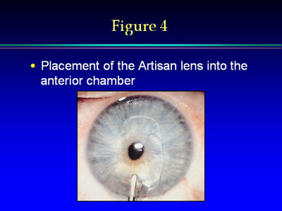
At this point in the procedure, the Verisyse lens is placed into the anterior chamber after a rinsing with balanced salt solution (BSS) (Slide 4). The lens may be centered with a microsurgical hook or manipulator. I use the cannula tip on the Healon because I can inject more Healon during the maneuvering of the implant if the chamber shallows. The lens is then grasped with Budo forceps (Ophtec, Groningen, The Netherlands). These forceps have a wider platform than most forceps for the purpose of providing stability to the lens as it is fixated to the anterior iris stroma.
It is important during fixation not to push posterior (and damage the crystalline lens) or anterior (and damage the corneal endothelium). The surgeon must be aware of staying in one plane with the implant and enclavation needle as iris fixation occurs.
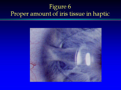
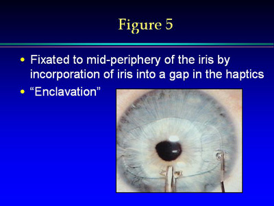
With regard to terminology, "iris fixation" and "enclavation" have the same meaning. They both refer to the step in which a knuckle of iris tissue is created with the enclavation needle and then pushed up into the claw of the haptic (Slide 3, Slide 5). The appropriate amount of enclavated iris tissue in the claw measures approximately 1 mm in all dimensions (Slide 6).
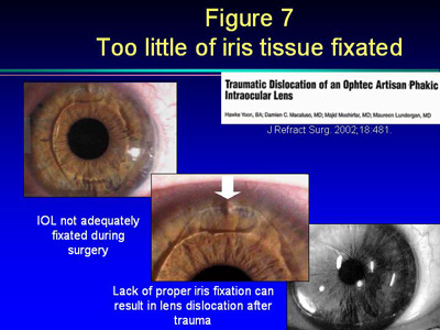
This amount of iris tissue in the haptic provides for stable fixation. If too little iris tissue is enclavated, then dislocation with trauma can occur (Slide 7). If too much iris tissue is enclavated, then posterior pressure of the optic on the iris can increase the chance of promoting posterior synechiae.
During enclavation of the first haptic, it is important to obtain horizontal centration of the optic over the pupil. After fixating the first haptic, the surgeon can place more viscoelastic between the optic and the corneal endothelium if desired. It is important to be cognizant of maintaining a safe distance between the corneal endothelium and the lens optic to protect the corneal endothelium. At this point, the other haptic is enclavated while concentrating on vertical centration.
During this step, the surgeon realizes, with experience, maneuvers that promote optimal centration. For instance, it is typical for the anterior chamber to shallow slightly during enclavation. When the chamber shallows, it is common for the implant to be in a position more inferior to the position it will be in when the chamber is deepened.
When both haptics are adequately enclavated and the surgeon deepens the anterior chamber, the implant will often move superiorly. Therefore, it can be helpful if after the second enclavation step the lens optic is slightly inferior. Then, when the chamber is deepened with BSS, optimal implant centration is obtained.
If at this point optimal implant centration is not obtained, more viscoelastic can be instilled, the optic can be regrasped with the Budo forceps, and the enclavation needle can be used to push the knuckle of iris tissue back through the claw to free it from the iris. Then enclavation can be performed at the new location for optimal centration. The fact that this lens can be re-fixated for optimal centration is an advantage.
After the surgeon is satisfied with lens centration, a peripheral iridectomy (PI) is performed to prevent pupillary block glaucoma. Many surgeons create a PI with a YAG laser preoperatively so this step does not need to be performed intraoperatively. I prefer preoperative YAG laser PI.
The wound is then sutured. Three interrupted 10-0 nylon sutures work well for this step. The remaining viscoelastic is removed with an irrigation and aspiration device before the last suture is tied. It is critical that the viscoelastic be completely removed from the anterior chamber to minimize the risk of a postoperative rise in IOP. Antibiotic drops are instilled and the eye is patched along with a shield.
Postoperative Care
The postoperative care begins immediately after the surgery is completed, which is signified by the removal of the lid speculum. After the speculum is removed, it is important to re-examine the IOP and make sure the pressure is normalized and the wound is watertight. The surgeon may occasionally need to re-pressurize the globe after removing the speculum if removing it significantly lowers the IOP, because the speculum was causing pressure on the globe. Sometimes this can be done without replacing the speculum because the drape is still holding the lashes open, and sometimes the speculum must be reinserted to safely re-pressurize the globe. BSS works well for this maneuver. Occasionally, the enclavation sites can leak and a suture then must be placed.
If the procedure is performed with peribulbar or retrobulbar anesthesia, then the surgeon can put on an antibiotic drop, tape the eyelid if desired (so it does not open under the patch), or place an eye patch and tape a metal shield on. The patient can remove the shield 5 hours postoperatively and begin topical drop therapy. In the case of topical anesthesia surgery, the surgeon can instill an antibiotic drop and tape on a clear shield. The patient is instructed to use the topical antibiotic drop six times per day for the first 3 days and then four times per day until instructed to do otherwise. A topical steroid drop is prescribed four times per day. Patients are instructed to wear the shield, except when putting in drops, until they see me the next morning. After that, the shield is used only when sleeping, for the first 2 weeks.
In the immediate postoperative period, if a patient has had peri- or retrobulbar anesthesia, then he or she is warned that when the shield, patch, and tape are removed, he or she will still have blur because the anesthesia also anesthetizes the optic nerve. Patients should also be warned that they may have some double vision if the extraocular muscle anesthesia has not worn off.
The first postoperative visit involves removing the shield and cleaning debris on the eyelids. We instruct patients how to keep their eyelid margins clean. The uncorrected vision is checked with and without pinhole. IOP is also measured and documented. If the IOP is elevated, then it could be retained viscoelastic, malignant glaucoma, or pupillary block glaucoma. These must all be kept in mind when treating a patient with a postoperative rise in IOP.
If the uncorrected vision is not 20/20, then I perform a refraction to see if the reduced vision is correctable by lenses. If a patient has blurry uncorrected vision, then a refraction can be comforting. In actuality, phakic IOL surgery is typically less traumatic than cataract surgery, and minimal corneal edema should be present. Thus, these patients will often have day one postoperative vision that has them excited about this whole process.
During the slit lamp examination, the examiner should look at the corneal wound and make sure it is watertight. Instilling fluorescein and making sure no leak is present is important. The sutures are then assessed for tension. After assessing the wound, a good evaluation of the cornea is performed starting from the surface epithelium and moving posteriorly.
After assessing the cornea, the anterior chamber is examined for depth and cellular reaction. Because all phakic IOLs are capable of pupillary block, the first day postoperative visit is important to make sure this has not happened by checking IOP and performing a quality slit lamp examination. A pressure elevation can occur after phakic IOL implantation from retained viscoelastic or pupillary block from a non-patent PI. PI with retro-illumination is important to make sure it is patent.
A laser PI is typically smaller than a surgical PI and assessing patency can be more challenging, but it is important that the red reflex be seen through the laser PI upon retro-illumination. If it cannot be documented that the PI is patent, then the PI should be made larger, or a laser PI should be created in a different location. Some surgeons will routinely place two PIs at separate quadrants in the superior iris to further minimize the risk of a non-patent PI creating an angle closure glaucoma attack.
After the depth and patency of the PI are assessed, the cellular (flare and cell) reaction is assessed and documented. Typically, not a lot of inflammation occurs with phakic IOLs compared to cataract surgery. The examiner can also focus further posteriorly and make sure there is no vitreous reaction.
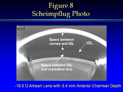
It is important to assess the distance from the anterior surface of the Verisyse implant to the posterior cornea. It is also important to assess the distance between the posterior surface of the IOL to the anterior surface of the crystalline lens. It has been shown that a safe distance between the Verisyse lens and the corneal endothelium and the crystalline lens is present. (Slide 8)
I then finish the slit lamp examination by looking at the crystalline lens to make sure it is clear with no evidence of trauma. If lenticular touch was present during surgery, then a localized opacity in the anterior lens can occur. These are typically localized and non-progressive and would be considered more common with a posterior chamber phakic IOL than with the Verisyse lens (in which the iris serves as a cushion between the implant and the crystalline lens).
In the Artisan lens FDA clinical trials, no cases of endophthalmitis (n=1179 implants) were present.4 Zero incidences of macular edema, pupillary blocks, hypopyon, or persistent raised IOP were reported during the study.
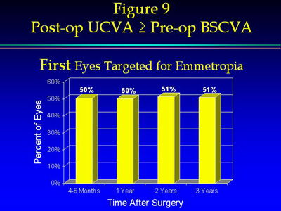
At the 1-week visit, a history is taken to see how the vision is doing and how comfortable the eye feels. It is also important to review the medication schedule and make sure patients are wearing their shields when they sleep. At this visit, the uncorrected vision is documented and a manifest refraction is performed. It is not unusual for the best-corrected visual acuity (BCVA) to actually improve after phakic IOL placement. This is due, at least in part, to image magnification.5 This was also noted in the U.S. clinical trials. At the 3-year visit, 51% of patients had uncorrected vision that was better than their preoperative BCVA (Slide 9).
Generally, everything that was performed at the slit lamp examination at the 1-day visit postoperatively is repeated at the 1-week visit postoperatively. The IOP is checked again to make sure no early steroid response occurs. It is expected that any cellular response that was noted at the 1-day visit is less at this visit. If an increase in anterior chamber reaction occurs and the vitreous is quiet with no other signs of infection, then the steroid drops are increased in frequency and the patient may be put on a stronger steroid drop.
If anything unusual is detected at the 1 week visit, then a repeat visit is scheduled to take place in the next few days or weeks to make sure it is not progressive. Otherwise, if the examination is routine at the 1-week visit, then the patient is instructed to use the antibiotic drops for 10 days postoperatively and to use the steroid drops in a gradual taper so they will be off the steroids at approximately 2 weeks. If the patient is blurry at this visit and is having a hard time driving or working, then a temporary lens prescription can be provided to help them function.
The 1-Month Postoperative Visit
At the 1-month postoperative visit, the patient is typically off all drops and a quality assessment can be done to gauge the near final outcome of the procedure. If residual cylinder is present and if the positive cylinder is in the axis of the sutures, then the selective suture removal process can begin. Surgeons should consider waiting a couple of weeks to see the full effect of the suture removal before deciding whether to remove another suture.
When to Enhance
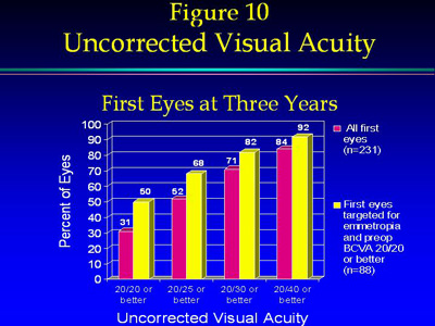
Many patients who have Verisyse lens implantation do not need laser enhancement because Verisyse lenses are so accurate (Slide 10).
If a patient has significant cylinder preoperatively, then an astigmatic keratotomy (AK) can be performed preoperatively, intraoperatively, or postoperatively. AK is effective in lessening astigmatism before or after phakic IOL placement. Surgeons can also place the incision at the time of lens implantation in the steep axis of the cylinder to somewhat lessen the astigmatism.
The time to perform an AK procedure is dependent on the approach taken for the IOL placement. If it has been 3 months after the sutures have been removed and the examination shows the refractive cylinder is not changing, then it is a reasonable time to consider AK.
If it is a spherical (with or without cylinder) laser enhancement with PRK or LASIK, then it may be worth waiting longer after the phakic IOL procedure.6 Six months postoperatively to perform PRK and 1 year to perform LASIK after a Verisyse procedure is reasonable.
Long-term Considerations
As studies continue to be run on the long-term risks of phakic IOLs, good predictability with a low risk of major complications seems to exist.4 As with any intraocular surgery, the long-term effects are important to consider. Patients with myopia are already at an increased risk of developing retinal detachment when compared to the general population. Patients with myopia are at an even greater risk of retinal detachment after intraocular surgery. In the US Verisyse
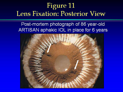
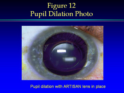
lens clinical trials, six patients of 1,179 patients who had Verisyse lens implantation experienced retinal detachment (0.51%). This is less than in studies looking at refractive lensectomy or cataract surgery in this same highly myopic patient population. It is also important to note that because the Verisyse lens fixates to the relatively immobile peripheral iris, dilated retinal examinations can be performed without complications (Slide 11). When the enclavation site is assessed posteriorly in post-mortem eyes, the quiet nature of undisrupted posterior uveal pigment helps one see why this lens does not cause long-term uveal inflammatory complications but rather allows for a quiet anterior chamber in a majority of patients (Slide 12).
Endothelial cell counts are also a subject of interest in the long-term. The results and analyses of endothelial cell counts in the U.S. clinical trials indicate that the endothelial cell counts remained relatively constant over the 3-year study period. The analyses of the percent change in endothelial cell counts data proved no significant differences
exist for any time period when compared to baseline, or between periods. We believe that endothelial cell counts should be performed preoperatively and checked postoperatively if clinically it seems necessary. The preoperative measurement will serve as a control for any postoperative endothelial cell count testing.
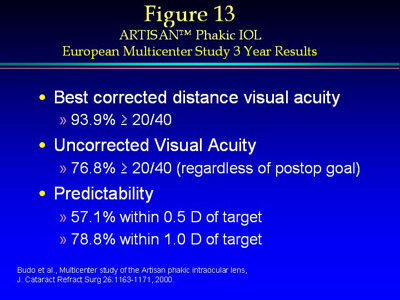
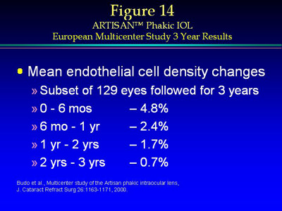
In the largest prospective Verisyse lens clinical trial in Europe, the results were similar to the FDA clinical trials.5 This study consisted of 518 patients with eyes implanted with Verisyse lenses at nine sites from 1991 to 1999 with preoperative myopia of 5 D to 20 D (5-mm optic only) with a 3-year follow-up reported on 249 patients. Visual acuity and endothelial cell data are summarized in Slides 13 and 14. Results demonstrated refractive stability and good predictability with a favorable risk:benefit ratio. Efficacy and safety were demonstrated in this clinical trial.
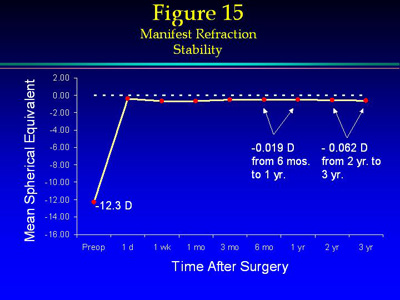
Finally, the stability of the refractive end result is a feature attractive to patients with higher refractive errors (Slide 15). This is especially impressive because this is in the same group of high refractive errors in which the predictability of LASIK falls off.7,8
Conclusion
Phakic IOLs have made many advances over the past 25 years in terms of safety and efficacy with improvements in implant design and surgical technique. With proper attention to details, including postoperative care, the risk:benefit ratio becomes acceptable for individuals desiring refractive surgery. By following the above principles for Verisyse phakic lens preoperative evaluation, surgical care, and postoperative care, these patients should do well. Verisyse phakic IOL surgery is now considered by many to be a mainstream option for patients seeking reduced dependency on optical devices in the form of a quality surgical vision correction.
References
Rad AS, Jabbarvand M, Saifi N. Progressive keratectasia after laser in situ keratomileusis. J Refract Surg. 2004; 20(Suppl):S718-S722.
Binder PS. Ectasia after laser in situ keratomileusis. J Cataract Refract Surg. 2003; 29:2419-3429.
Pop M, Payette Y. Refractive lens exchange versus iris-claw Artisan phakic intraocular lens for hyperopia. J Refract Surg. 2004; 20:20-24.
Artisan Phakic IOL for the Correction of Myopia Clinical Trial, PMA #P030028, Ophtec, USA. Presented at: FDA Ophthalmic Devices Panel; February 5, 2004; Boca Raton, Fla.
Budo C, Hessloehl JC, Izak M, et al. Multicenter study of the Artisan phakic intraocular lens. J Cataract Refract Surg. 2000; 26:1163-1171.
Guell JL, Vazquez M, Gris O, De Muller A, Manero F. Combined surgery to correct high myopia: Iris claw phakic intraocular lens and laser in situ keratomileusis. J Refract Surg. 1999; 15:529-537.
Davidorf JM, Zaldivar R, Oscherow S. Results and complications of laser in situ keratomileusis by experienced surgeons. J Refract Surg.1998; 14:114-122.
Holland SP, Srivannaboon S, Reinstein DZ. Avoiding serious corneal complications of laser assisted in situ keratomileusis and photorefractive keratectomy. Ophthalmology. 2000; 107:640-652.