Introduction
An anterior capsulotomy is the capsular incision necessary to allow removal of the nucleus and cortex. It should be capable of preserving the integrity of the capsular bag against the asymmetric forces that occur during phacoemulsification and intraocular lens (IOL) implantation. By maintaining symmetry of the capsular bag after IOL implantation, the anterior capsulotomy should confine the haptics and prevent IOL subluxation due to delayed contractile forces. The continuous curvilinear capsulorrhexis (CCC), developed concurrently by Gimbel and Neuhann in 1986,1 was a landmark advance because it achieved both of these goals. This development has lead to safer phacoemulsification and better IOL centration postoperatively.
The anterior capsule is a homogeneous, basement membrane that consists of collagen-like protein. It has elastic properties without being comprised of elastic fibers. The underlying epithelial cells are loosely adherent. Finally, the anterior capsule has a smooth surface contour except at the equator where the zonules attach. These properties of the anterior capsule allow a multidirectional CCC to be performed. However, these same characteristics can lead to misdirected and errant tears.
Management
To discuss the misdirected and errant tears, a surgeon must first understand the effects of capsulorrhexis size. A smaller diameter capsulorrhexis is easier to control than a larger one. For most patients, a capsulorrhexis diameter measuring 0.5 mm to 1.0 mm smaller than the IOL’s optic diameter is preferred. The IOL generally remains well centered postoperatively in this situation. However, in eyes that have weakened zonules or have an excessively hardened nucleus, a capsulorrhexis diameter that is 0.5 mm to 1.0 mm larger than the optic is preferred. This will allow for cataract removal techniques that minimize stress to the already weakened zonules. As the capsule contracts postoperatively, the IOL will remain centered. It is important to avoid making a capsulorrhexis equal to the optic diameter because the IOL can "pea-pod" off center as the capsulorrhexis and capsular bag contract postoperatively.
To detect weak zonules at the onset of the capsulorrhexis, a "zonular strength assessment" should be performed each time a capsulorrhexis is performed. The surgeon, with the instrument that is used to initiate the capsular puncture, should slightly "jiggle" the lens diaphragm to determine zonular strength. If the zonules appear strong, then the capsulorrhexis can be the standard size of 0.5 mm to 1.0 mm smaller than the optic diameter. If the assessment shows a generally loose lens diaphragm, then the capsulorrhexis should be 0.5 mm to 1.0 mm larger than the optic. Finally, if the zonular strength assessment shows an asymmetric weakness, the capsulorrhexis should still be 0.5 mm to 1.0 mm larger than the optic, but placed off-center and away from the possible dehiscence. The IOL optic will better align and center with the capsulorrhexis when the haptic is oriented toward the quadrant of zonular weakness.
A larger diameter capsulorrhexis is more difficult to control because the anterior capsular contour more dramatically slopes away from the surgeon’s instrument as the surgeon moves toward the periphery. As it spirals peripherally, the capsulorrhexis tear can suddenly encounter a zonular attachment that can act as a straight edge, sending the tear out to the capsular fornix and possibly around and through the posterior capsule.
Tears Run "Downhill"
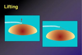
Once an errant tear begins to extend radially, the surgeon should appreciate that it becomes much harder to control as the tear approaches the periphery. This difficulty occurs because the anterior capsular dome, and thus the base of the tear, drops away from the tip of the tool (forceps) that is controlling it. This is much like giving a dog too much lead on its leash. An errant dog or the errant tear cannot be controlled when there is too much "leash" ahead of a surgeon’s grip (Slide 1).
Understanding the principle that errant tears run "downhill" and away from the forceps tips warrants the following preventive measures:
- Avoid lifting the capsular flap during the capsulorrhexis.
- Keep the capsule forceps low to the anterior lens surface and forward to the base of the tear.
- Lead the tear like a wagon is pulled in a circle by its handle. Think of the tearing motion as being more of a "peeling" action.
- Utilize capsulorrhexis forceps, such as the Connor Peeler (Rhein Medical Inc., Tampa, Fla.), that has forward facing tips. Tips with this orientation facilitate a peeling action, rather than the lifting motion that downward facing forceps tips promote.
- Regrasp frequently, at least at every quarter clock hour of circumference, to optimize control of the base of the flap.
Slide 1 demonstrates how a lifting movement can cause the base of the tear to advance downhill and out toward the periphery.
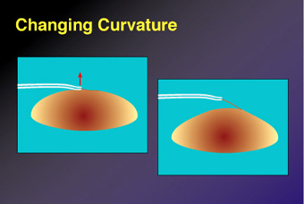
Patients may present with a so-called downhill challenge. These patients have an unusually steep anterior capsular dome that predisposes them to having an errant capsulorrhexis tear occur. Children, young adults, and hyperopes fall into this category. A swollen, intumescent lens or eyes with posterior pressure will be also be a challenge because of the presence of a steep anterior capsular dome.
Slide 2 shows how a steeper anterior lens curvature produces an unwanted effect similar to that of lifting the flap upward. The base of the tear runs downhill and away from the point of control.
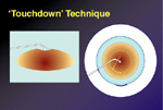
By anticipating this lens configuration, the downhill slope of the dome can be negated (i.e., a "touchdown" maneuver). The surgeon employs the fellow hand to flatten the steep anterior lens dome as the capsulorrhexis is being performed. A blunt-tipped instrument, such as a Connor Wand manipulator (Rhein Medical), is used to depress the central lens surface just under the initiated capsulorrhexis flap. This reverses the chamber shallowing caused by posterior pressure and helps flatten the anterior lens dome. The capsulorrhexis is then completed around this centrally placed tool (Slide 3).
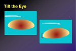
If the capsulorrhexis tear still migrates peripherally, despite taking the precautions, the following procedure can be performed. The first step is to deepen the anterior chamber with a cohesive viscoelastic. Next, the errant flap should be positioned into a peeling orientation. The eye can be tilted to grasp the flap at its base and from a forward position (Slide 4). If the misdirected tear does not turnabout, then a touchdown maneuver can be employed with the fellow hand. This technique of depressing the central lens surface with a wand manipulator to counter posterior pressure and flatten the anterior capsule contour will often allow the recovery of an errant capsulorrhexis.
When an errant tear cannot be rescued, the surgeon should not attempt to re-initiate the capsulorrhexis with a new tear proceeding in the opposite direction. This will produce a teardrop-shaped capsulotomy, in which the iris conceals the full extent of the radial tear. During phacoemulsification, this capsulorrhexis "split" may extend around the capsular fornix and through the posterior capsule, and result in a dropped nucleus. Instead, a cystotome should be used to offset the forces transmitted to this weakened area by making numerous relaxing incisions in the capsulorrhexis for 90°. These multiple puncture incisions should begin adjacent to the radial tear and as far out as can be visualized. After one-quarter clock hour, or 90°, the capsule can be regrasped and the capsulorrhexis can be finished. The capsular bag will be able to sustain the forces required to remove the cataract and allow the surgeon to insert the IOL into the bag.
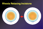
If the intact capsulorrhexis rim is inadvertently cut during phacoemulsification, the procedure should be stopped immediately, and the anterior chamber should be refilled with a thick, cohesive viscoelastic. Then, fine-tipped scissors should be used to make two additional relaxing incisions in the capsulorrhexis approximately 120° away from the tear (Slide 5). Elevate the nucleus out of the capsular bag by injecting either fluid or viscoelastic. Sandwich the nucleus with viscoelastic and complete the emulsification at the iris plane. The IOL may still be inserted into the salvaged capsular bag.
Additional Pearls
Surgeons may not be able to perform the touchdown technique to counteract a steep anterior capsular dome in children or patients with an intumescent lens because the nucleus is too soft. There may not be enough resistance for the wand to depress against to flatten the anterior lens surface. To prevent an errant tear from occurring in these situations, a two-stage capsulorrhexis technique can be used. The initial capsulorrhexis should be kept central and small (i.e., approximately 3 mm in diameter). An irrigation and aspiration tip is then used to remove anterior cortex and decompress the lens. Then, the anterior chamber is refilled with viscoelastic and the smaller diameter capsulorrhexis rim is incised with either scissors or a cystotome. The capsulorrhexis is enlarged to the standard 5-mm to 5.5-mm size before completing phacoemulsification.
Finally, if a posterior capsular rent occurs during phacoemulsification, whether centrally or as a result of a radiating anterior capsular cut, the principles described above can be used. It is important to remember that because tears always run downhill, a surgeon should make the posterior capsule concave with a thick, cohesive viscoelastic before attempting a posterior capsulorrhexis with long-tipped forceps. This step should be performed as soon as the posterior capsule defect is recognized and before continuing with phacoemulsification, cortex removal, or IOL implantation. Completing a posterior capsulorrhexis salvages the integrity and strength of the capsular bag, permitting in the bag IOL implantation.
Reference
- Gimbel HV, Neuhann T. Development, advantages, and methods of the continuous circular capsulorhexis technique. J Cataract Refract Surg. 1990;16:31-37.