Definition
Sympathetic ophthalmia is a rare, bilateral inflammatory process involving the uveal tract occurring secondary to penetrating trauma or ocular surgery. It is characterized by an insidious onset followed by a chronic, relapsing, progressive course. The etiology has been attributed to an autoimmune hypersensitivity response directed against residual uveal tissue in the traumatized eye. The traumatized eye has been called the "exciting" eye, and the nontraumatized eye is referred to as the "sympathizing" eye. Histopathologically, sympathetic ophthalmia is characterized by infiltration of the uveal tract with diffuse granulomatous inflammation consisting of lymphocytes, epithelioid cells, and giant cells. These histopathologic features characterize both the exciting and the sympathizing uveal tissue. Sympathetic ophthalmia generally occurs between 2 weeks to 1 year following penetrating or surgical trauma. It is believed to be prevented through enucleation of the initial, traumatized inciting eye. Once it occurs, it is treated through a combination of topical, depot and systemic steroids, in addition to systemic immunosuppressive and chemotherapeutic agents in rare instances. Visual outcome is generally poor.
History
The concept of sympathetic ophthalmia has evolved over the past seven centuries since its original conception in the 14th century.1,2 Interestingly, despite the increased incidence of eye injuries in wars subsequent to the 19th century, there have been no reported cases of sympathetic ophthalmia in the Korean, Vietnam, or Middle East Wars.1,2 With advances in surgical techniques, sterile procedures, antibiotics, and anti-inflammatory agents, the incidence of sympathetic ophthalmia appears to be declining. However, this is difficult to substantiate and has been widely debated in the literature. Some argue that advances in medical and surgical management are offset by more aggressive surgical management of severely traumatized eyes.
Incidence
In 1972, Liddy and Stuart reported a review of sympathetic ophthalmia during a 10-year period in Canada and found the incidence following penetrating trauma to be 0.19%, in contrast to the 0.007% incidence following all types of surgical procedures.3 Another survey of 6613 cases of ocular surgery (4540 cataract extractions; 1288 glaucoma procedures) revealed one case (an iris inclusion operation for glaucoma), resulting in a 0.015% incidence of postoperative sympathetic ophthalmia.4 The incidence is difficult to document since pathologic confirmation is available in only one third of suspected cases. In other cases, pathological diagnosis has been made when no clinical evidence of an inflammatory process was apparent.5
Epidemiology
In contrast to the disparity in data regarding the incidence of sympathetic ophthalmia, the data regarding epidemiology are more consistently reproducible. It has been observed that the incidence of sympathetic ophthalmia is much higher in males (approximately two thirds of cases), particularly following penetrating injuries to the globe.6 This observation has been attributed to the higher prevalence of penetrating injuries in this population. The incidence following surgical trauma is equivalent between males and females.6
A retrospective clinicopathologic review of 105 cases at the Massachusetts Eye and Ear Infirmary documented a racial predilection for the disease process with 90% of patients being white and 10% black.6 Interestingly, the course has been noted to be more fulminant in darker-skinned individuals, possibly related to a higher amount of pigmented uveal tissue.
The age distribution for sympathetic ophthalmia follows a bimodal distribution with peaks during the adolescent/younger adult years and again in the population older than the age of 60.6 This distribution directly reflects the higher incidence of trauma in the younger age group as well as the higher incidence of ocular surgical procedures in the population over the age of 60.6
The type of ocular insult preceding onset of sympathetic ophthalmia is highly variable. In the Massachusetts Eye and Ear study, the researchers found that 53.5% of cases followed penetrating ocular injuries and 40.4% of cases were attributed to intraocular surgical procedures.6 Most traumatic cases are documented as sequelae to penetrating ocular injuries (particularly those injuries involving uveal prolapse/incarceration). A few cases have been reported to follow contusion injuries with occult scleral ruptures and perforating corneal ulcers.7
Surgical cases predominantly include procedures complicated by injury to, or incarceration of, uveal tissue. Surgical procedures that have been associated with sympathetic ophthalmia include cataract extraction (particularly when characterized by iris entrapment into the wound), secondary intraocular lens placement, trabeculectomy, iridencleisis, contact and noncontact yttrium-aluminum-garnet (YAG) lasers, cryodestruction, proton beam and helium ion radiation for choroidal melanoma, combined cataract/glaucoma procedures, cataract extraction with implantation of an anterior chamber intraocular lens,8 laser cryophotocoagulation,9 and vitrectomy.6,10 The incidence of sympathetic ophthalmia following vitrectomy is documented to be approximately 0.01%.10
Clinical Presentation
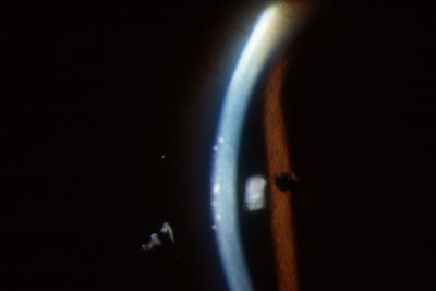
The diagnosis of sympathetic ophthalmia is a clinical diagnosis and requires consideration of a patient's history, and clinical signs and symptoms supplemented by histopathologic correlation when possible. The most common presenting symptoms include blurry vision, pain, increased lacrimation, and photophobia in the noninjured eye. Patients may also complain of periorbital tenderness. Symptom onset may be either insidious, or more rarely, rapid in development. A granulomatous uveitis ensues, characterized by mutton-fat keratic precipitates (conglomerates of lymphocytes, epithelioid cells and giant cells) on the endothelial surface of the cornea (Slide 1).
Patients with sympathetic ophthalmia have been documented to have abnormalities in intraocular pressure, including both glaucoma (related to inflammatory cell blockage of the trabecular meshwork) and hypotony (attributed to ciliary body shutdown).
Anterior segment findings include cell and flare in the anterior chamber, conjunctival injection, and keratic precipitates on the corneal endothelium. Infiltration of the iris may result in thickening of the iris and formation of posterior synechiae as the inflammation spreads over the anterior aspect of the lens capsule.
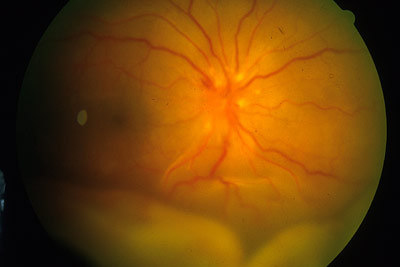
Additionally, these patients may have a variety of posterior segment findings, including exudative retinal detachments, papillitis, generalized retinal edema, vitreous inflammation and Dalen-Fuchs nodules (yellowish-white nodules beneath the retinal pigment epithelium [RPE]; most common in the periphery)(Slide 2).
Although sympathetic ophthalmia is a clinical diagnosis, clinical suspicion may be confirmed by use of fluorescein angiography. Angiography reveals multiple fluorescing dots at the level of the RPE appearing during the venous phase and persisting throughout the later phases of the angiogram.
Time of Onset
Time of onset of sympathetic ophthalmia varies greatly. Cases in the literature have been documented as short as 5 days after the trauma to as long as 66 years later.11,12 Generally, onset of sympathetic ophthalmia rarely occurs before 2 weeks, with 80% of cases occurring between 3 weeks and 3 months post-trauma. Ninety percent of cases occur within 1 year of injury.11
Etiology
The etiology of sympathetic ophthalmia has not yet been elucidated. The histopathologic findings are believed to be due to a delayed hypersensitivity response directed against a cell surface membrane antigen found on the RPE cells, choroidal melanocytes, and retinal photoreceptors.13 Other proposed etiologies include infectious agents and sensitivity to retinal S antigen or other retinal or uveal proteins. The factors determining time of onset after initial insult are unknown. Some have postulated a combined mechanism requiring both an antigen to incite the immune response and a concurrent infective agent to trigger the inflammatory cascade.
Histopathology
Six primary histopathologic features characterize both the exciting and the sympathizing eyes in sympathetic ophthalmia. These include the following:
- Diffuse granulomatous inflammation of the choroid characterized by lymphocytes, epithelioid cells, eosinophils, and giant cells. Few to no plasma cells are present and there is a characteristic absence of polymorphonuclear cells (Slides 4 and 5).
- Dalen-Fuchs nodules are present between Bruch membrane and the RPE
(Slide 6).
- Collections of epithelioid cells
- Characteristic, but not pathognomonic
- Not present in all cases (incidence varies from 25% to 35.5%)6,14
- Sparing of the choriocapillaris
- No granulomatous inflammation of the retina (Slide 7)
- Absence of necrosis
- Presence of epithelioid cells that contain phagocytosed uveal pigment
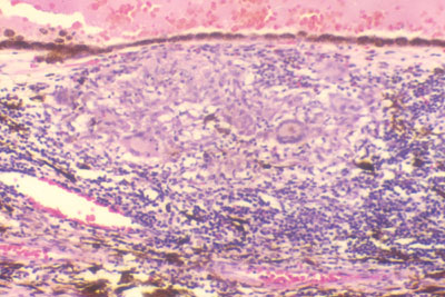
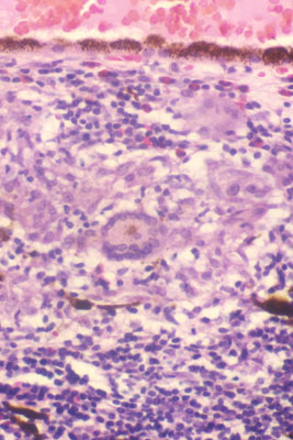
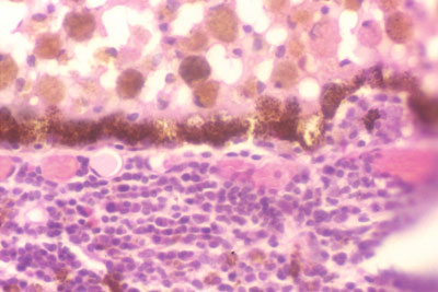
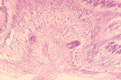
The lymphocytic infiltrate consists predominantly of T lymphocytes. Additionally, the severity of giant cell infiltration is directly correlated with final visual outcome.6
Differential Diagnosis
Sympathetic ophthalmia must be distinguished from other causes of granulomatous uveitis. Both fungal and bacterial infections may evolve into an anterior and posterior granulomatous uveitis. These entities may progress into a bacterial endophthalmitis. Post-traumatic iridocyclitis may also cause an inflammatory reaction and should be considered.
Vogt-Koyanagi-Harada syndrome is a bilateral granulomatous uveitis; its clinical presentation is similar to sympathetic ophthalmia. However, Vogt-Koyanagi-Harada syndrome is a systemic disease that affects the uveal tract as well as the meninges and thus may be characterized by meningeal signs and symptoms as well as ocular inflammation. Patients with Vogt-Koyanagi-Harada syndrome also have vitiligo, alopecia, and tinnitus. Vitiligo and alopecia may also be seen in sympathetic ophthalmia. Therefore, in some cases, differentiation is based on a history of ocular trauma or surgery.
Phacoanaphylaxis, or lens-induced granulomatous inflammation, is a condition that is closely related to, and may simulate, sympathetic ophthalmia in some cases. It is most commonly a unilateral condition, but it may occasionally be bilateral. Between 4% and 25% of cases of sympathetic ophthalmia are characterized by a component of phacoanaphylaxis.7 Histologically, it is characterized by a ruptured lens capsule with a central area of lens material surrounded by a zonal granuloma consisting of acute inflammatory cells, giant cells, and chronic inflammatory cells. When present, lens extraction is curative.
Treatment
Definitive treatment for sympathetic ophthalmia involves enucleation of the traumatized eye to prevent the devastating sequelae of this disease process. It is generally accepted that prompt wound closure or enucleation should be performed within 2 weeks of the initial traumatic event. This is an accepted time interval because of the extremely low incidence within this initial 2-week period. During surgery, exquisite attention must be placed on complete removal of all uveal tissue.
Controversy exists regarding the potential clinical benefits of removing the exciting eye once the inflammatory process has commenced. Winter and colleagues reviewed a series of 257 cases of sympathetic ophthalmia (histopathologically confirmed) in which the exciting eye was removed immediately prior to, concomitant with, or shortly following diagnosis of sympathetic ophthalmia.15 They found no benefit to enucleation of the exciting eye in prevention of sympathetic ophthalmia.15 Additionally, evisceration does not prevent the development of sympathetic ophthalmia, as uveal tissue found in the scleral canals and in the vicinity of the optic disc remains within the orbit following this surgical procedure.
Once the inflammatory process has begun, the mainstay of treatment is systemic steroids supplemented with sub-Tenon's injections of depot steroids and topical steroids. Prophylactic steroid administration has not been found to prevent development of sympathetic ophthalmia.
In rare cases in which the patient is intolerant to steroids or cases in which steroids are contraindicated, the patient may be treated with immunosuppressive agents including azathioprine, methotrexate, or chlorambucil.16,17
Disease Course and Prognosis
The disease process of sympathetic ophthalmia is characterized by a relapsing and remitting course that eventually becomes chronically progressive. With time, untreated chronic inflammation may eventually result in irreversible ocular damage and ultimately phthisis bulbi. Associated complications include glaucoma, cataract, retinal detachment, extensive chorioretinal inflammation, or scarring and optic atrophy.
The diagnosis of sympathetic ophthalmia has generally carried a grim prognosis associated with a significant loss of visual acuity. Recent advances in treatment with steroids and immunosuppressive agents have significantly improved the outlook for these patients. Makley and Azar found that in patients treated with steroids, 64% retained a visual acuity of 20/60 or better.18 Another long-term retrospective analysis of patients revealed that 50% of patients treated with steroids and immunosuppressive agents retain a visual acuity of 20/40 or better.19 Optimally, avoidance of this disease entity is at the forefront of its management; however, several treatment options are now available to assist in the management of this relentless, devastating disease process.
References
Albert DM, Diaz-Rohena R. A historical review of sympathetic ophthalmia and its epidemiology. Surv Ophthalmol. 1989;34:1-14.
Treisler G. Ocular casualties and the six year war. Am J Ophthalmol. 1969;68:669.
Liddy BL, Stuart J. Sympathetic ophthalmia in Canada. Canadian J Ophthalmol. 1972;7:157-159.
Allen JC. Sympathetic ophthalmia. A disappearing disease. JAMA. 1969;209:1090.
Goto H, Rao NA. Sympathetic ophthalmia and Vogt-Koyanagi-Harada syndrome. Int Ophthalmol Clin. 1990;30:279-285.
Lubin JR, Albert DM, Weinstein M. Sixty-five years of sympathetic ophthalmia: A clinicopathologic review of 105 cases (1913-1978). Ophthalmology. 1980;87:109-121.
Rao NA. Sympathetic ophthalmia. In: Ryan SJ, ed: Retina, 3rd ed. St. Louis, Mo: Mosby. 2001:1756-1761.
Lakhanpal V, Dogra MR, Jacobson MS. Sympathetic ophthalmia associated with anterior chamber intraocular lens implantation. Ann Ophthalmol. 1991;l23:139-143.
Bechrakis NE, Muller-Stolzenburg NW, Helbig H, Foerster MH. Sympathetic ophthalmia following laser photocoagulation. Arch Ophthalmol. 1994;112:80-84.
Gass JDM. Sympathetic ophthalmia following vitrectomy. Am J Ophthalmol. 1982;93:552-558.
Duke-Elder S, Perkins ES. Disease of the uveal tract. In: Duke-Elder S, ed: System of Ophthalmology, vol 9. St. Louis, Mo: Mosby; 1966.
Zaharia MA, Lamarche J, Laurin M. Sympathetic uveitis 66 years after injury. Can J Ophthalmol. 1984;19:240-243.
Albert DM, Diaz-Rohena R. A historical review of sympathetic ophthalmia and its epidemiology. Surv Ophthalmol. 1989;34:1-14.
Fuchs A. Sympathisierende entzudung sarkom in einem auge. Klin Monatsbi Augenheilkd. 1921:67:303.
Winter FC. Sympathetic uveitis: A clinical and pathologic study of the visual result. Am J Ophthalmol. 1855;39:340-347.
Tessler HH, Jennings T. High-dose short-term chlorambucil for intractable sympathetic ophthalmia and Behcet's disease. Br J Ophthalmol. 1990;74:353-357.
Andrasch RH, Pirofsky B, Burns RP. Immunosuppressive therapy for severe chronic uveitis. Arch Ophthalmol. 1978;96:247-251.
Makley TA Jr, Azar A. Sympathetic ophthalmia: a long-term follow-up. Arch Ophthalmol. 1978;96:257-262.
Chan CC, Roberg RG, Whitcup SM, Nussenblatt RB. 32 Cases of sympathetic ophthalmia: A retrospective study at the National Eye Institute, Bethesda, MD, from 1982-1992. Arch Ophthalmol. 1995;113:597-600.