Introduction
In 1985, 76% of the members of the American Society of Cataract and Refractive Surgeons (ASCRS), responding to an annual survey, indicated a preference for retrobulbar anesthesia with a supplemental facial nerve block for cataract surgery.1 We have come a long way in 16 years. This tutorial addresses the spectrum of regional anesthesia from retrobulbar to peribulbar to parabulbar/sub-Tenon. The common thread among these blocks is a combination of analgesia, akinesia, and some degree of visual/sensory block. This combination distinguishes regional anesthesia from topical/intracameral anesthesia, which will be covered in a separate tutorial.
Retrobulbar Anesthesia
Based on the ASCRS survey, 16% of respondents in 1985 preferred retrobulbar anesthesia without a facial block. Therefore, 92% of the ophthalmologists who identified themselves as cataract and refractive surgeons 16 years ago and were motivated to complete a detailed survey used a technique that Atkinson described in his classic monograph, Anesthesia in Ophthalmology, in 1955:
Good results will be obtained and orbital hemorrhage rarely occurs if the following directions are observed: an intradermal wheal is made. A 3.5-cm 23-gauge needle with a rounded point is introduced through this wheal at the inferior temporal margin of the orbit and the skin is moved upward with the needle until the point just clears the orbital margin. About 0.5 cc of the anesthetic solution is now injected so that pain will not be experienced when the orbital septum is pierced… A pause of a few moments allows the anesthesia to work during which time the patient is directed to look upward and away from the site of injection. This is done to move the inferior oblique muscle and fascia between the lateral and inferior rectus muscles forward and upward, out of the way. The needle is directed straight back close to the floor of the orbit, until the point is beyond the globe and fascia before directing it upward toward the apex of the orbit for a depth of 2.5 to 3.5 cm depending on the size of the orbit. When the needle has reached its proper depth, one should aspirate before injecting the anesthetic solution in order to determine whether or not the needle has entered a vessel... The anesthetic solution is slowly injected, the amount depending on the size of the orbit, the operation to be done, and whether or not hyaluronidase is added to the anesthetic solution. When hyaluronidase is not used, 1.5 cc [of anesthetic] is considered a safe amount for intraocular operations…more effective results are obtained for cataract extractions and larger injections may be given if hyaluronidase is added to the anesthetic solution. It may be safely injected until there is noticeable proptosis, which usually occurs after injection 2 to 3 cc in the average orbit. The hyaluronidase causes the solution to diffuse rapidly and the proptosis quickly subsides. Pressure over the eye, combined with moving it in the orbit for at least 5 minutes, produces greater diffusion of the anesthetic solution, enhances anesthesia and akinesia, and increases hypotony.2
Atkinson based both the depth and volume of his retrobulbar injection on his judgment of the size of the orbit. Perhaps his ability to make this judgment successfully allowed him to continue using this same technique for his entire career. However, the failure of others that followed to reliably judge orbital anatomy and tailor their retrobulbar block according to this judgment in order to avoid complications, has led to modifications in Atkinson's technique.
A major change in this technique as it is currently used is the direction of patient gaze. Unsold's 1981 computed tomography (CT) study of the placement of anesthetic needles in cadaver orbits is the driving force behind this change.3 In the study, a 3.5-cm 25-gauge needle was introduced into the cadaver orbit with the eye sutured either into the position described above or alternatively into one of infratemporal gaze. Atkinson's advised gaze led to needle placement extremely close to the optic nerve, with the CT demonstrating a path that crosses underneath stopping medial to the optic canal. The up and in gaze caused the optic nerve to rotate downward and outward, near the path of the needle, whereas the down and out gaze caused the needle to end up above the inferior rectus muscle, anterior to the inferior aspect of the orbital fissure. However, because a down and out approach causes the patient to look directly at the needle, most practitioners who use retrobulbar anesthesia have their patients look straight ahead (Slide 1).
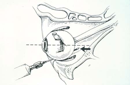
While the direction of gaze can be used to improve the safety of this technique, the length of the needle introduced into the orbit, if long enough, can obviously overcome this advantage. Such were the findings in a 1987 study performed by Nicoll and coworkers.4 In this study, the use of a 38-mm needle for 6,000 retrobulbar blocks was associated with a 1 in 375 incidence of central system spread. There was no significant difference between a group of patients who gazed straight ahead and one in which patients gazed up and in.
In 1989, Katsev and colleagues measured 120 human orbits and found that a 38-mm needle, introduced from Atkinson's traditional position of insertion6 and in needle design,7,8 have been proposed to improve the margin of safety of retrobulbar anesthesia. No modification, however, will substitute for an accurate judgment of orbital size and anatomy. The gradually waning popularity of retrobulbar anesthesia recently suggests that alternative techniques for which such judgment is less critical will continue to grow in usage. In Leaming's 1999 survey, only 29% of respondents preferred retrobulbar anesthesia either with or without a facial block.9 The success of assessing orbital anatomy from surface markings and refraction was studied by Birch and associates in 1995.10 In this ultrasound study of retrobulbar blocks, performed in the method described by Atkinson, 14 of 25 needles were seen indenting the glove, and none were found further than 3.3 mm from it.
Peribulbar Anesthesia
In 1986, Davis and Mandel reported on their use of peribulbar anesthesia.11 They actually gave credit to Kelman for first performing but not publishing the technique in the mid 1970s.12 Davis and Mandel made multiple injections, the first being a subcutaneous injection of 0.5 mL of lidocaine inserted a finger's width medial to the lateral canthus, through the lower lid just above the inferior orbital rim. Secondly, another injection of 0.5 mL of lidocaine was introduced into the orbicularis.
A third injection of 1.0 mL was injected through the upper lid in the same manner, beginning just below the superior orbital notch. They used a 1.25-cm (0.5 inch), 27-gauge needle for all these injections.
A more posterior injection was then made with a 23-gauge, 3.125-cm (1.25 inch) blunt retrobulbar needle, and 4 mL of 1% lidocaine and hyaluronidase was administered (Slide 2 and Slide 3). Next, an inferior injection was given 15 mm medial to the lateral canthus just above the inferior orbital rim, and 1 mL of anesthetic was injected just beneath the orbicularis muscle. Davis and Mandel go on to write, "the needle is then advanced along the inferior orbit to the equator of the globe." This statement tells us that successful and safe peribulbar anesthesia requires some of the same three-dimensional judgment that is required in Atkinson's original technique, because the position of the equator of the globe differs in relation to the axial length of the eye and the bony anatomy of the orbit. No mention of ultrasonic or other measures of these distances is made. Once the tip of the needle has reached the position on the orbital floor that parallels the equator of the globe, 1 mL of anesthetic solution is injected. Next, "the barrel of the needle is angled over the malar eminence, and the needle is advanced in a superior and medial direction to the full depth of the needle." There, another 1 mL to 2 mL is injected. These steps are repeated beneath the superior orbital notch anteriorly and "at the superonasal equator of the globe." Davis and Mandel describe the volumes delivered as approximate and, once again, dependent on the dimensions of the eye and orbit.
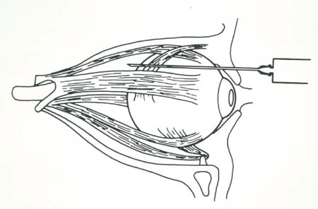
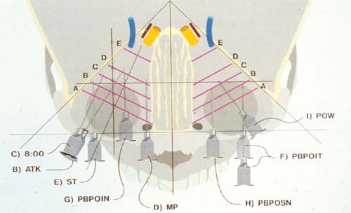
A later modification of their peribulbar technique involved using a shorter needle (2.19 cm or 7/8 inch), in response to reports of respiratory depression, optic nerve trauma, and glove perforation. A similar anterior peribulbar technique published by Bloomberg13 described the use of an 18-mm to 24-mm, 27-gauge needle to inject 8 mL to 10 mL of a half and half mixture of 2% mepivacaine and 0.75% bupivicaine, to which hyaluronidase and sodium bicarbonate (0.2mL per 10 mL of solution) are added. The needle is introduced in Atkinson's position and is directed toward the floor of the orbit. In this technique, as in Davis and Mandel's, 10 to 20 minutes of ocular compression follows the injections.
The use of shorter needles directed away from the globe is common to all later modifications of Davis and Mandel's technique.
A cluster of reports of inferior rectus muscle paresis and restriction paralleled the height of popularity of peribulbar anesthesia in the early to mid 1990s.14 Although there may have been other causative factors, clearly the use of more needles directed toward the extraocular muscles comes with a greater risk of extraocular muscle trauma and strabismus. The authors of one 1995 report of inferior oblique muscle trauma following retrobulbar and peribulbar blocks advised the adoption of sub-Tenon anesthesia as a way to avoid postoperative strabismus.15
Parabulbar (Sub-Tenon) Anesthesia
Dissections of the orbit have revealed that the anterior extension of the visceral layer of dura is in fact Tenon capsule.7(p52) The sub-Tenon space is, therefore, contiguous with the subdural space and is, in effect, an anatomic pathway from the limbus to the retrobulbar space. Because the sensory and motor nerves of the globe, and the four rectus muscles course through and can be easily accessed within this space,16 anesthetic fluid infused and kept within the sub-Tenon space can provide analgesia, akinesia, and a sensory block of the optic nerve with a single low volume infusion, as demonstrated in an magnetic resonance image (MRI) study published in 1997.8(p31)
Although the sub-Tenon space may be entered with a variety of needles,17,18 a small snip placed anteriorly, 2 mm or more behind the limbus, where Tenon capsule lies separate from conjunctiva, allows ready access with a cannula, eliminating the inherent risk of globe perforation.19 Metal cannulas have been developed and used by Fukusaku,20 Stevens,21 and others. These metal cannula techniques involve infusing anesthetic 20 mm to 30 mm posterior to the limbus into the retrobulbar space. No attempt is made to block the opening made in Tenon capsule for the introduction of the cannula, and variable to absent akinesia is provided.20,21
In an attempt to make sub-Tenon anesthesia simple, safe, and effective, a flexible cannula was designed to deliver an anterior sub-Tenon anesthetic infusion. This parabulbar technique is begun 2 mm to 3 mm posterior to the limbus (Slide 4, Slide 5, Slide 6, Slide 7, Slide 9, Slide 10, Slide 11, and Slide 12). A later modification by Markoff begins just anterior to the plica.22
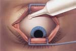
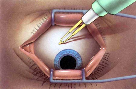
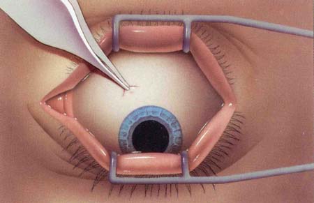
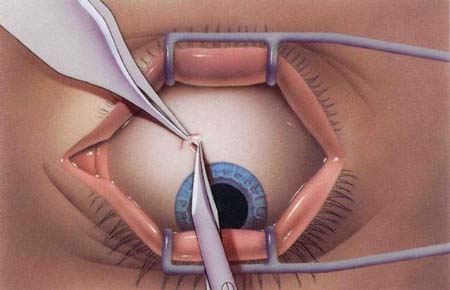
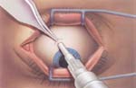
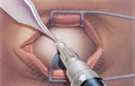
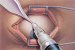
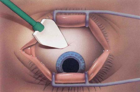
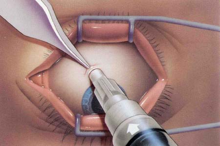
The cannula is 10 mm long, so that it infuses anesthetic near the point at which extraocular muscle fibers are innervated by branches of the oculomotor and abduscens nerves. A low volume infusion (Markoff uses 0.5 mL; I use 1.0 mL to 1.5 mL) is quickly performed, while the opening in Tenon capsule is closed by the hub of the cannula. A pressure head is created which hydraulically dissects anesthetic around the muscles into the retrobulbar space and into the anterior subdural space surrounding the optic nerve.8(p31) This flow of anesthetic from the most anterior to the most posterior extension of the sub-Tenon space creates reliable levels of analgesia, akinesia, and sensory block.
Because parabulbar anesthesia can provide a complete block without the inherent risks of globe perforation, extraocular muscle trauma, and optic nerve trauma caused by a needle, Stewart and Lambrou have advised a continued evolution of regional ophthalmic anesthesia away from needles and toward this "simple and safe technique."23
References
- Leaming DV. Practice styles and preferences of ASCRS members: 1993 survey. J Cataract Refract Surg. 1994;20:459.
- Atkinson WS. Anesthesia in ophthalmology. Springfield, Ill: Charles C Thomas; 1955.
- Unsold R, Stanley J, DeGroot J. The CT-topography of retrobulbar anesthesia. Arch Clin Ophthalmol. 1981;217:125.
- Nicoll JMV, Acharya PA, Ahlen K, Baguneid S, edge KR. Central nervous system complications after 6000 retrobulbar blocks. Anesth Analg. 1987;66:1298.
- Katsev DA, Drews RC, Rose BT. An anatomic study of retrobulbar needle path length. Ophthalmology. 1989;96:1221.
- Gills JP, Loyd TL. A technique of retrobulbar block with paralysis of the orbicularis oculi. Am Intraocular Implant Soc J. 1983;9:339.
- Gills JP, Hustead RF, Sanders DR, eds. Ophthalmic Anesthesia. 1993:138.
- Greenbaum S. Anesthesia for cataract surgery. In: Greenbaum S, ed. Ocular Anesthesia. Philadelphia, Pa: WB Saunders; 1997.
- Leaming DV. Practice styles and preferences of ASCRS members-1999 survey. J Cataract Refract Surg. 2000;26:913.
- Birch AA, Evans M, Redembo E. The ultrasonic localization of retrobulbar needles during retrobulbar block. Ophthalmology. 1995;102:824.
- Davis DB, Mandel MR. Posterior peribulbar anesthesia: An alternative to retrobulbar anesthesia. J Cataract Refract Surg. 1986;12:182.
- Kelman CD. Forward. In: Greenbaum S, ed. Ocular Anesthesia. Philadelphia, Pa: WB Saunders; 1997.
- Bloomberg L. Administration of peribulbar anesthesia. J Cataract Refract Surg. 1986;12:677.
- Greenbaum S. Anesthesia for cataract surgery. In: Greenbaum S, ed. Ocular Anesthesia. Philadelphia, Pa: WB Saunders; 1997:49.
- Hunter DG, Lam GC, Guyton DL. Inferior oblique muscle injury from local anesthesia for cataract surgery. Ophthalmology. 1995;102:501.
- Dutton JJ. Atlas of Clinical and Surgical Orbital Anatomy. Philadelphia, Pa: WB Saunders; 1994:94.
- Hansen EA, Mein CE, Mazzoli R. Ocular Anesthesia for cataract surgery: A direct subTenon’s approach. Ophthalmic Surg. 1990;21:696.
- Mein CE, Woodcock MG. Local Anesthesia for vitreoretinal surgery. Retina. 1990;10:47.
- Yanoff M, Redovan EG. Anterior eyewall perforation during subconjunctival cataract block. Ophthalmic Surg. 1990;21:262.
- Fukusaku H, Marron JA. Pinpoint anesthesia: A new approach to local ocular anesthesia. J Cataract Refract Surg. 1994;20:468.
- Stevens JD. A new local anesthesia technique for cataract extraction by one quadrant sub-Tenon’s infiltration. Br J Ophthalmol. 1992;76:670.
- Markoff DD. Parabulbar anesthesia for cataract surgery (abstract). Invest Ophthalmol Vis Sci. 1995;36:S809.
- Stewart M, Lambrou F. The management of preoperative, and postoperative complications of cataract surgery from the perspective of vitreoretinal surgeons. Adv Clin Ophthalmol. 1994;1:63.