Introduction
For the past 50 years, ophthalmic surgeons have witnessed enormous breakthroughs in the treatment of many ocular diseases and disorders, especially in the field of cataract surgery and its ability to correct preexisting refractive errors. We owe a great debt to Sir Harold Ridley and his perseverance in designing and implanting the first IOL in 1949. Since then, gradual, almost imperceptible improvements in IOL calculations, phacoemulsification, IOL designs and materials, phaco incisions, and astigmatism control have generated a procedure that can offer rapid improvement in uncorrected visual acuity. Today, patients who have cataracts have learned that not only can their surgeon restore their lost vision but also significantly reduce or eliminate their need for glasses.
Success with refractive cataract surgery depends on a number of important elements that fall into three categories: IOL power accuracy, astigmatism control, and outcomes analysis.
IOL Power Accuracy
To improve IOL calculations, the first step is to train a refractive cataract surgery team. These team members include anyone in your practice who performs pre- and postoperative refractions, biometry, IOL calculations, refractive data entry, or outcomes analysis. Assembling this team and stressing the value of the important role each of these individuals plays to achieve the best results is critical. The next step is to teach your team the basics about IOL calculations. I like to start by describing the five basic components of IOL calculations for most formulae: desired postoperative refraction, keratometry reading, axial length, anterior chamber (AC) depth, and lens constant.
Desired Postoperative Refraction
For monofocal lenses, surgeons have traditionally been aiming for –0.5 D to avoid hyperopic surprise. Some surgeons will target for monovision in
the other eye, thereby attempting to produce a low range of myopia in the fellow eye of –1.5 to –1.75. Our experience with multifocal IOLs, such as
the Array (Allergan, Irvine, Calif.) lens, suggests that plano to 0.5 D may be a more desirable target refraction for this type of IOL because there is less
likelihood of unwanted visual images postoperatively. Some Array surgeons target low myopia in the fellow eye, such as –0.5, to produce
micromonovision, thereby providing a broader range of intermediate and near vision for the bilateral Array patient.
Keratometry Reading
Many cataract practices still rely on manual keratometry for IOL calculations, particularly because mire clarity can be assessed and recorded.
This helps to determine the quality of the keratometry reading. Automatic keratometers have been improved and offer a reliable alternative.
Corneal topography is also gaining popularity, particularly if astigmatism surgery is anticipated. When measuring the corneal power, the most
challenging patients tend to be those who have undergone previous corneal refractive surgery, such as radial keratotomy, photorefractive keratectomy,
or laser in situ keratomileusis (LASIK). Useful methods to assess these patients are presented in the tutorial IOL Power calculation for the
Unusual Eye by Jack Holladay, MD.
Axial Length
Studies have shown that errors in axial length measurement tend to be the most common cause of an IOL power surprise.1
In eyes of standard length, a 0.1-mm error results in a 0.3-D error. In shorter eyes, this error is even more significant. As advocated by John Retzlaff, MD,
measurement accuracy should be within 0.15 mm with the same eye and within 0.3 mm in the fellow eye.
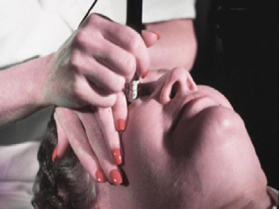
Currently, three methods to measure axial length are available: contact ultrasound, immersion ultrasound, and optical coherence biometry (OCB) with the IOLMaster (Zeiss Humphrey, Dublin, Calif.). Handheld contact ultrasound is the most popular method being used today. Unfortunately, it is also the least accurate. Corneal indentation varies depending on technician and technique and can influence IOL power calculation, especially with short eyes. Many cataract surgeons have transitioned to immersion ultrasound because it does not produce corneal indentation. Newer immersion methods, such as the use of Hanson shells, have improved this technique (Slide 1).
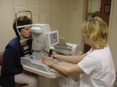
The most precise and technician friendly method for preoperative biometry is the use of the Zeiss Humphrey IOLMaster. This machine benefits from having the least technician variance. It is fast, noncontact and therefore technician and patient friendly (Slide 2). My colleagues and I have seen a measurable improvement in our IOL calculation accuracy since the introduction of the IOLMaster in our practice. Many surgeons have had the false impression that since the IOLMaster has so many advantages compared to ultrasound, it is not as dependent upon technician capability. In my experience, success with the IOLMaster can still vary with the technician's skill. This is particularly the case with difficult eyes such as those with dense nuclear sclerosis and posterior subcapsular cataract. There is truly an art to "mastering" the IOLMaster. New software upgrades have allowed for more accurate keratometry, particularly if the technician is aware of the importance of having an adequate tear film and having the eyelids opened as wide as possible. Axial length measurements require an understanding of signal:noise ratio and how best to manipulate the focus beam within the central circle on the video slit lamp. Even eyes that initially show error readings can many times be successfully measured once a seasoned technician understands how to properly perform this task.
AC Depth
When performing axial length measurements with contact methods, monitoring the anterior chamber depth is a way for the technician
to determine how accurate the scan might be, that is, the deeper the AC the more likely that there is less corneal indentation. Immersion scans
and OCB measurements are not as likely to show variations in the AC depth. One possible source of confusion among technicians is equating
AC depth with the "ACD," a term for the lens constant of the Holladay II and the Hoffer Q IOL formulae.
Personalized Lens Constants
There is much confusion in the area of lens constants regarding terminology and accuracy with many cataract biometry technicians.
Biometrists can be perplexed by the multitude of names that are given for the lens constant depending on the formula being used. The following
is an abbreviated list of these names.
| Lens Constants: | |
| SRK Formulas | A constant |
| Holladay I | Surgeon factor |
| Holladay II | ACD |
| Hoffer Q | ACD |
It is important to remind our technicians that these lens "constants" are really variables and they can vary significantly from the manufacturer’s lens constant to what has been termed the "surgeon’s lens constant." This is because the U.S. Food and Drug Administration requires many IOL manufacturers to "guesstimate" the lens constant, which is printed on the IOL container before it is actually implanted in the human eye. Because this constant, which is actually a variable, can vary even within the same practice, it is important to consider the value of personalization of lens constants.
There is a misconception that personalization is determined by the surgeon’s surgical technique. However, it is less likely to be dependent on the surgeon’s technique as on the biometry technician and the equipment being used to perform preoperative biometry. Technological advances such as the IOLMaster may reduce this variation. When personalizing constants, it is best to perform this assessment on at least 20 or more of the same IOLs implanted by the same surgeon at the same location. As an example, when implanting an SI40, we now have a separate personalized A constant for the IOLMaster (117.792) and contact ultrasound A scan (117.725). Using similar biometric techniques, many biometrists use a separate lens constant for the same IOL for a contact A scan versus the IOLMaster. Postoperative refractions must be measured when there is a stable refraction, which usually requires 1 month or more. There are a number of computer programs that are helpful in performing this task. The Holladay IOL Consultant, which utilizes the Holladay II formula, the Hoffer programs utilizing the Hoffer Q formula, and other formulas have been popular for ongoing personalization of lens constants. Computer programs such as these also allow the practice to perform outcomes analysis of all IOL calculations on an ongoing basis.
Outcome Analysis
For 10 years, we have had an outcomes analysis program, which we have termed "The Patient Satisfaction Program." The goal of
this program has been to improve results primarily in IOL calculation to the highest level possible in order to provide the highest level of patient
satisfaction. Our goal has been to be within ±0.50 D of the spherical target that is determined preoperatively, as often as possible. When we initiated
our program, we were at ±0.50 D approximately 45% of the time. We now regularly achieve ±0.50 D in 90% of patients, due to greater attention to
detail and a better understanding of the importance that each member of the refractive cataract surgery team plays in achieving the best results.
Another important area where analyzing results can be of great benefit is in control of astigmatism. James Gills, MD, has been a leader in surgical astigmatic correction to improve uncorrected acuities after cataract surgery and we agree with his preference for limbal relaxing incisions. He developed one of the first surgical nomograms for limbal relaxing incisions (LRIs). We have also developed a nomogram that relies on degrees of arc rather than millimeters. An LRI kit that is manufactured by Duckworth and Kent has been helpful. This astigmatic surgical system offers a simple and reproducible method for surgeons to perform this procedure. Thanks to "coupling," most LRIs will not influence IOL calculation accuracy. Other methods, such as laser vision correction, standard central astigmatic keratotomy, and toric IOLs, can also reduce problem astigmatism.
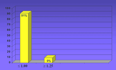
There are various methods to evaluate the influence that a surgeon’s incision has on the astigmatic outcome and these should be considered when beginning astigmatic correction. As I. Howard Fine, MD, suggests, how do we know what to correct when we do not know what our phaco incision i s already producing? Success in astigmatism control can benefit from outcomes analysis, similar to IOL calculations. The total amount of astigmatism is measured after surgery and if a patient is less than 1 D at any axis, the patient will generally enjoy good uncorrected acuity, assuming that the intended spherical result is achieved. We now include outcomes analysis for astigmatism in our Patient Satisfaction Program and have been successful in achieving 1 D or less of astigmatism in more than 90% of patients (Slide 3).
Any discussion on refractive cataract surgery should also include the importance of state-of-the-art phaco techniques and technology, proper phaco incision construction, and the value of avoiding complications and handling them properly if they arise. Today’s refractive cataract surgeon also must consider having adequate tools to offer postoperative enhancement for unanticipated postoperative refractive errors. These would include being able to perform limbal relaxing incisions, mini refractive keratotomy, laser vision correction, and, if needed, low impact IOL exchange.
Multifocal IOLs
There is a growing interest for better uncorrected visual acuity after cataract surgery due to the popularity of LASIK. This includes not just distance vision, but also good uncorrected near acuity. This will become even more popular with more widespread computer use, particularly in the "baby boomer" generation. Once your office has a well-informed and experienced refractive cataract surgery team, there will be a natural inclination to consider the use of multifocal IOLs.
Multifocal IOLs have become a catalyst in the field of refractive cataract surgery. However, there have been a number of concerns regarding the use of these lenses, particularly with regard to unwanted visual imagery, such as halos at night. I have found this phenomenon to be common after implantation of the Array lens, and I usually anticipate it. Fortunately, there is a significant amount of visual cortical adaptation potential, such that most patients can learn to ignore these halos, especially after bilateral implantation. My colleagues and I have found the Array to be most successful with hyperopes who desire reduced dependence on glasses and are aware of the likelihood of an adjustment to a new visual system. Patients implanted with the Array learn to use their near vision component and, during that process, learn to ignore the rings around lights at night.
In many ways refractive cataract surgery is not a destination but a journey. As we develop a better understanding of how multifocal vision works and the importance of visual cortical adaptation, it is likely that we will see an expanded role for multifocal IOLs for cataract and pre-cataract patients. As learned from our early experience with IOLs, we must keep an open mind and explore all modalities to restore multifocal vision for our presbyopic patients.
Reference
- Olson T. Sources of error in intraocular lens power calculation. J Cataract Refract Surg. 1992;18:125-129.