The authors have no financial or proprietary interest in any
product mentioned in this tutorial.
Supported in part by an unrestricted
grant from Research to Prevent Blindness, Inc, New York, NY.
Introduction
Opacification of the posterior capsule caused by postoperative proliferation of cells in the capsular bag remains the most frequent complication of cataract surgery. In addition to classic posterior capsule opacification (PCO), postoperative lens epithelial cell (LEC) proliferation is also involved in the pathogenesis of other entities. These entities include anterior capsule opacification (ACO) and interlenticular opacification (ILO), which is a recently described complication related to piggyback IOLs. Thus, there are three distinct anatomic locations within the capsular bag where clinically significant opacification may occur postoperatively (Slide 1). In this tutorial, the etiopathogenesis, clinical manifestations, and pharmacological, surgical, and implant-related factors for prevention of PCO are discussed. Interested readers may consult the published articles for other modalities of capsular bag opacification including ACO and ILO.1,10-12,90,91,94,95,97-100,103-106
Background and Significance
Posterior capsule opacification (PCO, secondary cataract) has been recognized since the introduction of extracapsular cataract surgery (ECCE) and was noted by Sir Harold Ridley in his first intraocular lens (IOL) implantations.3,8 PCO was particularly common and severe in the early days of IOL surgery when the importance of cortical cleanup was less appreciated. In the 1980s and early 1990s, the incidence of PCO ranged from 25% to 50%.7,8
Improvements in cataract surgical technique have led to a gradual, but steady, decrease in the incidence of this complication. Our data show that through a combination of modern techniques and IOL designs, the incidence of Nd:YAG laser posterior capsulotomy is now decreasing into single digits.4-6,80-82
Why Eradicate PCO?
Although cataract is the most common cause of blindness in the world, after-cataract is an extremely common cause, as well. Jan G.F. Worst, MD, has stated:
The most meaningful development in intraocular implant research in the next five years will be effective prevention of secondary cataract formation (International Intraocular Implant Club Report, Vol. 1, No. 2, January 1999)
Eradication of PCO following ECCE has major medical and financial implications:
- An Nd:YAG laser secondary posterior capsulotomy can be associated with significant complications including IOL damage, postoperative intraocular pressure (IOP) elevation, cystoid macular edema, retinal detachment, and IOL subluxation (Slide 2).36,37,40,41
- Dense PCO and secondary membrane formation is particularly common following pediatric IOL implantation.75 A delay in diagnosis can cause irreparable amblyopia.
- PCO represents a significant cost to the U.S. healthcare system. The cost of Nd:YAG laser treatments for approximately 1 million patients per year is $250 million annually.
- A posterior capsulotomy can increase the risk of posterior segment complications in high myopes and patients with uveitis, glaucoma, and diabetic retinopathy.
- PCO of even a mild degree can decrease near acuity through a multifocal IOL and may interfere with the function of accommodating IOL designs.
- A significant incidence of PCO means that cataract surgery alone may not restore lasting sight to the 25 million people worldwide who are blind from cataract.
Etiopathogenesis
Postoperative proliferation of LECs in the capsular bag is central to the pathogenesis of PCO. Proliferation of these cells can also lead to ACO and ILO.14,25-29,49,54,72,73
In the normal crystalline lens, the epithelial cells are confined to the anterior surface at the equatorial region and the equatorial lens bow. This single row of cuboidal cells can be divided into two different biological zones (Slide 1):
- The anterior-central zone (corresponding to the zone of the anterior lens capsule) consists of a monolayer of flat cuboidal, epithelial cells with minimal mitotic activity. In response to a variety of stimuli, the anterior epithelial cells ("A" cells) proliferate and undergo fibrous metaplasia. This has been called "pseudofibrous metaplasia" by Font and Brownstein.24
- The second zone is important in the pathogenesis of "pearl" formation. This layer is a continuation of anterior lens cells around the equator, forming the equatorial lens bow ("E" cells). Unlike within the A cell layer, cell mitoses, division, and multiplication are active in the second zone. New lens fibers are continuously produced in this zone throughout life.
Most cases of clinical PCO are caused by the proliferation of remnant or regenerated lens epithelial cells left in the capsular bag following cataract surgery (Slide 3 and Slide 4). Although both types of cells (from the anterior central zone and from the equatorial lens bow) have the potential to produce visually significant opacification, most cases of classic PCO are caused by proliferation of the equatorial cells. The term posterior capsule opacification is actually a misnomer. It is not the capsule that opacifies. Rather, an opaque membrane develops as retained cells proliferate and migrate onto the posterior capsular surface.
The opacification usually takes one of two morphologic forms. One form consists of capsular pearls that may consist of clusters of swollen, opacified epithelial "pearls" or clusters of posteriorly migrated E cells (bladder or Wedl cells). It is probable that both lens epithelial cell types can also contribute to the fibrous form of opacification. A cells most likely are important in the pathogenesis of fibrosis PCO, since the primary type of response of these cells is to undergo fibrous metaplasia. Although the preferred type of growth of the E cells is in the direction of bloated, swollen, bullous-like bladder (Wedl) cells, these also may contribute to formation of the fibrous form of PCO by undergoing a fibrous metaplasia. This is a particularly common occurrence in cataracts in under developed settings in which cataract surgery has been delayed for many years and cases in which posterior subcapsular cataracts have turned into fibrous plaques.
E cells are also responsible for formation of a Soemmering's ring. The Soemmering's ring, a dumb-bell or donut shaped lesion that often forms following any type of rupture of the anterior capsule, was first described in connection with ocular trauma. The pathogenetic basis of a Soemmering's ring is rupture of the anterior lens capsule with extrusion of nuclear and some central lens material. The extruded cortical remnants then transform into Elschnig pearls. It is not widely appreciated that a Soemmering's ring forms virtually every time any type of ECCE is performed (manual, automated or with phacoemulsification) (Slide 5). This material is derived from proliferation of the E cells of the equatorial lens bow. We have noted that these cells have the capability to proliferate and migrate posteriorly across the visual axis, thereby opacifying the posterior capsule. Because the Soemmering's ring is a direct precursor to PCO, surgeons should strive to prevent its formation.
Cell types other than lens epithelial cells may be involved in PCO. As ECCE is always associated with some breakdown of the blood-aqueous barrier, inflammatory cells, erythrocytes, and many other inflammatory mediators may be released into the aqueous humor. The severity of this inflammatory response may be exacerbated by the IOL. This foreign body elicits a three-stage immune response that involves many different cell types, including polymorphonuclear leukocytes, giant cells, and fibroblasts. Collagen deposition onto the IOL and onto the capsule may cause opacities and fine wrinkles to form in the posterior capsule. In most cases, however, this inflammatory response is clinically insignificant. Iris melanocytes also have been shown to adhere to and migrate over the anterior surface of the posterior capsule.
Clinical Manifestations
The interval between surgery and PCO varies, ranging anywhere from 3 months to 4 years after the surgery. Although the causes of PCO are multifactorial as reported in several studies, there is an inverse correlation with age.7,18,20,-23,35 Young age is a significant risk factor for PCO, and its occurrence is a virtual certainty in pediatric patients.107
Visual symptoms do not always correlate with the observed amount of PCO. Some patients with significant PCO on slit lamp examination are relatively asymptomatic, whereas others have significant symptoms with mild apparent haze, which is reversed by capsulotomy.17
Prevention of PCO
PCO prevention has been an active research interest of ours since 1982. Based upon our work, we can review the principles of PCO prevention. These measures can be divided into two categories. One strategy is to minimize the number of retained/regenerated cells and cortex (including the Soemmering's ring) through thorough cortical cleanup. The second strategy is to prevent the remaining cells from migrating posteriorly. The edge of the IOL optic is critical in the formation of such a physical barrier.
We have identified three surgery-related factors and three IOL-related factors that are particularly important in the prevention of PCO (Slide 6).
Surgery-Related Factors to Reduce PCO
Hydrodissection-Enhanced Cortical Cleanup. An important and
underrated surgical step is hydrodissection (Slide 7).
I. Howard Fine, MD, perfected and popularized this technique and coined the
term "cortical cleaving hydrodissection."22 Until recently, many
surgeons had a rather fatalistic attitude regarding removal of lens cortex and
cells during manual or automated ECCE or with phacoemulsification. A common
opinion was that removing all or even most equatorial cells from the bag is
impossible. PCO was therefore considered an inevitable complication. This
conclusion arose, in part, because PCO occurred in up to 50% of cases. We now
know from autopsy and experimental studies that thorough cortical and cellular
cleanup can be accomplished in most cases. By freeing and rotating the lens
nucleus, hydrodissection facilitates lens removal without zonular-capsular
rupture.
Recently, we have shown an important additional long-term advantage of hydrodissection. Proper hydrodissection will allow more efficient removal of cortex and cells, which in turn reduces PCO. A successfully performed cortical cleaving hydrodissection provides an easy way to extract the entire lens cortex as well as nucleus. Occasionally, this can even occur without the need for cortical aspiration with a separate irrigation and aspiration (I&A) instrument. The necessary tenting up of the anterior capsule is best achieved by using a cannula with a bent tip allowing a flow of fluid toward the capsule to efficiently separate capsule from cortex (see link to hydrodissection modules by Chang and by Packer).
In-The-Bag (Capsular) Fixation. The hallmark of modern cataract surgery is the achievement of consistent and secure in-the-bag (capsular) fixation (Slide 6). An advantage of in-the-bag fixation is the accomplishment of good optic centration and sequestration of the IOL from adjacent uveal tissues. Numerous other advantages have been described elsewhere. However, it is not often appreciated that this is also extremely important in reducing the amount of PCO.
One desired goal of in-the-bag fixation is enhancing the IOL optic barrier effect, which is functional and maximal when the lens optic is fully in-the-bag with direct contact with the posterior capsule. In case one or both haptics are not placed in the bag, a potential space is created, allowing an avenue for cells to grow posteriorly toward the visual axis. The reader may recall the barrier ridge IOL design devised by Kenneth Hoffer in the 1980s, which did not function sufficiently at the time. The reason was not a problem with the concept or the IOLs, but rather that only approximately 30% of posterior chamber IOLs were implanted inside the bag at the time.
With non-phaco ECCE, in-the-bag fixation of IOLs occurs approximately 60% of the time. One explanation is that many cases combine rigid design IOLs with can-opener anterior capsulotomies. Secure and permanent in-the-bag fixation only occurred in approximately 60% of cases. However, when considering modern foldable lens implantation, the number rapidly rises to more than 90%. It is not the foldable IOL itself, or even the small incision that provides this positive result, but rather the fact that successful foldable IOL insertion generally requires meticulous surgery, with the necessity of performing a continuous curvilinear capsulorrhexis (CCC) and securing implantation of both IOL loops in the bag.
Capsulorrhexis Edge on IOL Surface. A less obvious, but significant addition to precise in-the-bag fixation, is creating a CCC diameter slightly smaller than that of the IOL optic. For example, if the IOL optic were 6 mm, the capsulorrhexis diameter would ideally be slightly smaller, perhaps 5 mm to 5.5 mm. This places the cut anterior capsule edge on the anterior surface of the optic, providing a tight fit (analogous to a "shrink wrap") and helping to sequester the optic in the capsular bag from the surrounding aqueous humor (Slide 6). This mechanism may support protecting the milieu within the capsule from at least some potentially deleterious factors within the aqueous, especially some macromolecules, and some inflammatory mediators. The concept of capsular sequestration based on the CCC size and shape is subtle, but more and more surgeons appear to be applying this principle and seeing its advantages.
Three IOL-Related Factors to reduce PCO.
In addition to the three surgery-related factors, three IOL-related factors also play an important role in the eradication of PCO.
IOL Biocompatibility. Lens material biocompatibility (Slide 6, Slide 10, Slide 11, and Slide 12) is an often-misunderstood term. It can be defined by many criteria, for example, the ability to inhibit stimulation of epithelial cellular proliferation. The lesser the cell proliferation, the lower the chance for secondary cataract formation. Alcon's original three-piece AcrySof IOL scored well with respect to Soemmering's ring formation, PCO and anterior capsule opacification. In addition, the amount of cell proliferation is greatly influenced by surgical factors such as copious cortical clean-up. Furthermore, the duration of the implant in the eye plays a role. Long-term studies are required to assess the overall role of biocompatibility in the pathogenesis of PCO.
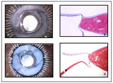
Maximal IOL Optic-Posterior Capsule Contact. Other contributing factors in reducing PCO are posterior angulation of the IOL haptic and posterior convexity of the optic (Slide 6). This is due to the creation of a shrink wrap, which is a tight fit of the posterior capsule against the back of the IOL optic. The relative "stickiness" of the IOL optic biomaterial helps produce an adhesion between the capsule and IOL optic. There is preliminary evidence that the hydrophobic acrylic IOL biomaterial provides enhanced capsular adhesion, or "bioadhesion."46-48,58 This will require further study.
Barrier Effect of the IOL Optic. The IOL optic barrier effect (Slide 6) plays an important role as a second line of defense against PCO, especially in cases in which retained cortex and cells remain following ECCE. The concept of the barrier effect goes back to the original Ridley lens. If accurately implanted in the capsular bag, the Ridley lens provided an excellent barrier effect, with almost complete filling of the capsular bag and contact of the posterior IOL optic to the posterior capsule (no space, no cells). A lens with one or both haptics out-of-the-bag has much less of a chance to produce a barrier effect. The IOL optic's barrier function has been one of the main reasons that posterior chamber IOLs implanted after ECCE throughout the decades did not produce an unacceptably high incidence of florid PCO. Actually, the barrier effect has enabled the success of IOL implantation after ECCE during the past decades.
A subtle difference between classic optics with a round tapered edge and optics with a square truncated edge became evident recently (Slide 6). The effect of a square-edge optic design as a barrier was first discussed by Nishi and colleagues in articles related to PCO.59-71 In a clinicopathological study, our laboratory was the first to confirm this phenomenon in human eyes.76,77 We reported results of a large histopathological analysis covering the IOL barrier effect, with special reference to the efficacy of the truncated edge. A truncated, square-edged optic rim appears to cause a complete blockade of cells at the optic edge, preventing epithelial ingrowth over the posterior capsule.42,43,50,51,55,56,85,88,89 The enhanced barrier effect of this particular edge geometry provides another supplemental factor that has significantly diminished the overall incidence of clinical PCO.
Our past studies demonstrated that the original, three-piece AcrySof MA60 IOL (Alcon) successfully combined these three IOL-related factors (Slide 6) in a way that produced a major PCO advantage. Other manufacturers have begun to incorporate these PCO preventing features, such as a sharp or squared posterior edge. The Cee-On 911 silicone IOL (Pharmacia, Peapack, NJ) was the first silicone IOL to feature a squared edge. The acrylic Sensar IOL (Allergan, Irvine, Calif.) and silicone Clariflex IOL (Allergan) now feature a sharp posterior edge, combined with a rounded anterior edge.
A major disadvantage of the truncated edge is the production of clinical visual aberrations such as glare, halos, and crescents. However, subtle changes in manufacturing are now helping alleviate these complications. For example, the OptiEdge IOL (Allergan) has a squared posterior edge and a round anterior edge. Therefore, optical dysphotopsias are minimized, while the PCO benefit of a squared posterior edge is retained.
Clinical Studies
Three studies confirm the advantage of applying one or more surgical- or IOL-related factors to prevent or delay PCO formation. In a large clinical study, Ram, Pandey, Apple, Werner and associates81 confirmed previous pathological studies demonstrating the importance of in-the-bag fixation of posterior chamber IOLs (both rigid and foldable) in reducing the incidence of PCO. This is true for both standard ECCE and phacoemulsification. This study was performed in Chandigarh, India, and was a combined effort of the Postgraduate Institute of Medical Education and Research (PGIMER), Chandigarh, India, and the Center for Research on Ocular Therapeutics and Biodevices, Storm Eye Institute, Charleston, South Carolina, USA.
The study comprised 278 eyes of 263 patients receiving ECCE and 318 eyes of 297 patients having phacoemulsification with posterior chamber IOL implantation. PCO leading to a decrease in Snellen visual acuity of 2 lines or more was considered visually significant. The presence of PCO and IOL haptic fixation were evaluated postoperatively using slit lamp biomicroscopy. Haptic position was noted as in-the-bag (B-B), one haptic in the bag and one in the sulcus (bag-sulcus [B-S]), or both haptics out of the bag (sulcus-sulcus [S-S]). In addition, the rate of visually significant PCO was compared among three IOL biomaterials: polymethylmethacrylate (PMMA), silicone, and hydrophobic acrylic. Visually significant PCO occurred in 42.45% of eyes having ECCE and 19.18% of eyes having phacoemulsification (P<.001, Chi-square test) after a mean follow-up of 2.4 years +/-0.7 (SD). In both groups, visually significant PCO was significantly less in eyes with B-B fixation than in those with B-S or S-S fixation (P<.001). The rate of visually significant PCO in all eyes in the phacoemulsification group with B-B fixation was low (11.90%) and was significantly lower in eyes with a hydrophobic acrylic IOL (2.22%; P<.05, Chi-square test).
The results of this study suggested that in-the-bag posterior chamber IOL fixation reduces the incidence of PCO. Thorough removal of lens substance, including hydrodissection-assisted cortical cleanup, and in-the-bag posterior chamber IOL fixation seem to be the most important surgical factors in reducing PCO, regardless of surgical procedure or IOL type used. IOL biomaterial and design also help prevent PCO.
Ravalico and associates83 attempted to determine the ideal capsulorrhexis size for minimizing the incidence of PCO. The researchers retrospectively evaluated 107 patients who had ECCE with capsulorrhexis and capsular bag IOL implantation. The PCO site (central, paracentral, or peripheral) and degree (mild, moderate, or severe) were evaluated in relation to the capsulorrhexis edge location relative to the IOL optic. Patients were divided into three groups. Group 1 included patients with a capsulorrhexis-free edge located on the IOL optic for 360°. In group 2, patients had a capsulorrhexis-free edge located asymmetrically on and peripherally to the IOL optic. Group 3 included patients with a capsulorrhexis-free edge located peripherally to the IOL optic for 360°. Each group was divided into two subgroups; one received poly-Hema IOLs and the second, PMMA IOLs. The results of this study demonstrated that in groups 1 and 2, the capsular transparency was higher than in group 3 (P<.04). The percentage of central opacification was lower in group 1 than in groups 2 and 3 (P<.04). No statistically significant differences between the poly-Hema and the PMMA subgroups were seen. The researchers concluded that capsulorrhexis with a slightly smaller diameter than the IOL optic appears to be better than a large-size capsulorrhexis in reducing the incidence of PCO.
T. Akahoshi, MD, reported his experience of three-piece AcrySof IOL implantation in more than 17,000 human eyes in Japan. The incidence of Nd:YAG capsulotomy was 1.19% (207 out of 17,329) after 75 months of follow-up. Among the Nd:YAG treated cases, 81.2% had an eccentric and incomplete coverage of the lens optics by the anterior capsulorrhexis margin. In 8.2% of the cases, the anterior capsule margin was on the optic edge and in 10.6%, it was completely outside. Long-term follow-up revealed that the incidence of after-cataract formation in patients with three-piece AcrySof IOLs is extremely low. The size and position of the CCC, however, seem to be the most important factors to reduce the Nd:YAG capsulotomy rate.
Pharmacological and/or Mechanical Prevention of PCO
Several experimental studies have also been performed in animal models using intraocular infusion of drugs such as colchicine, methotraxate, retinoic acid, and 5-fluorouracil (5-FU) in attempt to prevent PCO.19,78,79,84,85,93,94,97
We designed an intracapsular ring to prevent capsular bag contraction and inhibit LEC proliferation and metaplasia by sustained release of 5-FU. We prospectively studied the effects of the intracapsular rings on the prevention of PCO by analyzing postmortem ocular specimens macroscopically and histologically. We also evaluated the toxic effects of 5-FU on the corneal endothelium, capsular bag, and retina of rabbits.74 Seventeen rabbits were divided into three groups. The first group consisted of six rabbits (6 eyes) that had phacoemulsification (PE) only (control group); the second, six rabbits (6 eyes) that had PE with implantation of an open loop hydrogel intracapsular ring; and the third, five rabbits (5 eyes) that had PE with implantation of a ring with sustained release of 0.25 µg/hour of 5-FU for 9 days. All eyes in the three groups were followed for 8 weeks before enucleation. Capsular bag shrinkage, positioning of the intracapsular ring, and evaluation of central and peripheral PCO for intensity and area were assessed by stereomicroscopy from a posterior (Miyake-Apple) view.2,53 Residual equatorial LECs were counted by the same observer in histological sections. Transmission electron microscopy (TEM) analysis was performed in the cornea, capsular bag, and retina to evaluate the toxicity of 5-FU.
No significant difference was seen in the degree of capsular bag shrinkage in the three groups. Decentration of the intracapsular ring was seen in two eyes (one each in groups 2 and 3, respectively). A statistically significant difference (P<.05, student t-test) was demonstrated between the control group and the group with intracapsular rings only, concerning the area and intensity of central PCO. No difference was seen between the latter and the group with rings releasing 5-FU. No evidence of toxicity of 5-FU to intraocular structures (cornea, capsular bag, and retina) was demonstrated with TEM analysis. Our study revealed that implantation of an intracapsular ring may prevent central PCO after cataract surgery by mechanically blocking migration of LECs toward the central visual axis. The potential pharmacological affect of 5-FU for PCO prevention was not demonstrated in this experimental study.74
The use of intracapsular tension rings to prevent PCO and PCO-related capsular bag shrinkage has recently been reported by several researchers.44,45,52 Kugelberg and coworkers44 examined after-cataract development and eye growth in lensectomized newborn rabbits implanted with open PMMA capsular tension rings of two different sizes (7 mm and 10 mm). The wet mass of the after cataract was measured at 3 months after surgery in eyes with and without a ring. A significant reduction in the wet mass of the after cataract was seen in the eyes with 10-mm rings in comparison to those with the 7-mm rings or the aphakic eyes.
Nishi and colleagues63,64 investigated the inhibitory effect of a discontinuous capsular bend created by an IOL with a band-shaped loop or a capsular tension ring on migrating LECs. The round form of the open-circular loop of a PMMA IOL was changed to a band-like shape, 1 mm wide and 0.2 mm thick. A capsular tension ring of the same shape with a diameter of 14 mm was also made. They implanted the IOL or the ring into the capsular bag in five rabbit eyes after cataract surgery. The same IOL with an unmodified haptic or a conventional capsular tension ring was implanted in the contralateral eye as a control. Gross and histopathological examinations were performed in the enucleated eyes after 8 weeks. LEC accumulation was seen at the equatorial corner outside the IOL haptic or the ring, with inhibition of LEC migration toward the center. In the control eyes, LECs accumulated inside the haptic or ring, forming a Soemmering's ring and showing markedly less inhibition of LEC migration.
Hara and colleagues30,31 determined the efficacy of an equator ring in maintaining the circular contour of the capsular bag equator and the transparency of the posterior capsule after crystalline lens extraction in 12 rabbit eyes. A flexible silicone ring was implanted in the capsular bag in seven rabbit eyes. A 13-mm Sinskey-style posterior chamber IOL was also implanted inside the ring. Three other eyes received only equatorial rings and two eyes received only IOLs. All eyes were followed for 3.5 months. The investigators reported equatorial distortion and severe PCO in two eyes that received only the IOL. In nine (90%) of the 10 eyes implanted with the rings, the circular contour of the equator was preserved, and seven (70%) of these 10 eyes had transparent posterior capsules.
Hashizoe and colleagues33 also evaluated the efficacy of a flexible, silicone equator ring in maintaining capsular bag integrity at the equator and transparency of the posterior capsule, but using cynomolgus monkeys. The eyes were followed for an average of 5.9 months. The ring effectively maintained the circular contour of the capsular bag and posterior capsule transparency. The researchers concluded that the equator ring is a promising device for maintaining capsular bag integrity and minimizing PCO after cataract surgery.
Nagamoto and Bissen-Miyajima57designed four prototypes of rings for supporting and preserving the postoperative integrity of the capsular bag, independently of IOL implantation following CCC. An open, PMMA ring, inserted experimentally in cadaver eyes through a 3.5-mm incision, adjusted well to various capsular bag sizes and could be implanted with common IOL types. Although some capsular shrinkage occurred, generally, the roundness of the capsular bag equator was preserved. The control specimens with the IOL alone had capsular shrinkage in areas that were not in contact with the haptics, thus the capsular bag was deformed. The capsular bags of the control group (no ring, no IOL) folded and lost roundness.
An open-loop intracapsular ring has the advantage of being implantable through a small incision. We used intracapsular rings manufactured from a hydrogel biomaterial, that is, copolymer of hydroxypropylmethacrylate (HPMA)-methylmethacrylate (MMA). Recently, flexible closed-loop equator rings manufactured from silicone elastomer have also been investigated by some authors.30,31 No study has compared the efficacy of open-loop, intracapsular hydrogel rings and silicone rings in preventing PCO and capsular bag shrinkage.
Intraocular application of pharmacologic agents has also been investigated by several authors as a means to prevent PCO.15,16,32,34,38 The idea is to selectively destroy the LECs and avoid toxic side effects on other intraocular tissues such as the sensitive corneal endothelium. Pharmacologic agents being investigated include cancer chemotherapeutic drugs (e.g., antimetabolites: methotraxate, mitomycin, daunomycin, 5-FU, colchicine, and daunorubicin), anti-inflammatory substances, hypo-osmolar drugs, and immunological agents.
Published studies on the pharmacological prevention of PCO have usually been based on cell culture and in vitro experiments, but some animal data have been reported. Hartmann and colleagues32 and Sourdille and Ducournau94 reported the first clinical studies in patients using the antimetabolite daunorubicin for PCO prevention. However, longer follow-up is necessary to evaluate the long-term effect of this drug. Pharmacologic agents in combination with a sustained drug delivery system (SDDS) have been used by some authors.32,44 The idea is to extend the intraocular delivery time to a longer period and thereby avoid toxic intraocular drug levels by release of a constant therapeutic drug concentration.
Solomon and colleagues have shown in a rabbit model that slow release of 5-FU prevents toxic drug levels. The effect of this antimetabolite on PCO reduction was significant compared to the controls. Legler and colleagues17 performed a similar study with colchicine. Significant PCO reduction was observed with no toxic side effects in the group with the lowest colchicine concentration. In this study,44 the SDDS had the shape of a wafer and was inserted in the capsular bag without an IOL.
Our current in vivo study tested 5-FU, and we found that the shrinkage of the capsular bag diameter was less in groups 2 and 3 when compared to group 1. However, this difference was not statistically significant. This can probably be correlated with the degree of peripheral PCO. There was no significant difference in the intensity and area of peripheral PCO among the three groups. Also, in our study the eyes were followed for only 8 weeks. Due to the small number of specimens in each group, we were unable to conclude that the open intracapsular ring was ineffective in preventing the shrinkage of the capsular bag.
Gross evaluation of enucleated eyes using the Miyake-Apple posterior view revealed that implantation of an intracapsular ring prevented central PCO when compared to control group (Slide 7, Slide 8, and Slide 9). This correlates well with the results of Kugelberg and coworkers44 concerning after cataract and eye growth in lensectomized newborn rabbits and the study by Nishi and colleagues63,64 investigating the inhibitory effect of a discontinuous capsular bend on migrating LECs.
In our aforementioned rabbit study,74 we also counted the equatorial LECs in histological sections as reported by Ruiz and colleagues.84 We noticed that LECs were significantly less in eyes with an intracapsular ring (groups 2 and 3) when compared to the control group (Slide 7, Slide 8, and Slide 9). This can be explained by the presence of the ring in the capsular bag that prevented the central migration of the LECs, creating a barrier effect (no space, no cell theory). Although in our study the intracapsular ring with a round edge presented a barrier effect, it was not able to completely prevent the central migration of the residual regenerative cortex in some eyes. Slide 8 shows an example of migration of regenerative/retained cortex in one of the eyes in group 2 which underwent implantation of the intracapsular ring. Future studies may be helpful to precisely address the effect of a squared-edge intracapsular ring on the hindrance of PCO as several recent studies demonstrated that square-edge IOL optics offer a better barrier effect, when compared to round edged IOL optics.
The antimetabolite 5-FU specifically acts on mitotic cells by inhibiting the enzyme thymidylate synthetase. Therefore, the postmitotic tissues of the anterior and posterior chambers of the eye should not be affected by the action of this antimetabolite and only the proliferative activity of the LECs of the anterior capsule could be affected by the sustained release of this drug. We could not find any evidence of toxicity of 5-FU to the corneal endothelium or retina with TEM analyses. We are aware that the corneal endothelium in rabbit corneas, in contrast to human corneas, has a tremendous regenerative capacity. As reported by Van Horn and coworkers,103 even if 50% of the central corneal endothelial cells are destroyed by transcorneal freezing, within 10 days sufficient cell division and migration occur to completely repopulate the area of Descemet's membrane denuded of cells, and the corneal thickness returns to normal. Thus, results regarding the toxicity of 5-FU to corneal endothelial cells must be interpreted with caution. In human eyes, cells that do not rapidly replicate, such as those of corneal endothelium, iris, ciliary body, and retina, are supposed to be less sensitive to the drug. Except for the lens epithelium, all structures within the anterior and posterior chambers are post mitotic.
Our experimental study did not demonstrate an additional effect of 5-FU for prevention of PCO when compared to the mechanical effect provided by the presence of the intracapsular ring. The 5-FU drug delivery properties of the biomaterial were dependent on the hydration of the polymer.15 The rate of 5-FU release was calculated by the manufacturer (Corneal Laboratories) according to a preliminary (in vitro) study15 and the peak of 5-FU concentration (nontoxic) for the intraocular tissues as studied by Rootman and associates.84 Based on these studies, 5-FU concentration in the anterior chamber and capsular bag of the rabbits was maintained at 1 µg/mL for 7 days. According to the manufacturer, the ring can be modified to contain larger amounts of 5-FU, allowing a better effect on prevention of PCO. Nevertheless, additional studies are necessary to determine the effects of this modification on ocular structures, with regard to 5-FU toxicity.
According to our macroscopic and histological evaluations, the presence of an intracapsular ring seems to prevent central PCO after cataract surgery by a mechanical effect. An open-loop intracapsular ring can be implanted in the eye through a small incision and offers a barrier effect preventing the central migration of the LECs toward the posterior capsule. Associated release of 5-FU may have an additional impact on the inhibition of PCO, at least on central PCO, although this could not be demonstrated statistically in our study. Concerning the toxicity of the 5-FU release to the corneal endothelium, no definitive conclusion can be drawn from this study because of the proliferation capacity of rabbit corneal endothelial cells. This fact raises concerns on the applicability of this concept to the human eye.
New Concept for PCO eradication: Sealed Capsule Irrigation
PCO is the major cause for vision loss following cataract surgery and a major problem in pediatric cataract surgery, where the incidence approaches 100%. Reducing or eliminating PCO relies on removing cells from the anterior and equatorial regions of the lens capsule after cataract removal. This is very difficult to do, and many methods have been described, including CO2 laser, preoperative Nd:YAG laser, postoperative Ar laser, Heparin drops, gene transduction, manual vacuum removal, electrical removal, thermal, and other means. The most effective methods employed have been pharmacologic agents. However, all pharmacologic methods in an unsealed system risk toxicity to surrounding important intraocular structures. Various delivery systems have been tested in an effort to reduce the effect of toxicity to surrounding tissues, including polymer matrix wafers, polyglycolic acid disk, intracapsular rings, binding to IOL's viscoelastic containing toxic chemicals, inhibition of cell adhesion, adenovirus-mediated gene transfer, and antibody-linked toxin. No mechanical system has yet been described for restricting contamination of toxin to the lens capsule.
Since the advent of the continuous circular capsulorhexis (CCC) described by Gimbel and Neuhann, 27 the physical properties of the CCC have been found to be very strong, with capability of stretch of the capsulorhexis up to 62% before induction of a radial tear.11 The uniform opening of the CCC allows for sealing compared with a can opener anterior capsulotomy. Sealing of a microcapsulorhexis with a silicone plug has been previously described. However, no sealed irrigation system has been considered for the lens capsule nor for elsewhere in the eye. Dr. Anthony Maloof has developed a new concept in irrigation of the human lens capsule following lens surgery called Sealed Capsule Irrigation (SCI), which may allow the isolated safe delivery of pharmacologic agents into the capsular bag following cataract surgery (Slide 13). SCI is a form of Sealed Irrigation System applied to the internal eye and may be applied elsewhere to the body. In the eye, the technique of capsular bag irrigation may be used with pharmacologic agents to target lens epithelial cells and reduce PCO. SCI should meet the following requirements: it should be minimally invasive, easy to use, fit through a small incision, be relatively inexpensive, provide a repeatable seal with the lens capsule, and not add significantly to the duration of routine cataract surgery.
The intact human lens capsule is functionally a separate compartment within the eye. Once breached, the intralenticular compartment becomes continuous with the anterior chamber and the rest of the eye, allowing exposure to foreign lens antigens. Until now, lens capsule irrigation occured during lens removal and cleanup of the capsular bag. However, using the elastic properties of the capsule and providing an intact capsulorhexis, we devised a technique to reseal the capsular bag following lens removal. By resealing the capsular bag, we recompartmentalized the lens and allowed for the selective irrigation of the internal contents of the capsular bag.
We have developed a device called Perfect Capsule (Milvella Pty. Ltd., Sydney, Australia), which is made from biomedical grade soft silicone and allows the surgeon to reseal the capsular bag. The device consists of a rounded plate containing a suction ring that abuts the anterior capsule and an extension arm that passes through a phacoemulsification wound. This extension arm carries a vacuum channel which supplies vacuum to the suction ring and a combined irrigation and aspiration channel. The irrigation and aspiration channels allow for communication between the sealed capsular bag and the external eye. We have performed initial testing of a first-generation device on postmortem porcine lens capsules and demonstrated its effectiveness for sealed capsule irrigation. We have further refined the device to its current third-generation form to incorporate changes that will allow it to be used in small-incision cataract surgery and address the potential risk of pseudosuction, which would result in loss of sealing of the capsular bag. We considered the properties of the adult capsule to be less elastic than the pediatric capsule and less prone to pseudosuction. The device was modified to contain a vacuum manifold within the suction ring to ensures that no focal occlusion of the suction ring is possible at any point and that the vacuum is evenly distributed to the entire ring.
In performing product validation of this third-generation device, 13 randomly chosen devices were subject to testing on a pig capsule. In all cases, the devices sealed the capsule using vacuum generated by a 20 mL lockable syringe, resulting in a maximal vacuum pressure of greater than 700 mm Hg on application with no evidence of pseudosuction with less than 2.5% reduction in vacuum pressure over a 1-minute period. One of these devices was then selected for repeat testing for a period of 10 minutes with less than 5% reduction in vacuum at 10 minutes. SCI also has been performed with the third-generation device in human eyes using trypan blue to irrigate the capsular bag. In 12 patients undergoing cataract surgery, SCI was performed following cataract removal, using trypan blue to selectively irrigate the capsular bag. In all 12 subjects, there was no visible leakage of trypan blue into the anterior chamber following SCI.
In a study on rabbits, we have been able to selectively irrigate the capsular bag with mitomycin 0.02% with an exposure time of 1 minute without adverse damage to any intraocular structure. Ordinarily, exposure of the internal rabbit eye to mitomycin 0.02% will lead to significant toxicity to the retina and cornea, but this did not occur when SCI was performed.
We are continuing to demonstrate that selective capsular bag irrigation can be performed in animals and humans. Using this technique, targeting of lens epithelial cells to prevent PCO can be conducted safely using precise delivery of known doses of pharmacologic agents with less fear of toxicity to surrounding intraocular structures. This method may be utilized to eliminate or modulate lens epithelial cell activity after cataract surgery, which may lead to less postoperative inflammation and a theoretical reduction in the risk of postoperative cystoid macular edema, reduced anterior, and posterior capsule opacification. It may also allow for definitive implantation of multifocal and accommodative lenses so that the treatment of presbyopia may finally become a reality (Slide 14). The definitive agent to be utilized with SCI remains unknown, but it may not necessarily require high toxicity. The ultimate aim may be to induce apoptosis in the lens epithelial cells or to deactivate it rather that inducing immediate cell death.
Analysis of Nd:YAG Posterior Capsulotomy Rates
Complete analysis of our large series of postmortem eyes has helped us develop the above-mentioned six factors that we believe contribute to the reduction of PCO. An analysis of Nd:YAG laser posterior capsulotomy rates among eight commonly used IOL models has led us to the optimistic conclusion that the incidence of PCO is rapidly diminishing, at least in the industrialized world. Slide 15 shows the ranking of the Nd:YAG laser posterior capsulotomy rates (%) for eight lens designs as of December 2000, starting with the lens showing the lowest percentage at the top and the highest rate at the bottom (Slide 10).
The four lenses with the lowest rates ranging between 3.3% and 20.7% are modern designs, mostly implanted after 1992. This is in contrast to the four lenses with the higher rates ranging between 23.3% and 33.7%, which were all older designs, implanted before 1992. The difference in the Nd:YAG laser rates between the acrylic IOLs and the other IOL types was found to be statistically significant (P<.05, for all comparisons, Chi square test). The Nd:YAG capsulotomy rate for all six foldable IOLs, 15.3% (170/1109), was significantly lower than the rate of the rigid IOLs (Slide 11, Slide 12, and Slide 13) (32.3%; 1722/5316; P<.05, Chi square test). If one removes the AcrySof IOL from the group, the rate noted among the other foldable IOLs studied increases to 158/748 (21.1%). To evaluate the influence of lens quality vs. the influence of the surgical technique on the PCO and Nd:YAG laser posterior capsulotomy rates, it is useful to follow a trend time-line over a long-term period. Under optimal conditions, but not possible in this analysis, the information should be viewed considering the age and the duration of each implant. However, the dates of implantation or the time between implantation and death were difficult to determine, due to ethical considerations. These variables are going to factor out over time as larger numbers are obtained and the trend "time-line" is extended.
Tracking the trend timelines for each lens design is required to rule out other factors in addition to the duration of each implant in the eye to properly assess the differences among the IOLs (Slide 16). Various surgeons' criteria for Nd:YAG laser capsulotomy (e.g., aggressive, conservative) also play a role in the rate. Nevertheless, surgeons' criteria, surgical technique, and implant duration will become standard as the number of accessions and the duration of the study increases.
Conclusions
Nd:YAG laser capsulotomy rates have been reduced to single digits through a combination of improved surgical technique and modern IOL designs and foldable materials - at least in the industrialized world (Slide 17). This will be a significant benefit to patients in achieving improved long-term results and reducing Nd:YAG laser capsulotomy complications. Declining Nd:YAG laser rates will greatly benefit the U.S. Medicare system economically, since, historically, Nd:YAG capsulotomy is the second most commonly performed procedure. The major trend toward fewer Nd:YAG capsulotomies has occurred since approximately 1992, which coincides with the expanding use of capsulorrhexis and foldable IOLs. We believe that analyzing human pseudophakic globes obtained postmortem is an important way to understand how different IOL designs and characteristics relate to PCO. To date, one cannot precisely determine the proportional importance of IOL design versus surgical techniques in the documented decrease of Nd:YAG laser rates. However, follow-up and analysis of increasing numbers of pseudophakic autopsy eyes will be beneficial.
PCO represents the final, common complication of cataract surgery. Now, approximately 50 years after Ridley's first IOL implantation, we have the technology, surgical techniques, skills, IOLs, and understanding necessary to eradicate PCO.
References
-
Alfaiate M, Leite E, Mira J, Cunha-Vaz JG. Prevalence and surgical complications of pseudoexfoliation syndrome in Portuguese patients with senile cataract. J Cataract Refract Surg. 1996;22:972-976.
-
Apple D, Lim E, Morgan R, et al. Preparation and study of human eyes obtained postmortem with the Miyake posterior photographic technique. Ophthalmology. 1990; 97:810-816.
-
Apple DJ, Auffarth GU, Peng Q, Visessook N. Foldable Intraocular Lenses. Evolution, Clinicopathologic Correlations, Complications. 2000.
-
Apple DJ, Peng Q, Visessook N, et al. Eradication of posterior capsule opacification. Documentation of a marked decrease in Nd:YAG laser posterior capsulotomy rates noted in an analysis of 5416 pseudophakic human eyes obtained postmortem. Ophthalmology. 2001;108:505-518.
-
Apple DJ, Peng Q, Visessook N, et al. Surgical prevention of posterior capsule opacification. Part I. Progress in eliminating this complication of cataract surgery. J Cataract Refract Surg. 2000;26:180-187.
-
Apple DJ, Ram J, Foster A, Peng Q. Elimination of cataract blindness: A global perspective entering the new millennium. Surv Ophthalmol. 2000;45:S70-S99.
-
Apple DJ, Solomon DK, Tetz MR, et al. Posterior capsule opacification. Surv Ophthalmol. 1992;37:73-116.
-
Apple DJ. Harold Ridley, MA, MD, FRCS. A golden anniversary celebration and a golden age. Arch Ophthalmol. 1999;117:827-828.
-
Apple DJ. Intraocular lens biocompatibility. J Cataract Refract Surg. 1992;18:217-218.
-
Arshinoff S. Classifying capsulorhexis complications. J Cataract Refract Surg. 1994;20:475.
-
Assia E, Apple D, Barden A, et al. An experimental study comparing various anterior capsulectomy techniques. Arch Ophthalmol. 1991;109:642-647.
-
Ayaki M, Ohara K, Ibaraki N, et al. The outgrowth of lens epithelial cells onto the anterior capsule after intraocular lens implantation. Am J Ophthalmol. 1993;115:668-669.
-
Barman SA, Hollick EJ, Boyce JF, et al. Quantification of posterior capsule opacification in digital images after cataract surgery. Invest Ophthalmol Vis Sci. 2000;41:3882-3892.
-
Bertelmann E, Kojetinsky C. Posterior capsule opacification and anterior capsule opacification. Curr Opin Ophthalmol. 2001;12:35-40.
-
Bougaran R, Jallet V, Villain F, Colin J. A capsular ring to prevent complications after cataract surgery. ARVO abstract 703. Invest Ophthalmol Vis Sci. 1997;38 (Suppl):144.
-
Brown JD, Pearson PA, Blandford D, et al. Controlled release 5-FU delivery systems: Release studies and HPLC assay. ARVO abstract 3063. Invest Ophthalmol Vis Sci. 1991;32(Suppl):1293.
-
Cheng CY, Yen MY, Chen SJ, et al. Visual acuity and contrast sensitivity in different types of posterior capsule opacification. J Cataract Refract Surg. 2001;27:1055-1060.
-
Choung HK, Lee JH. Comparison of the short-term clinical results of silicone and acrylic intraocular lens in patients with diabetes mellitus. Korean J Ophthalmol. 2001;15:15-21.
-
Chung HS, Lim SJ, Kim HB. Effect of mitomycin-C on posterior capsule opacification in rabbit eyes. J Cataract Refract Surg. 2000;26:1537-1542.
-
Davidson MG, Morgan DK, McGahan MC. Effect of surgical technique on in vitro posterior capsule opacification. J Cataract Refract Surg. 2000;26:1550-1554.
-
Davison JA. Capsule contraction syndrome. J Cataract Refract Surg. 1993;19:582-589.
-
Fine IH. Cortical cleaving hydrodissection. J Cataract Refract Surg. 1992;18:508-512.
-
Flach AJ, Dolan BJ. Incidence of postoperative posterior capsular opacification following treatment with diclofenac 0.1% and ketorolac 0.5% ophthalmic solutions: 3-year randomized, double-masked, prospective clinical investigation. Trans Am Ophthalmol Soc. 2000;98:101-105.
-
Font RL, Brownstein S. A light and electron microscopic study of anterior subcapsular cataracts. Am J Ophthalmol. 1974;78:972-984.
-
Gayton JL, Apple DJ, Peng Q, et al. Interlenticular opacification: Clinicopathological correlation of a complication of posterior chamber piggyback intraocular lenses. J Cataract Refract Surg. 2000;26:330-336.
-
Gayton JL, Sanders VN. Implanting two posterior chamber intraocular lenses in a case of microphthalmos. J Cataract Refract Surg. 1993;19:776-777.
-
Gimbel H, Neuhann T. Development, advantages and methods of the continuous circular capsulorhexis technique. J Cataract Refract Surg. 1990;16:31-37.
-
Gonvers M, Sickenberg M, van Melle G. Change in capsulorhexis size after implantation of three types of intraocular lenses. J Cataract Refract Surg. 1997;23:231-238.
-
Hansen SO, Tetz MR, Solomon KD, et al. Decentration of flexible loop posterior chamber intraocular lenses in a series of 222 postmortem eyes. Ophthalmology. 1988;95:344-349.
-
Hara T, Hara T, Sakanishi K, Yamada Y. Efficacy of equator rings in an experimental rabbit study. Arch Ophthalmol. 1995;113:1060-1065.
-
Hara T, Hara T, Yamada Y. Equator ring for maintenance of completely circular contour of the capsular bag equator after cataract removal. Ophthalmic Surg. 1991;22:358-359.
-
Hartman C, Wiedemann P, Gothe K, et al. Prevention of secondary cataract by intracapsular administration of daunomycin. Ophthalmology. 1990;4:102-106.
-
Hashizoe M, Hara T, Ogura Y, et al. Equator ring efficacy in maintaining capsular bag integrity and transparency after cataract removal in monkey eyes. Graefe's Arch Clin Exp Ophthalmol. 1998;236:375-379.
-
Haus CM, Galand AL. Mitomycin against posterior capsular opacification: An experimental study in rabbits. Br J Ophthalmol. 1996;80:1087-1091.
-
Hayashi K, Hayashi H, Nakao F, Hayashi F. Changes in posterior capsule opacification after poly(methyl methacrylate), silicone, and acrylic intraocular lens implantation. J Cataract Refract Surg. 2001;27:817-824.
-
Holweger RR, Marefat B. Intraocular pressure change after neodymium:YAG capsulotomy. J Cataract Refract Surg. 1997;23:115-121.
-
Hu CY, Woung LC, Wang MC. Change in the area of laser posterior capsulotomy: 3 months follow-up. J Cataract Refract Surg. 2001;27:537-542.
-
Inan UU, Ozturk F, Kaynak S, et al. Prevention of posterior capsule opacification by intraoperative single-dose pharmacologic agents. J Cataract Refract Surg. 2001;27:1079-1087.
-
Ismail MM, Alio JL, Moreno JMR. Prevention of secondary cataract by antimitotic drugs: Experimental study. Ophthalmic Res. 1996;28:64-69.
-
Javitt JC, Tielsch JM, Canner JK, et al. National outcomes of cataract extraction. Increased risk of retinal complications associated with Nd:YAG laser capsulotomy. The Cataract Patient Outcomes Research Team. Ophthalmology. 1992;99:1487-1497.
-
Koch D, Liu J, Gill P, Parke D. Axial myopia increases the risk of retinal complications after neodymium-YAG laser posterior capsulotomy. Arch Ophthalmol. 1989;107:986-990.
-
Kruger AJ, Schauersberger J, Abela C, et al. Two year results: Sharp versus rounded optic edges on silicone lenses. J Cataract Refract Surg. 2000;26:566-570.
-
Kucuksumer Y, Bayraktar S, Sahin S, Yilmaz OF. Posterior capsule opacification 3 years after implantation of an AcrySof and a MemoryLens in fellow eyes. J Cataract Refract Surg. 2000;26:1176-1182.
-
Kugelberg U, Zetterstrom C, Lundgren B, et al. After cataract and ocular growth in newborn rabbit eyes implanted with a capsule tension ring. J Cataract Refract Surg. 1997;23:635-640.
-
Legler UFC, Apple DJ, Assia EI, et al. Inhibition of posterior capsule opacification: The effect of colchicine in a sustained drug delivery system. J Cataract Refract Surg. 1993;19:462-470.
-
Linnola RJ, Werner L, Pandey SK, et al. Adhesion of fibronectin, vitronectin, laminin and collagen type IV to intraocular lens materials in human autopsy eyes. Part I: histological sections. J Cataract Refract Surg. 2000;26:1792-1806.
-
Linnola RJ, Werner L, Pandey SK, et al. Adhesion of fibronectin, vitronectin, laminin and collagen type IV to intraocular lens materials in human autopsy eyes. Part II: explanted IOLs. J Cataract Refract Surg. 2000;26:1807-1818.
-
Linnola RJ. Sandwich theory: bioactivity-based explanation for posterior capsule opacification. J Cataract Refract Surg. 1997; 23:1539-1542.
-
Macky TA, Pandey SK, Werner L, et al. Anterior capsule opacification. Int Ophthalmol Clin. 2001;41:17-31.
-
Meacock WR, Spalton DJ, Boyce JF, Jose RM. Effect of optic size on posterior capsule opacification: 5.5 mm versus 6.0 mm AcrySof intraocular lenses. J Cataract Refract Surg. 2001;27:1194-1198.
-
Meacock WR, Spalton DJ, Holick EJ, et al. The effect of polymethylmethacrylate and AcrySof intraocular lenses on the posterior capsule in patients with a large capsulorhexis. Jpn J Ophthalmol. 2001;45:348-354.
-
Menapace R, Findl O, Georgopoulos M, et al. The capsular tension ring: Designs, applications, and techniques. J Cataract Refract Surg. 2000;26:898-912.
-
Miyake K, Miyake C. Intraoperative posterior chamber lens haptic fixation in the human cadaver eye. Ophthalmic Surg. 1985;16:230-236.
-
Miyake K, Ota I, Miyake S, Maekubo K. Correlation between intraocular lens hydrophilicity and anterior capsule opacification and aqueous flare. J Cataract Refract Surg. 1996;22(Suppl):764-769.
-
Nagamoto T, Eguchi G. Effect of intraocular lens design on migration of lens epithelial cells onto the posterior capsule. J Cataract Refract Surg. 1997;23:866-872.
-
Nagamoto T, Hara E. Postoperative membranous proliferation from the anterior capsulotomy margin onto the intraocular lens optic. J Cataract Refract Surg. 1995;21:208-211.
-
Nagamoto T, Miyajima HB. A ring to support the capsular bag after continuous curvilinear capsulorhexis. J Cataract Refract Surg. 1994;20:417-420.
-
Nagata T, Minakata A, Watanabe I. Adhesiveness of AcrySof to a collagen film. J Cataract Refract Surg. 1998;24:367-70.
-
Nishi O, Nishi K, Akura J, Nagata T. Effect of round-edged acrylic intraocular lenses on preventing posterior capsule opacification. J Cataract Refract Surg. 2001;27:608-613.
-
Nishi O, Nishi K, Fujisawa T, et al. Effects of the cytokines on the proliferation of and collagen synthesis by human cataract lens epithelial cells. Br J Ophthalmol. 1996;80:63-68.
-
Nishi O, Nishi K, Fujisawa T, Shirasawa E, Ohmoto Y. Effects of the cytokines on the proliferation of and collagen synthesis by human cataract lens epithelial cells. Br J Ophthalmol. 1996;80:63-68.
-
Nishi O, Nishi K, Mano C, et al. Inhibition of migrating lens epithelial cells by blocking the adhesion molecule integrin: A preliminary report. J Cataract Refract Surg. 1997;23:860-865.
-
Nishi O, Nishi K, Mano C, et al. The inhibition of lens epithelial cell migration by a discontinuous capsular bend-shaped circular loop or a capsule-bending ring. Ophthalmic Surg Lasers. 1998;29:119-125.
-
Nishi O, Nishi K, Menapace R, Akura J. Capsular bending ring to prevent posterior capsule opacification: 2 year follow-up. J Cataract Refract Surg. 2001;27:1359-1365
-
Nishi O, Nishi K, Morita T, et al. Effect of intraocular sustained release of indomethacin on postoperative inflammation and posterior capsular opacification. J Cataract Refract Surg. 1996;22:806-810.
-
Nishi O, Nishi K, Ohmoto Y. Synthesis of interleukin-1, interleukin-6, and basic fibroblast growth factor by human cataract lens epithelial cells. J Cataract Refract Surg. 1996;22:852-858.
-
Nishi O, Nishi K. Intraocular lens encapsulation by shrinkage of the capsulorhexis opening. J Cataract Refract Surg. 1993;19:544-545.
-
Nishi O, Nishi K. Preventing posterior capsule opacification by creating a discontinuous sharp bend in the capsule. J Cataract Refract Surg. 1999;25:521-526
-
Nishi O. Posterior capsule opacification. Part 1: Experimental investigations. J Cataract Refract Surg. 1999;25:106-117.
-
Nishi O. Removal of lens epithelial cells by ultrasound in endocapsular cataract surgery. Ophthalmic Surg. 1987;18:577-580.
-
Nishi O, Nishi K, Menapace R, Akura J. Capsular bending ring to prevent posterior capsule opacification: 2 year follow-up. J Cataract Refract Surg. 2001;27:1359-65.
-
Obstbaum SA. The anterior capsulotomy revisited. J Cataract Refract Surg. 1998;24:143-144.
-
Ohmi S, Uenoyama K. Decentration associated with asymmetric capsular shrinkage and intraocular lens design in a rabbit model. J Cataract Refract Surg. 1995;21:293-296.
-
Pandey SK, Cochener B, Apple DJ, et al. Intracapsular ring sustained 5-fluorouracil delivery system for prevention of posterior capsule opacification in rabbits: a histological study. J Cataract Refract Surg. 2002;28:139-148.
-
Pandey SK, Wilson ME, Trivedi RH, et al. Pediatric cataract surgery and intraocular lens implantation: Current techniques, complications and management. Int Ophthalmol Clin. 2001;41:175-196.
-
Peng Q, Apple DJ, Visessook N, et al. Surgical prevention of posterior capsule opacification. Part II. Enhancement of cortical clean up by focusing on hydrodissection. J Cataract Refract Surg. 2000;26:188-197.
-
Peng Q, Visessook N, Apple DJ, et al. Surgical prevention of posterior capsule opacification. Part III. Intraocular lens optic barrier effect as a second line of defense. J Cataract Refract Surg. 2000;26:198-213.
-
Power WJ, Neylan D, Collum LMT. Daunorubicin as an inhibitor of human lens epithelial cell proliferation in culture. J Cataract Refract Surg. 1994;20:287-290.
-
Rakic JM, Galand A, Vrensen GFJM. Lens epithelial cell proliferation in human posterior capsule opacification. Exp Eye Research. 2000;5:489-494.
-
Ram J, Apple DJ, Peng Q, Visessook N, Auffarth GU, Schoderbek RJ, Ready EL. Update on fixation of rigid and foldable posterior chamber intraocular lenses. Part II. Choosing the correct haptic fixation and intraocular lens design to help eradicate posterior capsule opacification. Ophthalmology. 1999;106:891-900.
-
Ram J, Pandey SK, Apple DJ, Werner L, Brar GS, Singh R, Gupta A. Effect of in-the-bag intraocular lens fixation on the prevention of posterior capsule opacification. J Cataract Refract Surg. 2001;27:1039-1046.
-
Ram J. Kaushik S, Brar GS, Gupta A. Neodymium:YAG capsulotomy rates following phacoemulsification with implantation of PMMA, silicone, and acrylic intraocular lenses. Ophthalmic Surg Lasers. 2001;32:375-382.
-
Ravalico G, Tognetto D, et al. Capsulorhexis size and posterior capsule opacification. J Cataract Refract Surg. 1996;22:98-103.
-
Rootman J, Tisdall J, Gudauskas G, Ostray A. Intraocular penetration of subconjunctivally administered 14C-flurouracil in rabbits. Arch Ophthalmol. 1979;97:2375-2378.
-
Ruiz JM, Medrano M, Alio JL. Inhibition of posterior capsule opacification by 5-Fluorouracil in rabbits. Ophthalmic Res. 1990;22:201-208.
-
Scaramuzza A, Fernando GT, Crayford BB. Posterior capsule opacification and lens epithelial cell layer formation: Hydroview hydrogel versus AcrySof acrylic intraocular lenses. J Cataract Refract Surg. 2001;27:1047-1054.
-
Schaumberg DA, Dana MR, Christen WG, Glynn RJ. A systematic overview of the incidence of posterior capsular opacification. Ophthalmology. 1998;105:1213-1221.
-
Schmidbauer JM, Vargas LG, Peng Q, Escobar-Gomez M, Werner L, Arthur SN, Apple DJ. Posterior capsule opacification. Int Ophthalmol Clin. 2001;41:109-31.
-
Schmack, Gerstmeyer K. Long-term results of the foldable CeeOn Edge intraocular lens. J Cataract Refract Surg. 2000;26:1172-1175.
-
Schmidbauer JM, Vargas LG, Apple DJ, et al. Influence of surgery-related factors on the Nd: YAG laser capsulotomy rates of 3-piece silicone IOLs - analysis of 457 pseudophakic human globes obtained postmortem. Klin Montasbl Augenheilkd. 2001;218:523-537.
-
Scorolli L, Martini E, Scalinci SZ, et al. Capsule contraction after continuous curvilinear capsulorhexis. J Cataract Refract Surg. 1996;22:1245-1246.
-
Shammas HJ. Relaxing the fibrosed capsulorhexis rim to correct induced hyperopia after phacoemulsification. J Cataract Refract Surg. 1995;21:228-229.
-
Solomon KD, VanMeter WS, Pearson PA, et al. Sustained release drug delivery systems in extracapsular cataract surgery. ARVO abstract 1724. Invest Ophthalmol Vis Sci. 1990;31(Suppl):351.
-
Sourdille P, Ducournau Y. Effect of daunomycin on epithelial cells of the crystalline lens. Ophtalmologie. 1990;4:107-108.
-
Spang KM, Rohrbach JM, Weidle EG. Complete occlusion of the anterior capsular opening after intact capsulorhexis: clinicopathologic correlation. Am J Ophthalmol. 1999;127:343-345.
-
Tanaka Y, Ibaraki N, Hongoh M. Histopathological study of rabbit anterior capsule opacification after IOL surgery. Nippon Ganka Gakkai Zasshi. 1993;97:672-677.
-
Tetz MR, Ries MW, Lucas C, et al. Inhibition of posterior capsule opacification by an intraocular-lens-bound sustained drug delivery system: An experimental animal study and literature review. J Cataract Refract Surg. 1996;22:1070-1078.
-
Toldos JJM, Roig AA, Benabent EC. Total anterior capsule closure after silicone intraocular lens implantation. J Cataract Refract Surg. 1996;22:269-271.
-
Trivedi RH, Izak A, Werner L, Macky TA, Pandey SK, Apple DJ. Interlenticular opacification of piggyback intraocular lenses. Int Ophthalmol Clin. 2001;41:47-62.
-
Tsuboi S, Tsujioka M, Kusube T, Kojima S. Effect of continuous circular capsulorhexis and intraocular lens fixation on the blood-aqueous barrier. Arch Ophthalmol. 1992;110:1124-1127.
-
Ursell PG, Spalton DJ, Pande MV. Anterior capsule stability in eyes with intraocular lenses made of poly(methyl methacrylate), silicone, and AcrySof. J Cataract Refract Surg.1997;23:1532-1538.
-
Van Horn DL, Sendele DD, Seideman S, Buco PJ. Regenerative capacity of the corneal endothelium in rabbit and cat. Invest Ophthalmol Vis Sci. 1977;16:697-613.
-
Werner L, Apple DJ, Pandey SK, et al. Analysis of elements of interlenticular opacification. Am J Ophthalmol. 2002;133:320-326.
-
Werner L, Apple DJ, Pandey SK. Postoperative proliferation of anterior and equatorial lens epithelial cells: A comparison between various foldable IOL designs. In: Buratto L, Osher R, Masket S, eds. Cataract Surgery in Complicated Cases. 2000:399-417.
-
Werner L, Pandey SK, Apple DJ, et al. Anterior capsule opacification: Correlation of pathological findings with clinical sequelae. Ophthalmology. 2001;108:1675-1681.
-
Werner L, Pandey SK, Escobar-Gomez M, et al. Anterior capsule opacification: A histopathological study comparing different IOL styles. Ophthalmology. 2000;107:463-467.
-
Wilson ME, Pandey SK, Werner L, et al. Pediatric cataract surgery: Current techniques, complications and management. In: Agarwal A, Agarwal S, Fine H, Sachdeva M, Agarwal A, eds. Phacoemulsification, Laser Cataract Surgery and Foldable IOLs. 2000:369-388.
-
Akahoshi T. Clear corneal cataract surgery and AcrySofä implantation. Presented in the ASCRS Symposium on Cataract, IOL and Refractive Surgery, Boston, MA, April 28, 2001.