Introduction
Runge opened and obliterated the frontal sinus in 1750. Lanbeck first described the clinical presentation of frontal sinus mucoceles in 1818. In 1884, Ogston created a trephine hole through the anterior table and drained the sinus into the nose through the anterior ethmoidal cells.1,2 Despite these early reports, the cause of mucocele is still not fully understood. Over the years, many different surgical approaches have been proposed to treat mucocele. However, it is difficult to compare the surgical results due to the variability of pathology, numerous surgical approaches used, and the slow rate of regrowth of mucoceles.
Anatomy
The paranasal sinuses are air-filled extensions of the nasal cavity and are named according to the bones in which they are located. Relative to the orbit, the frontal sinus lies superiorly, the maxillary sinus inferiorly, the ethmoid sinus medially, and the sphenoid sinus posteriorly. The membrane lining the sinuses secrete mucous into the nasal cavity through the ostia of the sinuses. The sinuses vary considerably both in size and form. Many of the sinuses are absent at birth, though there are usually a few ethmoidal cells and tiny maxillary sinuses present. The frontal and sphenoid sinuses develop during childhood and adolescence. Growth of these sinuses is important because they alter the size and shape of the face during infancy and childhood and add resonance to the adolescent voice.3 The frontal sinuses are paired, irregularly parameatal cavities situated within the squamous portion of the frontal bone above the orbits. The thickest wall of the sinus is in the anterior table, with cancellous bone between its cortical plates. The orbital plate of the frontal bone forms the inferior and, generally, the thinnest wall. The posterior table lies in direct contact with the dura of the anterior cranial fossa.1 The frontal sinuses may have septations dividing it into several compartments. A separate nasofrontal duct exists for both the left and right frontal sinus. Usually, the frontal sinus drains into the infundibulum, which opens into the hiatus semilunaris of the middle meatus on the lateral nasal wall. Branches of the supraorbital nerves innervate the frontal sinuses.3
Pathophysiology
Mucocele is a dilatation of the sinus cavity caused by the accumulation and retention of mucoid materials within the sinus. It is thought to be the result of continuous or intermittent obstruction of the sinus ostium.4,5,6 With further expansion, the sinus walls become attenuated and are eventually destroyed. The expansion may extend into the intracranial space or into the orbit. The center of the mucocele typically contains yellow mucoid material, and occasionally the mucocele can precipitate a purulent infection.1,5 The pathogenesis of mucoceles is uncertain and controversial, though two general theories exist. In one theory, a retention cyst that enlarges is believed to develop from obstruction of one or more ducts of the mucous glands into the sinus submucosa. The other theory is that obstruction at or near the sinus outlet causes retention of the mucous secretions and subsequent mucocele formation. The obstruction is most commonly secondary to trauma, osteoma formation, or mucosal thickening associated with chronic sinusitis.5,6,7 Intrinsic abnormalities in the microanatomy of the respiratory epithelium may also play a role as the condition has been associated with cystic fibrosis, which can produce mucoceles during infancy and childhood.5 Paranasal sinus mucoceles are rare in children and instead are mostly found in adults ranging in age from 18 to 30 years old. Mucoceles occur with equal frequency in men and women and are most common in the frontal sinus. Sites of involvement in order of decreasing frequency are the frontal (65%), ethmoid (30%), sphenoid, and maxillary sinuses.1,2
Clinical Findings
Presentation of the mucocele depends on the anatomic area involved. The first symptoms of frontal mucocele may be intermittent headache with superior nasal orbital fullness and signs of a superior nasal orbital mass, proptosis, or diplopia (Slide 1). The floor of the frontal sinus or the orbital roof is gradually thinned, and the expansion of the mucocele is in the direction of least resistance.2 A soft-tissue swelling may appear above the medial canthus and at the superior medial aspect of the orbit (Slide 2). The mass may be cystic and have an eggshell sensation on palpation. A frontal sinus mucocele tends to displace the globe inferiorly, laterally, and anteriorly. As a result, diplopia is a common complaint.5,6 Visual decrease is generally not pronounced with sinus mucoceles except in rare cases where a mucocele occupying the posterior orbit may exert pressure on the optic nerve and optic chiasm, causing progressive loss of vision or even blindness. More commonly, extraocular motility problems may be present, along with retinal striae from indentation of the globe.5 Displacement of the cavernous portion of the carotid artery laterally may create transient or permanent palsies of the oculomotor nerves.1,8
Ethmoidal sinus mucoceles may cause decalcification in the intra-sinus septum. The lamina papyracea is frequently displaced laterally in the orbit, resulting in proptosis or lateral displacement of the globe. Diplopia is often the presenting complaint. The lacrimal system may be affected and obstructed, and the patient may complain of epiphora. An expanding frontal ethmoid sinus mucocele can expose the dura either by erosion of the ethmoidal roof or dehiscence of the posterior wall of the frontal sinus.
Diagnostic Features
Mucoceles are often recognized by their radiographic appearance. The findings on plain film and computed tomography are diverse, but sinus opacification with thinning and expansion of the sinus walls is a relatively constant feature. The characteristic opacification is the result of the trapped mucous that eventually displaces the air in the sinus. The mucoperiosteal margin decalcifies and fades, causing a diminution of the normal scallop borders. The computed tomography (CT) scan is the most useful in evaluating the orbit and intracranial extent of the lesion.1,5,9 The typical mucocele appearance is a smooth, expanded oval or round area of bone erosion with radiolucency (Slide 3). The affected adjacent bone is osteolytic, but there is often a zone of sclerosing osteitis around the margins of the lesion. In more advanced cases, the central mucoid contents of the lesion may resemble a tumor from the paranasal sinuses invading the orbit. The appearance on magnetic resonance imaging demonstrates highly variable signal intensities. Some have a low intensity T1 weighted range that becomes hyperintense on T2 weighted sequence. Others may be hyperintense on both T1 and T2 weighted images.5
Treatment
Mucoceles are managed by reestablishing normal sinus drainage with marsupialization of the mucocele or removal of the lining of the cyst. Occasionally, obliteration of the frontal sinus with fat is needed. Preoperative computed tomography scans, including coronal and axial views of the orbit or, when indicated, magnetic resonance imaging scans, are obtained to locate the mass and determine its relationship to important orbital structures. Proper preoperative localization enables the surgeon to obtain appropriate consultations such as neurosurgical or otolaryngologic consultations.
Frontal sinus mucoceles can be approached by a superior nasal orbitotomy or an osteoplastic flap with sinus exposure, removal of the entire lining of the cyst, and obliteration of the sinus cavity with abdominal fat.5,10 Osteoplastic frontal sinusotomy uses a hinged osteoperiosteal flap of the anterior frontal sinus wall, creating a direct frontal sinusotomy approach (Slide 4). This was first reported by Schonborn in 1894 and Brieger in 1895. Bergara and Tato and colleagues described their approach with accompanying fat obliteration in the 1940s. Advantages of the osteoplastic flap technique include direct visualization of both frontal sinuses, access to the supraorbital ethmoids, absence of cosmetic deformity if approached coronally, elimination of the need for the nasal frontal duct for drainage, and obliteration of the sinus cavity. Disadvantages include its extensive nature with larger incisions and greater blood loss, the need to remove all mucosal remnants within the sinus, complications such as abnormal healing of the bone flap, scar camouflage and nerve injuries, and difficult follow-up using CT scans of the sinuses due to fat obliteration.11,12
More recent endonasal endoscopic techniques have been used to enter each of the involved sinuses. For complicated pathology, like mucoceles adjacent to the orbit, these intranasal endoscopic sinus techniques are often combined with computer assisted surgery. This uses special computer software and electromagnetic tracking to help produce axial, coronal, and sagittal views intraoperatively. Instruments are linked to the computer intraoperatively that identify and localize the normal anatomy and abnormal pathology. The mucocele can be marsupialized and the sinus ostia opened to establish drainage. Intranasal endoscopic surgery dramatically reduces morbidity. Because there is no sinus obliteration as with the osteoplastic frontal sinusotomy, the structural anatomy is preserved for follow-up radiographic visualization.
Analysis of the causes of failure in the surgical management of frontal sinus disease show that numerous factors play a role. These include1,11:
- Closure of the nasal frontal duct
- The presence of sinus septae that may obstruct drainage
- Narrow extensions of the sinus that may form pockets of infection
- Persistent islands of mucosa that may develop into cysts
- The presence of supraorbital ethmoidal sinuses
- Reinfection from infected and obstructed ethmoid and sphenoid sinuses
- The presence of resistant organisms
- Foci of osteomyelitis.
Thus, the same anatomic features that predispose an individual to frontal sinus disease limit the effectiveness of any surgical management.
Presently, it appears that endoscopic sinus surgery is the best approach to mucoceles of the orbit. The goals of this approach are to do a minimally invasive procedure by working with an endoscope in a nasal cavity while opening the involved sinuses efficiently to eliminate obstruction and pathology. Technique is mucosa sparing, with virtually all of the renewed mucosa derived from the ethmoid sinuses. The common drainage pathway for frontal ethmoid or maxillary sinuses is known as the ostiomeatal complex. Minimal disease in this area can obstruct the normal mucociliary activity that clears the sinuses and can cause either acute or chronic sinus disease. Endoscopy is based on the principle that extremely limited procedures directed towards the ostiomeatal complex anterior ethmoidal sinus correct extensive disease in the adjacent sinuses.13 When mucosa is stripped from the sinus cavity, healing is slow and scarring is significant. Regenerated mucosa may never redevelop normal ciliary density. Thus, the goal of endoscopic sinus surgery is to open diseased sinuses at the natural ostia and, whenever possible, completely remove the underlying bone partitions adjacent to the chronic sinus disease. Surgeons must be familiar with managing complications of disease, such as CSF leak and exposed dura, because the mucocele may have eroded the roof of the sinus. There is an additional risk of penetrating the orbit through the same thin walls of the ethmoidal sinus. Penetration of the orbit and dissection of orbital fat can injure the extraocular muscles or the optic nerve, resulting in diplopia or blindness. The more common problem is indirect injury by an expanding orbital hematoma. If the increased pressure is not immediately recognized, loss of vision can occur.13
There are several advantages of endoscopic sinusotomy, including eliminating the need for external incision, its use as an outpatient procedure, and the ability to follow frontal sinus disease endoscopically and radiographically (Slide 5). The disadvantage is an extensive learning curve associated with angled endoscopes in the confines of critical anatomic structures.
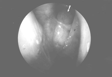
In recent years, there has been a reduction in the number of osteoplastic procedures due to improved technology and improved surgical skill in performing endoscopic sinus surgery. Advanced technology has included the use of improved angled instruments for reaching into the frontal sinus, improved endoscopes in digital enhancement, cross table fluoroscopes for obtaining internal images of the frontal sinus and its outflow intraoperatively, and recent use of image-guided computer assisted triplanar intraoperative guidance systems.
References
-
Lawson W. Frontal sinus. In: Blitzer A, Lawson W, Friedman WH. Surgery of the Paranasal Sinuses. Philadelphia, PA: WB Saunders Co.; 1991:183-218.
-
Kennedy DW, Josephson JS, Zinreich J, et al. Endoscopic sinus surgery for mucoceles: A viable alternative. Laryngoscope. 1980;99:885-895.
-
Moor KL. Clinically Oriented Anatomy. 3rd ed. Baltimore: Williams & Wilkins; 1992:637-782.
-
Tasman W, Jaeger EA. Duane's Foundations of Clinical Ophthalmology. Vol. 2. New York: JB Lippincott Company; 1991:26.
-
Jakobiec FA, Jones IS. Orbital inflammations. In: Jakobiec FA, Jones IS, eds. Diseases of the Orbit. New York: Harper & Row; 1979:187-256.
-
Dortzbach RK, Kronish JW. Orbital disease. In: Dortzbach RK, ed. Ophthalmic Plastic Surgery: Prevention and Management of Complications. New York: Raven Press; 1994:307-357.
-
Gilbard SM, Della Rocca RC. Paranasal sinus disease and the orbit. In: Smith BC, ed. Ophthalmic Plastic and Reconstructive Surgery. St. Louis: CV Mosby Co.; 1987:1124-1147.
-
Ehrenpreis SJ, Biedlingmaier JF. Isolated third-nerve palsy associated with frontal sinus mucocele. Neuro-Ophthalmology. 1995;15:105-108.
-
Roth M, Lanza DC, Zinreich J, et al. Advantages and disadvantages of three-dimensional computed tomography intraoperative localization for functional endoscopic sinus surgery. Laryngoscope. 1995;105:1279-1286.
-
Spoor TC. Anterior orbitotomy. In: Levine MR, ed. Manual of Oculoplastic Surgery. Boston: Butterworth-Heinemann; 1996:281-290.
-
Levine HL, May M. Endoscopic Sinus Surgery. New York: Thieme Medical Publishers; 1993:195-243.
-
Shore JW, Westfall CT, Leone CR, Shagets FW. Enophthalmos and upper eyelid retraction following osteoplastic frontal sinusotomy. Ophthalm Plast Reconstr Surg. 1987;3:13-15.
-
McCaffrey TV. Functional endoscopic sinus surgery: An overview. May Clinic Proc 1993;68:571-577.