Introduction
Modern cataract surgery has advanced to offer unprecedented patient benefits and has attained a level of successful outcomes where it may now be viewed as a refractive procedure. It is important to recognize, however, that the greatest benefit derived from contemporary small-incision surgery is the enhanced safety of the procedure. Even when a posterior capsule ruptures, modern instrumentation and techniques offer a level of control over the intraocular environment and allow for anatomic and visual results that rival an uncomplicated case.
Each case involving a broken posterior capsule is unique; however, several fundamental surgical principles apply universally. When followed, these principles will allow surgeons to safely and thoroughly remove all lens material, to remove presenting vitreous without imparting unnecessary retinal traction forces, and to avoid further enlargement of the posterior capsular tear.
Management
The first step in managing a broken posterior capsule is to immediately recognize its occurrence. Standard teaching has emphasized the importance of sudden deepening of the anterior chamber, which is likely to herald a breach in capsular integrity. A more subtle and earlier sign is deepening of the posterior chamber. In many cases, the first sign of trouble may be a slight increase in space between the anterior lens capsule and the posterior surface of the iris.
Generally, any increase in space or depth of the anterior segment should elicit suspicion of a broken posterior capsule. Another important finding is a change in the fluidic environment. Specifically, an alteration in followability or holdability may be due to a defect in the capsule, resulting in altered flow. Other important signs include a change in cutting characteristics and a sudden inability to rotate the nucleus. Finally, an experienced surgeon should always sense the tactile rebound tension or trampoline-like quality that the capsular and zonular network normally impart. Any change in this “feeling” must be investigated.
Once a problem is suspected, a surgeon must have the discipline to immediately stop working. This does not necessarily mean removal of instruments from within the eye, because abrupt shallowing of the anterior chamber may extend the tear. Rather, filling the anterior chamber with an ophthalmic viscosurgical device (OVD) through the sideport incision may permit removal of the phaco tip or irrigation and aspiration (I&A) instrument without incurring sudden hypotony. At the same time, an OVD is used to tamponade the anterior hyaloid face and stabilize any remaining lens material. This will allow ample time for careful assessment of the anatomy, which will dictate subsequent surgical strategy.
Knowledge of the characteristics of different OVDs is important because the OVD that is required when a complication occurs will likely be different from an OVD for routine use. A less cohesive, highly dispersive agent with low viscosity forms an effective plug in a capsular break to tamponade the anterior hyaloid face, particularly at higher shear rates. Newer viscoadaptive agents such as Healon 5 (sodium hyaluronate 2.3%, Pharmacia & Upjohn) may prove helpful in these challenging situations.
To avoid enlarging the capsular tear once it has been detected, the surgeon must maintain a truly closed-chamber environment. Although further maneuvers may be aided by small, self-sealing incisions, the clinician must use truly watertight incisions to minimize, or avoid, infusion. Unnecessary infusion, like hypotony, is a nemesis when a posterior capsule breaks.
If significant nuclear material is present, a decision must be made regarding further phacoemulsification. This decision depends upon a surgeon's experience and the anatomic particulars of the case; however, if conversion is necessary, the incision should be enlarged generously. Astigmatic concerns and consequences of enlarging the incision may be addressed at a later time. Care should also be taken to avoid pressure on the globe when removing lens material; viscodissection and instrument-aided removal are preferred. A modified lens glide (e.g., Visitec, Sarasota, Fla.) may be used both to support and to aid in removal. Alternatively, the lens glide may be used as a pseudoposterior capsule, as described by Marc Michelson, MD, allowing further phacoemulsification to be performed. However, phacoemulsification must be continued in a low-flow state, and an OVD must be used generously.
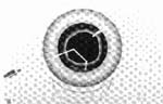
For residual cortex, I advocate a low-flow removal technique (or at least mobilization) with automated I&A performed only after the intraocular lens (IOL) has been implanted and the pupil is constricted. If an I&A handpiece is used, the high flow associated with the I&A handpiece will cause extension of the tear. Alternatively, if the anterior hyaloid face is broken, cortex may be removed using a vitrectomy instrument. Bimanual, separate infusion and aspiration instruments placed through watertight paracenteses afford enhanced control and better access to residual lens material. A unimanual, nonautomated technique may offer the most control. First, viscodissection is performed between residual cortex and the posterior capsule. A double-angled 27-gauge cannula placed on a TB syringe filled with balanced salt solution is then inserted through various paracentesis incisions to gain 360° access to lens material (Slide 1). Gentle infusion first loosens cortex; then, with the sensitivity afforded by this manual technique, cortical strands may be stripped from the capsular fornix and brought up into the anterior chamber. Complete removal may be deferred until after the IOL is positioned and the pupil is miotic.
If vitreous is present, a vitrectomy is mandated. Many authors have expounded a bimanual, two-port vitrectomy. However, most cataract surgeons rely on what is familiar and seemingly easier to use - a unimanual, coaxial vitrectomy. Unfortunately, this approach is inefficient, potentially more dangerous, and more likely to lead to enlargement of the capsular rent.
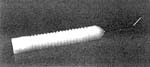
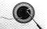
By separating the infusion line from the vitrectomy instrument, a more controlled and effective vitrectomy may be performed. Various anterior chamber maintainers are available. Self-maintaining cannulas have the advantage of freeing-up one hand, but I prefer to maintain bimanual control over the eye and have found that a blunt-end, 21-gauge infusion cannula (e.g., Storz #E4421-S21; Slide 2) is ideal. The standard infusion line connects easily to this cannula, and is then placed through a sideport incision. The infusion rate is lowered to a level that maintains volume as material is removed. A microvitreoretinal blade is then used to make a separate stab incision at the limbus to allow placement of a 20-gauge vitrectomy cutter (Slide 3). The incisions must be snug and watertight. If the phaco incision is not self-sealing, it should be hydrated or sutured. Vitreous removal should be performed at low vacuum power (i.e., 50 mm Hg to 100 mm Hg) with high cutting rates (i.e., 400 to 1500 cuts per minute). If lens material must be removed, the cutting rate should be reduced and the vacuum power gradually increased.
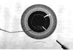
Alternatively, the cutting instrument may be placed through the pars plana (Slide 4). This placement offers an advantage of accessing the subincisional area where residual lens material often resides, permits efficient removal of anterior chamber vitreous by drawing it down posteriorly, and often limits the total amount of vitreous that is removed. Although placing an incision though the pars plana may be an undesirable technique to anterior segment surgeons, the technique may be accomplished safely and easily: after a small, conjunctival peritomy, calipers are used to carefully measure 3.5 mm posterior to the limbus. An MVR blade is then used to make a stab incision keeping the blade perpendicular to the eye wall. The metal tip of the blade should be visualized through the pupil to ensure that entry is complete. Once the vitrectomy is completed, the incision is freed of any remaining vitreous and closed with a suture.
Following IOL placement, a miotic should be instilled gradually. The OVD is removed with the vitrectomy instrument, with meticulous attention directed toward the pupil and wounds to ensure that all vitreous has been removed. Shallowing of the anterior chamber must be avoided to prevent further vitreous prolapse; temporary air injection followed by a gradual fluid-air exchange may be useful. Rapid stromal hydration of limbal incisions is also helpful to prevent hypotony. Watertight wounds should be confirmed. Postoperatively, vigorous steroid and nonsteroidal anti-inflammatory drugs should be administered, and cycloplegic and antihypertensive agents considered.
Summary
Phaco surgeons may take stock in the fact that despite a breech in the posterior capsule, the control rendered through closed-chamber surgical techniques may allow a surgical outcome that is indistinguishable from an uncomplicated case.