Introduction
It has been more than one-half of a century since the first intraocular lens (IOL) was implanted by Harold Ridley on November 29, 1949, at St. Thomas Hospital in London.1 Dr. Ridley replaced the cataractous natural crystalline lens of a patient with an artificial lens made of PMMA and initiated a significant change in the practice of ophthalmology.
Since then, millions of cataract surgeries have been performed and more than 100 million IOLs have been implanted with remarkable success. The IOL is one of the most successful long-term implants of the twentieth century and has made a difference in the quality of life of cataract patients. Meanwhile, cataract surgery has been transformed from a major surgery with 1 week of hospitalization to an outpatient procedure with an actual surgery time of less than 10 minutes for skilled surgeons. This improvement in the practice of cataract surgery has been achieved as a result of continuous improvements in surgical technique, instrumentation, and IOLs.
Foldable Intraocular Lenses
The advent of phacoemulsification and microsurgical techniques has revolutionized cataract surgery.2 Cataract surgeons are now able to remove a cataractous lens through a much smaller incision as compared to extracapsular cataract removal techniques. This evolution was not complete without a lens that could be implanted through this significantly smaller incision. Thus, the idea of a foldable IOL was conceived. A foldable IOL is made of a soft material that can be folded and implanted through a 2.8-mm to 3.5-mm incision.
Initial foldable lenses were made, as were many other new medical devices and implants, from readily available materials with appropriate properties and biomedical histories. Polyhydroxyethylmethacrylate (polyHEMA), hydrogel, and polydimethyl siloxane elastomer were the initial materials selected. However, the desire for thin profile lenses required higher refractive index optic materials.3,4 As a result, HEMA copolymer acrylic hydrogels and dimethyl siloxane-diphenyl siloxane copolymer silicone elastomers were introduced.
Another approach was to build on the success of the PMMA IOLs and specifically design a new synthetic acrylic foldable IOL biomaterial. Thus, phenylethyl acrylate-phenylethyl methacrylate copolymer acrylic elastomer was created to meet the demanding requirements of small-incision cataract surgery and in-the-bag lens implantation.5
Hydrophobic Acrylic Intraocular Lenses
AcrySof The AcrySof MA60BM (Alcon, Ft. Worth, Texas) three-piece IOL with PMMA haptics was first implanted in a human in December 1990, as part of the global clinical investigation. In December 1994, the U.S. Food and Drug Adminstration (FDA) approved the PMMA for this product.
The AcrySof IOL material was the first approved material specifically designed and developed for IOL application. It belongs to the acrylic family of polymers that have been a part of IOL history from inception, and the material is well trusted by ophthalmologists for long-term ocular implant use.
The AcrySof lens material embodies most of the desirable attributes required for a foldable IOL including high refractive index, high damping factor, chemical stability, absence of leachable monomers, and biocompatibility. Because of its high refractive index, these lenses have the thinnest optic for a given power and optic size. Chemical stability, purity from leachable monomers and thin optic profile, which minimizes iris touch, are expected to contribute to its exceptional biocompatibility. Strong adhesion of the AcrySof lens to the capsular bag (Neuhann), in vivo observations of (LEC) cell retraction on the lens surface (Spalton), and low posterior capsule opacification (PCO) rates have been some of this material’s clinically found benefits.
Hydrophobic acrylic IOL materials are crosslinked polymers or copolymers of acrylic esters. In addition to a major acrylic ester monomer, they may include other acrylic or methacrylic ester comonomers. They owe their excellent optical properties, chemical stability, and physical properties to a saturated, all-carbon backbone and polar ester-containing side groups. They are crosslinked either using multifunctional crosslinking monomers or other secondary crosslinking processes such as peroxide curing. The primary monomers used in the synthesis of the AcrySof IOL material are 2-phenylethyl acrylate and 2-phenylethylmethacrylate.
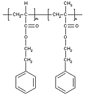
The synthesis mechanism of the AcrySof lens material is a free radical addition polymerization reaction initiated by thermal decomposition of a peroxide initiator. As a result, chains of 2-phenylethyl acrylate and 2-phenylethyl methacrylate are formed with a carbon-to-carbon backbone. The bifunctional crosslinks with the formulation, 1,4-butanediol diacrylate, interlinks the polymer chains, forming an infinite network structure.
The bondable ultraviolet (UV) absorber molecules, 2-(2'-hydroxy-3'-methallyl-5'-methylphenyl) benzotriazole, react with growing chain-end free radicals through their allyl functional group, and become a part of the crosslinked network structure. The final lens material is a crosslinked random quadpolymer of 2-phenylethyl acrylate, 2-phenylethyl methacrylate, 1,4-butanediol diacrylate and 2-(2'-hydroxy-3'-methallyl-5'-methylphenyl) benzotriazole.
The AcrySof IOL is manufactured using an advanced direct lens forming technology that is specifically developed for producing the highest quality IOL implant. The highest purity monomers are used in the synthesis of the AcrySof IOL, and monomers that have not reacted and initiator byproducts are removed as a result of an exhaustive solvent extraction process, which ensures that the lens is free from potentially leachable chemicals. Overall, this advanced lens manufacturing process is believed to play a critical role in producing the unique surface properties and superior biocompatibility of the AcrySof IOL.
The AcrySof IOL is currently available as both three-piece (Table 1) and single-piece lenses (Table 2).
Table 1. AcrySof Multi-Piece Foldable IOL Characteristics| Model | Dioptric Range | Optic Diameter | Overall Length | Haptic Angle |
|---|---|---|---|---|
| MA30BA | +10.0 to +30.0 | 5.5 mm | 12.5 mm | 5° |
| MA30AC | +10.0 to +30.0 | 5.5 mm | 12.5 mm | 5° |
| MA60BM | +6.0 to +30.0 | 6.0 mm | 13.0 mm | 10° |
| MA50BM | +6.0 to +30.0 | 6.5 mm | 13.0 mm | 10° |
| MA60MA | -5.0 to +5.0 | 6.0 mm | 13.0 mm | 5° |
| Model | Dioptric Range | Optic Diameter | Overall Length | Haptic Angle |
|---|---|---|---|---|
| SA30AL | +10.0 to 30.0 | 5.5 mm | 12.5 mm | 0° |
| SA30AT | +10.0 to +30.0 | 5.5 mm | 13.0 mm | 0° |
| SA60AT | +6.0 to +34.0 | 6.0 mm | 13.0 mm | 0° |
Sensar The Sensar AR40 (Allergan Surgical, Irvine, Calif.) is the second hydrophobic foldable acrylic IOL approved by the FDA. In 2000, the FDA approved the Sensar three-piece foldable IOL, which comprises an acrylic crosslinked terpolymer optic and two extruded blue core PMMA haptics, for distribution in the United States. The terpolymer optic is composed of ethyl acrylate (EA), ethyl methacrylate (EMA), and 2,2,2-trifluoroethyl methacrylate (TFEMA). The crosslinker is ethylene glycol dimethacrylate (EGDMA).
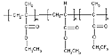
The Sensar also contains an acrylic bondable benzophenone UV absorber, 2-hydroxy-4-ethyloxyacryloxy benzophenone. Benzophenones are not the optimal UV absorbers for IOLs because their long wavelength UV cut-off is in the low 360 nanometers (nm) at concentrations below 1%. Consequently, significant loadings (less than 1%) of benzophenones are necessary to achieve UV wavelength cut-offs of more than 370 nm.
The Sensar lens is believed to be manufactured using conventional lathe cutting technology. Because this material’s glass transition temperature is below room temperature, this process must be done cryogenically. The surface smoothness and round edges of the Sensar lens indicate that it is most likely tumble polished. The Sensar refractive index is 1.47 D, as expected for a standard acrylic because its material composition includes fluorine.
Structural Differences Although both AcrySof and the Sensar are categorized as hydrophobic foldable acrylic IOLs, significant differences in the chemical structures and physical properties separate them (Table 3). The AcrySof material is a copolymer of 2-phenylethyl acrylate and 2-phenylethyl methacrylate crosslinked with 1,4-butanediol diacrylate. It also contains a chemically bonded benzotriazole UV absorber. The Sensar material is a terpolymer of ethyl acrylate, ethyl methacrylate and 2,2,2-trifluoroethyl methacrylate (TFEMA) crosslinked with ethylene glycol dimethacrylate. The Sensar material contains a bondable benzophenone UV absorber.
A key structural feature of the AcrySof IOL material is the aromatic chemical groups. These groups are responsible for the high refractive index of the AcrySof IOL material. In fact, the AcrySof refractive index of 1.55 D significantly exceeds that of any other approved IOL material.
The most notable chemical structure feature of the Sensar IOL material is its fluorinated methacrylate component. The role of this component is to decrease its surface energy and the IOL’s tackiness to itself and to surgical instruments. The water contact angle data (Table 3) confirm that the Sensar material is more hydrophobic; however, its tackiness is still clinically observed. Another consequence of this fluorinated chemical structure is a relatively low refractive index, 1.47 D, for an acrylic material. This value is significantly lower than the 1.55 D of the AcrySof IOL.
Another key difference between the AcrySof and the Sensar is in the area of UV light absorbency. The AcrySof’s incorporation of a more effective benzotriazole UV absorber leads to its ability to exclude almost all radiation below 400 nm (UV radiation). In contrast, the Sensar’s incorporation of benzophenone UV absorber is clearly evident in that it demonstrates 1% UV/Vis radiation transmittance at 372 nm. By comparison, the 1% threshold for AcrySof is 393 nm.
Table 3. Chemical and Physical Properties of AcrySof and Sensar IOLs
| AcrySof | Sensar |
|---|---|
| Aromatic acrylate-methacrylate copolymer | Aliphatic acrylate-methacrylate and fluoromethacrylate terpolymer |
| Pure acrylate-methacrylate copolymer | Fluorinated acrylate-methacrylate terpolymer |
| High refractive index: 1.55 | Low refractive index: 1.47 |
| Water contact angle: 72° | Water contact angle: 88° |
| Full size optic (5.5 mm, 6.0 mm and 6.5 mm) | Varying size optic (less than lens diameter) with constant center optic thickness |
| Available as multi-piece and single-piece lenses | Available only as multi-piece lens |
| Benzotriazole UV Blocker | Benzophenone UV Blocker |
| Manufacured using advanced direct lens optic casting technology | Manufactured using conventional lathe cut tumble polishing technology |
Clinical Aspects of Hydrophobic Acrylic IOLs
The clinical success of an IOL depends not only on the lens material, but also on the design of the device. The capsular bag is a delicate environment with which the IOL must be compatible.
Factors that contribute to this biocompatibility rapidly evolved through clinical experiences with foldable IOLs during the 1990s. These experiences established that the IOL surface properties must be optimized to achieve the desired interfacial interactions with the capsular bag, and that physical attributes and biomechanics of the IOL facilitate biocompatibility and achieve the optical objectives of the implant. An ideal biocompatible foldable IOL is well accepted by the surrounding ocular tissues, is biocompatible with the capsular bag, and provides satisfactory visual acuity during the patient’s lifetime without additional secondary surgical interventions.1 Hydrophobic acrylic IOLs have been proven to be among the most biocompatible IOLs currently available.
The AcrySof family of IOLs is the hydrophobic acrylic lens with which cataract surgeons have the most experience. It was released to the market early in 1995 and was the only hydrophobic acrylic lens available until February 2000, when Sensar successfully gained FDA approval.
Before AcrySof was released to the market, a study was performed at the Medical College of Georgia to investigate the interaction of this new IOL material to medications such as dexamethasone, pilocarpine, gentamicin, and norepinephrine.2 The study demonstrated that the AcrySof IOL did not act as a reservoir for drugs that penetrate into the eye. The soft acrylic material has a glass transition temperature (Tg) of 11°C, which allows it to be folded at operating room temperature and inserted through a small incision. The lens then unfolds slowly and gently as it warms inside the capsular bag. Early studies of acrylic IOLs investigated the possibility of damaging the IOL during the folding and insertion procedures and the effects this damage had on the optical quality of the device.
One study conducted in cooperation between the University of Tokyo and Chiba University assessed the effect of folding procedures on the modulation transfer function (MTF) and resolving power (RP) of the IOL.3 The study concluded that the optical quality of the device was not easily affected unless extreme nonphysiological manipulations were applied.
Another study conducted by the Center SaintVictor, Amiens Cédex, France, investigated the frequency and cause of scratches or marks on the IOL.4 The investigators concluded that the acrylic IOL was more fragile than silicone IOLs, but that alterations caused by folding did not affect visual acuity. These early complications of marks and scratches created on the lens during folding and insertion became moot with the advent of the Monarch Injector (Alcon) and later, the Monarch II Injector. This device now facilitates the insertion of an AcrySof IOL into the capsular bag through approximately a 3.0-mm to 3.2-mm incision and drastically decreases the possibility of damaging the material surface.
Additionally, in a recent study, Kei Müller-Jensen, MD, and Björn Barlinn, MD, concluded that AcrySof lens implantation is useful for patients with spherical corneas because of avoidance of postoperative astigmatism.18
Continued implantation of the device revealed the most notable attribute of the new acrylic IOL — the reduction of PCO. Tetsuro Oshika, MD, and colleagues reported less frequent PCO 2 years after surgery in 64 eyes with acrylic lenses.5
Paul G. Ursell, MD, and colleagues reported significantly reduced degrees of PCO with the acrylic lens, compared with silicone and PMMA lenses, 2 years postoperatively.6
Emma J. Hollick, MD, and colleagues reported significantly reduced PCO rates and lower YAG rates 3 years following the implantation of polyacrylic lenses.7 In another study, Dr. Hollick and colleagues demonstrated that the presence of LECs on the posterior capsule was considerably lower with polyacrylic IOLs than with PMMA or silicone IOLs, and also that LEC regression occurred more frequently.8
The PCO rate for the AcrySof IOL is approximately 11%, whereas the PCO rates for other common silicone or PMMA lenses range from 35% to 65%.
Observations with regard to decreased capsule opacification associated with acrylic lenses are not limited to the posterior capsule. Liliana Werner, MD, and colleagues noted in a comparative autopsy tissue study involving a wide variety of IOL designs and materials that the incidence of anterior capsule opacification (ACO) was least with the acrylic lenses.19
Another issue related to the anterior capsule concerns the stability of the relationship between the anterior capsule and the IOL. During the months following surgery, the anterior capsule can contract and lead to capsular phimosis syndrome or retract and lead to decentration of the optic or PCO. Decentration is a common cause of silicone IOL explantation.
Dr. Ursell and colleagues demonstrated that implantation of the AcrySof lens produces significantly less anterior capsule movement than PMMA or silicone lenses, and thus is likely to cause less decentration and capsular phimosis.21
Low rates of PCO and ACO and a stable anterior capsule with regard to the IOL are characteristics of a biocompatible implant. Dr. Hollick and colleagues demonstrated this biocompatibility by comparing the macrophage foreign-body response seen on the acrylic IOL with that seen on the PMMA and silicone IOLs. The macrophage response consists of small fibroblast-like cells and giant cells. The investigators determined that the silicone IOL had significantly higher small cells than the PMMA and acrylic lenses, and that the acrylic lenses had significantly fewer giant cells than either the silicone or PMMA lenses. These results indicate that the AcrySof IOL may produce good results in eyes with preexisting blood-aqueous barrier damage.22
Also, in their 2-year follow-up of patients, Dr. Oshika and colleagues reported that the aqueous flare intensity of eyes implanted with a soft acrylic IOL was less than that of eyes implanted with silicone or PMMA IOLs. The investigators hypothesized that this was a result of the thinner profile of the high refractive index acrylic lens, as well as the adherence of the acrylic material to the capsular bag.5
As observations concerning decreased incidences of PCO and ACO and increased indications of biocompatibility became commonplace in the industry, focus turned toward examining the details of the interaction between the acrylic IOL and the capsular bag. Reijo Linnola, MD, derived a theory he termed the “sandwich theory.”9 This theory states that biocompatible materials can be either bioinert or bioactive, and that bioactive materials are necessary for an IOL to prevent PCO.
A bioinert material does not bond to the LECs as they migrate and proliferate between the posterior capsule and the IOL. A bioactive material, however, forms a bond with a single layer of LECs, which are also bound to the posterior capsule. The tight bonds between both the LECs and the posterior capsule and the LECs and the IOL prevent any further LEC migration behind the IOL. With only a single layer of LECs behind the IOL, the visual axis remains clear. Over time, some of the cells in this single sandwiched layer die, demonstrating a regression of LECs. A bioactive bond then forms directly between the IOL and the posterior capsule.
Dr. Linnola and colleagues continued to pursue this theory.10-13 The roles of fibronectin, vitronectin, laminin, and collagen type IV in the adherence of hydrogel, PMMA, silicone and acrylic materials to LECs and the capsular bag were examined. The sandwich structure of IOL/1 cell layer/capsule was observed with a significantly higher incidence in association with the AcrySof IOL, and fibronectin was identified as the extra-cellular protein involved in the formation of this structure.
Robert L. Johnston, MD, and colleagues performed an in vitro study and demonstrated that PMMA bound significantly greater quantities of albumin than the acrylic material, and that the acrylic material bound significantly greater quantities of fibronectin.14
In addition, Nagata and colleagues demonstrated in vitro that a collagen film, derived from an extract of cow skin and a biomaterial having properties similar to those of the lens capsule surface, formed a stronger bond to the surface of an AcrySof IOL than a PMMA IOL.15
Similarly, Dr. Oshika and colleagues performed both an in vitro study examining the adhesive force between a collagen film and the IOL surface and an in vivo rabbit study examining the adhesion of the IOL to the capsule 3 weeks following implantation. In both cases, the acrylic IOL demonstrated significantly higher adhesion force than the PMMA and silicone IOLs. The investigators concluded that these differences seemed to play a role in preventing LECs from migrating and forming PCO.20
Another equally important characteristic of the AcrySof lens is the design of the device. The AcrySof IOL has a sharp, keen, rectangular edge design, whereas the edges of other common silicone and PMMA IOLs are rounded.
Okihiro Nishi, MD, and colleagues investigated the effect that the shape of the AcrySof IOL edge had on creating decreased incidence of PCO. The study demonstrated that there was a discontinuous bend in the capsular bag at the edge of the AcrySof IOL, and that this discontinuous bend inhibited the proliferation of LECs behind the IOL. The investigators concluded that the edge design of the AcrySof IOL is a significant contributing factor in decreasing PCO, and that acrylic material properties, such as the adhesiveness of the material to the capsular bag, also played in a role in creating this discontinuous bend, and, therefore, in decreasing PCO.16
Recently, an additional design option for the AcrySof IOL has been approved and introduced to the market. Rather than the traditional three-piece design, in which the PMMA haptics are attached to the acrylic optic, the lens is now also available as a one-piece, open-loop design, in which the optic and haptics are a continuous structure fabricated out of the acrylic material. The soft acrylic haptics place significantly less stress on the capsular bag than PMMA haptics, and this low applied force is constant over time, showing no force decay. The design of the haptic elbow permits a broad area of haptic contact with the capsular bag under varying compression forces, allowing for increased biointegration of the lens with the capsule. This newest advancement in the IOL industry is expected to have a significant effect in decreasing posterior capsule striae following cataract surgery.
In addition to the recent renovations of the AcrySof hydrophobic, acrylic IOL, a new representative of this material class, the Sensar IOL, has been introduced to the market. During a clinical study, the Sensar IOL demonstrated good clinical performance characteristics. One-year postoperatively, 85% best-corrected distance visual acuity of 20/40 or better was reported, with 39% of 20/20 or better.17
References
- Karakelle M, Scott R, Weinschenk J, Sheets JW. Biocompatibility of Foldable Intraocular Lenses.
- Chapman JM, Cheeks L, Green K. Drug interactions with intraocular lenses of different materials. J Cataract Refract Surg. 1992;18:456-459.
- Oshika T, Shiokawa Y. Effect of folding on the optical quality of soft acrylic intraocular lenses. J Cataract Refract Surg. 1996;22:1360-1364.
- Milazzo S, Turut P, Blin H. Alterations to the AcrySof intraocular lens during folding. J Cataract Refract Surg. 1996;22:1351-1354.
- Oshika T, Suzuki Y, Kizaki H, Yaguchi S. Two year clinical study of a soft acrylic intraocular lens. J Cataract Refract Surg. 1996;22:104-109.
- Ursell PG, Spalton DJ, Pande MV, Hollick EJ, Barman S, Boyce J, Tilling K. Relationship between intraocular lens biomaterials and posterior capsule opacification. J Cataract Refract Surg. 1998;24:352-360.
- Hollick EJ, Spalton DJ, Ursell PG, Pande MV, Barman SA, Boyce JF, Tilling K. The effect of polymethylmethacrylate, silicone, and polyacrylic intraocular lenses on posterior capsule opacification 3 years after cataract surgery. Ophthalmolology. 1999;106:49-55.
- Hollick EJ, Spalton DJ, Ursell PG, Pande MV. Lens epithelial cell regression on the posterior capsule with different intraocular lens materials. Br J Ophthalmol. 1998;82:1182-1188.
- Linnola RJ. Sandwich theory: Bioactivity-based explanation for posterior capsule opacification. J Cataract Refract Surg. 1997;23:1539-1542.
- Linnola RJ, Salonen JI, Happonen RP. Intraocular lens bioactivity tested using rabbit corneal tissue bultures. J Cataract Refract Surg. 1999;25:1480-1485.
- Linnola RJ, Sund M, Ylönen R, Pihlajaniemi T. Adhesion of soluble fibronectin, laminin, and collagen type IV to intraocular lens materials. J Cataract Refract Surg. 1999;25:1486-1491.
- Linnola RJ, Werner L, Pandey SK, Escobar-Gomez M, Znoiko SL, Apple DJ. Adhesion of fibronectin, vitronectin, laminin, and collagen type IV to intraocular lens materials in pseudophakic human autopsy eyes, Part 1: Histological Sections. J Cataract Refract Surg. 2000;26:1792-1806.
- Linnola RJ, Werner L, Pandey SK, Escobar-Gomez M, Znoiko SL, Apple DJ. Adhesion of fibronectin, vitronectin, laminin, and collagen type IV to intraocular lens materials in pseudophakic human autopsy eyes, Part 2: Explanted Intraocular Lenses. J Cataract Refract Surg. 2000;26:1807-1818.
- Johnston RL, Spalton DJ, Hussain A, Marshall J. In vitro protein absorption to 2 intraocular lens materials. J Cataract Refract Surg. 1999;25:1109-1115.
- Nagata T, Minakata A, Watanabe I. Adhesion of AcrySof to a collagen film. J Cataract Refract Surg. 1998;24:367-370.
- Nishi O, Nishi K, Sakanishi K. Inhibition of migrating lens epithelial cells at the capsular bend created by the rectangular optic edge of a posterior chamber intraocular lens. Ophthalmic Surg Lasers. 1998;29:587-594.
- Neuhann TH, Weber S. Klinische 12-Monatsergebnisse der Faltbaren, Hydrophoben Acryllinse AMO AR40NB. Klin Monatsbl Augenheilkd. 1999;215:329-333.
- Müller-Jensen J, Barlinn B. Corneal refractive changes after AcrySof lens versus PMMA lens implantation. Ophthalmologica. 2000;214:320-323.
- Werner L, Pandey SK, Escobar-Gomez M, Visessook N, Peng Q, Apple DJ. Anterior capsule opacification: a histopathological study comparing different IOL styles. Ophthalmolology. 2000;107:463-471.
- Oshika T, Nagata T, Ishii Y. Adhesion of lens capsule to intraocular lenses of polymethylmethacrylate, silicone, and acrylic foldable materials: An experimental study. Br J Ophthalmol. 1998;82:549-553.
- Ursell PG, Spalton DJ, Pande MV. Anterior capsule stability in eyes with intraocular lenses made of poly(methylmethacrylate), silicone, and AcrySof. J Cataract Refract Surg. 1997;23:1532-1538.
- Hollick EJ, Spalton DJ, Ursell PG, Pande MV. Biocompatibility of poly(methylmethacrylate), silicone, and AcrySof intraocular lenses: randomized comparison of the cellular reaction on the anterior lens surface. J Cataract Refract Surg. 1998;24:361-366.