Introduction
Patients with ocular adnexal trauma often have other significant multisystem injuries. Therefore, awareness of establishing an adequate airway, controlling excessive bleeding, and stabilizing patients' circulatory status is imperative. Ophthalmic examination should be delayed until serious systemic problems, such as cervical spinal injury, closed head injury, and occult chest and abdominal trauma, have been addressed.1 If adnexal injury is significant with contamination, intravenous antibiotics should be administered, as well as tetanus toxoid if tetanus immunization has not been given within 5 years. Pain can be severe in the traumatized patient; however, care should be used with regard to type and dosage of pain medication, especially in regard to the possibility of closed-head trauma.
When treating adnexal trauma, the goals should include preserving the eye and vision, maintaining function, and restoring cosmesis. These goals are accomplished through a well-executed primary repair with the wound being closed as soon as possible. However, delayed primary wound closure may be appropriate if the patient is unstable, intoxicated, or uncooperative. Wound closure can be delayed for up to 3 to 4 days with satisfactory surgical outcomes. In the case of an open globe, the globe must be repaired as soon as possible and should precede soft tissue repair.
Clinical Findings
An ocular examination should be performed to rule out an injury to the globe. In a conscious patient, visual acuity, pupillary response, and intraocular pressure should be evaluated. Also, a slit lamp fundus examination and pupillary examinations should be performed. Even in an unconscious patient, a complete eye examination is usually possible. The adnexal exam should delineate the extent and depth of the laceration and whether the septum, canaliculus, or lid margins are involved. Lid position and levator function should be assessed. Specific signs of canalicular injury are a laceration medial to the punctum and lateral punctal displacement. If there is a possibility of canalicular laceration, the involved canaliculus should be irrigated and probed. The orbital evaluation tests the extraocular muscle function including range of motion and diplopia in all fields of gaze, as well as hypesthesia of the cheek, side of the nose, and upper lip and gum region.
Palpation of the bony orbital rim should be performed. Significant tightness in the orbit on palpation should raise the suspicion of compression of the optic nerve. An orbital rim step, facial asymmetry, or flattening is suggestive of a trimalar fracture.2
Diagnostic Features
If a fracture or foreign body is suspected, facial films or computed tomography (CT) scans should be obtained (Slide 1, Slide 2, and Slide 3). A CT scan is usually the diagnostic tool of choice in acute trauma settings. Direct axial and coronal views with 3-mm cuts should be obtained. Facial films delineate fractures poorly, but can be helpful in identifying foreign bodies. A CT scan allows complete evaluation of the bony orbit for both soft tissue and bony defects, and may demonstrate the position of the foreign body (Slide 4, Slide 5, Slide 6, and Slide 7). A magnetic resonance imaging (MRI) scan may be useful to identify optic nerve injuries, but should be used with caution if an intraorbital foreign body is suspected.
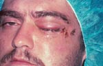
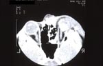
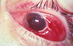
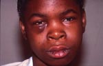
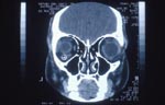
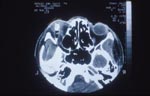
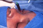
Canalicular laceration and nasolacrimal duct destruction secondary to facial fractures and lid trauma may be identified with lacrimal probing and irrigation.
Treatment
The majority of adnexal injuries may be treated with monitored anesthesia, augmented nerve blocks, and infiltrative anesthesia. During surgery, the first priority is to establish the extent of the injury. All wounds should be explored to detect deep orbital penetration, foreign bodies, or occult injury to the globe. Only devitalized tissue must be debrided. Also, it is important to minimize removal of tissue when revising ragged wound margins. All foreign bodies must be removed because retained foreign bodies can lead to chronic infection or abscess. Small fragments may spontaneously extrude for months after the initial injury.
Once the wound has been completely explored, consideration is given to the various components of the eyelid, including the anterior lamella, posterior lamella, and levator complex.
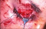
An anterior lamellar repair, which includes skin and orbicularis, results from shearing forces of the skin and orbicularis. Orbicularis contraction may falsely enlarge the superficial defects. The key to wound closure is to identify landmarks to reapproximate (Slide 9, Slide 10, and Slide 11). Wound margins should be trimmed of ragged edges or devitalized tissue until free bleeding occurs. Undermining of tissue with little to no tension is important. Accurate approximation of the muscle tears avoids wide scars. The eyelid skin is closed with 6-0 mild chromic, 6-0 silk, or 6-0 nylon sutures. These sutures should be removed after 4 to 6 days to avoid suture cysts and tunnels. The closure of larger areas of tissue loss is individualized and depends on the location and extent of injury. Care must be taken not to shorten the anterior lamella to cause lid retraction and lagophthalmus. Techniques of laterally and medially based advancement flaps are helpful with incisions made in the eyelid crease and incisions parallel with the lid margin; flaps advance medially and laterally. Sometimes, however, full thickness postauricular skin grafts, adjacent pedicle flaps, or advancement flaps may be required to maintain the integrity of the eyelid and protect the globe.
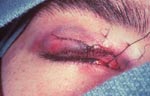
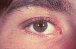
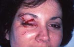
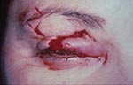
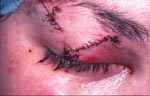
Meticulous closure of lacerations of the eyelid margin is essential to achieve correct anatomic repair and minimize postoperative complications. Failure to achieve these goals can lead to notching of the lid margin, poor contour of the lid, and lagophthalmus with exposure keratitis. What appears to be a small anterior eyelid laceration may have a more extensive posterior lamellar component. Irregular, angulated marginal edges should be squared off in the anterior posterior plane of the lid. In injuries with a small partial loss of tarsus, attempted primary closure without wound edge modification may lead to wound dehiscence or notching. The entire vertical portion of the tarsus must be removed corresponding to the length of the defect. Care must be taken to ensure that the tarsal plate excision is perpendicular to the lid margin. Vertical skin removal should not cross the upper eyelid crease because incisions more superior can result in unsightly scars (Slide 12, Slide 13, Slide 14, and Slide 15).
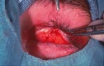
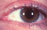
Marginal lacerations are closed using 6-0 silk sutures passed through the tarsal plate and
exiting the meibomian gland orifices at 1.5 mm to 2 mm from the wound edge and at the anterior and
posterior lash line.2,3 The tarsal plate is closed using a 6-0 Vicryl suture placed through the
anterior approach at 90% depth so as not to abrade the cornea. Three to four sutures are placed in
the upper tarsus and two sutures are placed in the lower tarsus in addition to the marginal repair.
The long arms of the marginal suture are tied beneath the first skin suture so that the knots do
not abrade the cornea. The marginal sutures should be removed in 10 to 12 days; earlier removal
can lead to wound separation and notching. In children, 6-0 to 7-0 Vicryl sutures can be used at
the margin and spontaneously absorb.
When tissue loss is too significant for primary closure, horizontal relaxation of the lid is required.
This can be achieved with a lateral canthotomy and cantholysis of the superior or inferior crux of
the lateral canthal tendon (Slide 16).
If further anterior lamellar tissue is required, a modified
Tenzel semicircular flap can be created.4 For defects with greater tissue loss, a Hughes tarsoconjunctival
flap for the lower lid and a Cutler-Beard composite flap for the upper lid can be used.
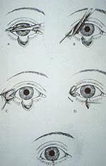
If the orbital septum and the upper lid are disrupted and preaponeurotic fat is exposed, the wound should be explored, and the levator complex should be identified. Injury of the levator aponeurosis or levator muscle should be repaired at the time of the primary wound closure. The original wound may require extension or a separate superior lid crease incision to adequately reexplore the levator complex. When these steps are required, it is most preferable to perform under attended local anesthesia as intraoperative patient cooperation for lid position maximizes a good outcome. Traumatic dehiscence of the levator muscle or aponeurosis is repaired by suturing the levator to the mid tarsal plate with a 6-0 silk or 6-0 nylon suture. Care should be taken not to place the sutures full thickness through the tarsus to avoid corneal abrasion. The placement of the affected lid is 1 mm to 1.5 mm higher than the normal side, as the eyelid will fall 1 mm to 1.5 mm once orbicularis tone returns and the effect of epinephrine wears off (Slide 17, Slide 18), Slide 19, Slide 20, and Slide 21).
References
- Gossman DM, Berlin JA. Management of acute adnexal trauma. In: Steward WB, ed. Surgery of the Eyelid, Orbit and Lacrimal System. American Academy of Ophthalmology; 1993(1):170-185.
- Cepela MA, Kulwin DR, Levine MR. Management of ocular adnexal trauma. In: Nesi FA, Lisman RD, Levine MR, eds. Smith's Ophthalmic Plastic and Reconstructive Surgery. St. Louis, Mo: CV Mosby; 1998:176-194.
- Nesi FA. Lid lacerations. In: Levine MR, ed. Manual of Oculoplastic Surgery, 2nd ed. Boston, Mass: Butterworth-Heinemann; 1996:29-32.
- Tenzel RR, Stewart WB. Eyelid reconstruction by the semicircle flap technique. Ophthalmology. 1998;85:1164-1168.