Introduction
When a lesion is to be resected, complete excision can be performed in a variety of techniques. Mohs resection is usually worthwhile for recurrent lesions, extremely invasive, large lesions or tumors involving the media canthal/canalicular system. Otherwise, resection with frozen section margin control is appropriate. Repair of the defects obviously depends on the size, location, and degree of eyelid/cheek laxity.
Reconstruction of the defect is determined by grasping the cut edges and attempting to pull them together. If they can be approximated primarily, then direct closure can be performed. If the edges leave a gap 3 mm or less, then a lateral canthotomy with inferior cantholysis should be adequate for repair. If the defect is more than 3 mm with the edges pulled together, then a more advanced reconstruction will be necessary.
Primary Repair
If the lateral release is necessary, this is to be performed initially. A standard lateral canthotomy and inferior cantholysis are performed. At this point, direct closure is performed. This can be best accomplished by first placing a 6-0 nylon suture at the grey line; this approximates the lid and tarsus. At this stage, the tarsus is reapproximated with 5-0 Vicryl interrupted sutures. A 6-0 nylon suture is then placed at the lash line, reapproximating the lid margin. The subcuticular tissue and skin edges can then be reapproximated with 6-0 Vicryl and 6-0 nylon, respectively.1
Tenzel Semicircular Flap Repair
This technique can be used for upper or lower eyelid reconstruction for defects involving 50% to 60% of the medial or central eyelid. This technique cannot be used for lateral lid/canthal or larger defects.
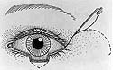
A semicircle directed inferiorly for upper lid defects or superiorly for lower lid reconstruction is used. The area to be incised is marked with a skin marker, starting at the lateral canthus and making the semicircle approximately 2 cm. The skin and orbicularis is then incised and undermined (Slide 1). A lateral canthotomy and cantholysis is then performed with sharp dissection (Slide 2). At this stage, the flap is mobilized medially allowing the lateral cut edge to be reapproximated. The orbital septum frequently needs to be incised to release the flap for appropriate repair.
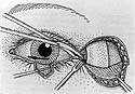
The lid defect is then repaired in the same manner as noted under Primary Repair. The lateral canthus is then reformed in a multilayer fashion. The deep tissue is plicated to the lateral periosteum with 5-0 Vicryl in a buried interrupted fashion. The orbicularis muscle, subcuticular tissue, and skin edges are then reapproximated with 5-0 Vicryl, 6-0 Vicryl, and 6-0 nylon, respectively.2
Tarsoconjunctival Advancement Flap (Hughes Procedure)
Tarsoconjunctival flaps can be used to reconstruct large defects of the lower eyelids. The tarsoconjunctival graft provides the internal lamella of the graft, and either a full-thickness skin graft or mobilization of a skin flap provides the anterior lamella.
The upper and lower eyelids are infiltrated with an anesthetic solution. The edges of the lower eyelid defect are grasped with forceps and brought together to measure the size of the defect. A 4-0 silk traction suture is placed through the margin of the ipsilateral upper eyelid. The upper eyelid is everted over a Desmarres retractor. A horizontal incision is made 3 mm inferior to the lid margin through tarsus and is equal in length to the lower eyelid defect. The tarsus and conjunctiva are grasped with a forceps and vertical incisions are made superiorly into the superior fornix. The dissection is carried sufficiently far superiorly to allow the tarsoconjunctival flap to rest in the lower lid defect. Align the superior edge of the tarsal graft with the lower eyelid margin. Suture the tarsal graft to the lower eyelid tarsus with 6-0 Vicryl sutures. The lower eyelid retractors and conjunctiva are sutured to the inferior aspect of the tarsal graft with a running 6-0 Vicryl suture (Slide 3). A myocutaneous advancement flap is created making vertical incisions through the skin and dissecting inferiorly. This flap can than be mobilized superiorly to cover this defect (Slide 4). The superior border of the skin flap is sutured to the tarsoconjunctival flap at the superior border of the tarsus with interrupted 6-0 Vicryl sutures (Slide 5). Alternatively, a free skin graft may be used in place of the advancement flap.
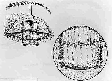
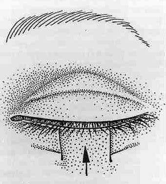
The conjunctival bridge is opened in 6 weeks. The graft is severed 1 mm above the desired lid margin. The resulting lid margin is sculpted smooth. The remaining conjunctival stump is trimmed to avoid irritation and vertical shortening of the upper eyelid.
Lower Eyelid Bridged Advancement Flaps in Upper Eyelid Reconstruction
(Cutler-Beard Procedure)
Lower eyelid advancement flaps can be used to reconstruct large defects of the upper eyelid. A marking pen and caliper are used to make a horizontal line 4 mm inferior to the lower eyelid margin. The horizontal width of the wound defect should be marked on the lower eyelid tarsus. A bone plate is placed posterior to the lower eyelid. A #15 Bard Parker scalpel blade is used to make a horizontal incision through the skin, at a level approximately 4 mm inferior to the lower eyelid margin (Slide 6). The lower eyelid is everted over a Desmarres retractor and an incision is made 4 mm inferior to the lower eyelid margin. This incision is connected to the previously made skin incision with Steven tenotomy scissors. Full thickness vertical incisions are made at each end of the horizontal incisions down to the level of the inferior fornix. This allows the lower eyelid flap to be mobilized under the lower eyelid margin bridge and into the upper eyelid defect. The conjunctiva and lower eyelid retractors are sutured to conjunctiva, Müller muscle, and levator aponeurosis of the upper eyelid with multiple interrupted 6-0 Vicryl sutures (Slide 7). An ear cartilage graft can be harvested and cut to approximate the size of the tarsal defect. The cartilage graft can be sutured superiorly to levator aponeurosis with multiple interrupted 6-0 Vicryl sutures. The skin-muscle layer is closed with interrupted 6-0 silk sutures (Slide 8). The lower eyelid advancement flap is opened after 6 to 8 weeks. A grooved dissector is placed under the flap and the flap is incised with a #15 Bard Parker scalpel blade. The lower edge of the lower eyelid bridge is denuded and the skin and orbicularis muscle of the advancement flap is re-approximated to the lower eyelid bridge.
References
- American Academy of Ophthalmology, Monographs 8. Surgery of the Eyelid, Orbit and Lacrimal System. 1993; Volume 1.
- Levine MR. Manual of Oculoplastic Surgery. New York, NY: Churchill Livingstone; 1988.