Introduction
Evisceration has been an important technique in ophthalmology for almost 200 years. According to Meltzer it was first described by Bear in 1817, who used the procedure after an iridectomy for acute glaucoma was complicated by an expulsive hemorrhage. Mules, in 1884, described a hollow glass sphere that was introduced into the scleral cavity after evisceration.1 Evisceration offers a more functional and cosmetically acceptable orbit compared with enucleation. Evisceration causes less disturbance in the orbital tissue and the physiologic dynamics of muscle function; and, the orbital volume can be maintained close to its original state.
Timothy, Freilich, and Linberg surveyed 114 board-certified ocularists in the United States regarding their experience with evisceration versus enucleation in terms of final function, cosmesis, and complications.2 Survey questions included:
- Which procedure provided the best final ocular motility?
- Enophthalmos and/or deep superior tarsal sulcus deformity is more common with which procedure?
- Orbital implant migration is more common with which procedure?
- Implant exposure and infection is more common with which procedure?
- Which procedure provides the best cosmetic results?
- Which procedure would be your first choice for a patient who requires removal of an eye?
Eighty-five ocularists responded to the survey, 82% of whom reported that the best ocular motility was obtained with evisceration. Eighty-two percent of the ocularists reported that the best overall final cosmesis was achieved by evisceration. Enophthalmos and a deep superior tarsal sulcus deformity were negligible with evisceration. The ocularists believed that orbital implant migration and exposure were significantly more common with enucleation than evisceration (92% and 58%, respectively). Finally, according to 92% of the ocularists, evisceration would be their first choice for a patient who required eye removal.
Evisceration, however, has several disadvantages including the possible dissemination of an unsuspected tumor, inadequate pathologic material, and inadequate volume if the eye is phthisical, and the risk of sympathetic ophthalmia, especially in penetrating injuries.
Incidence of Sympathetic Ophthalmia
The incidence of sympathetic ophthalmia is unknown. During the past 50 years, rates of occurrence for penetrating ocular injury ranged from 0.28% in Allen's series to 1.9% in Liddy's and Stuart's series, as stated by Albert and Diaz-Rohena.3 The incidence of sympathetic ophthalmia after intraocular surgery over the same time ranges from 0.015% in Allen's series to 0.05% in Liddy's and Stuart's series. Marak estimated that sympathetic ophthalmia is 30 to 50 times more frequent after trauma than after surgery.4 Therefore, if evisceration is a more functional and cosmetically acceptable orbit compared with enucleation, are we doing our patients an injustice by recommending evisceration over enucleation when direct exam or B scan ultrasound rules out unsuspected tumors and adequate scleral volume is present? Do we heed to Dr. Frederick Blodi's caveat of 1963 as stated by Bilyk, "whether we want to take the risk of this rare, but certainly severe, complication (sympathetic ophthalmia) against an alleged cosmetic advantage is up to the conscience of every ophthalmologist."5
In 1960, Berens reported 230 cases and in 1966, Strampelli and Valvo reported 1,000 cases of evisceration without incidence of sympathetic ophthalmia, as stated by Meltzer.1 Ruedemann reviewed his experience from 1947 to 1962. Of the 506 eviscerations, there were no cases of sympathetic ophthalmia.6 Conversely, in more modern times, Green et al, in 1972, reported four cases of sympathetic ophthalmia after evisceration.7 The number of evisceration specimens reviewed was not provided. Since 1972, to our knowledge, no cases of sympathetic ophthalmia following evisceration have been reported.
Methods
My coworkers at the Mt. Sinai Medical Center and University Hospitals of Cleveland and I reviewed our experience with evisceration from 1980 through 1996 and survey data from the American Society of Ophthalmic Plastic and Reconstructive Surgery (ASOPRS), the Uveitis Society and the Eastern Ophthalmic Pathology Society.8
Results
The Mt. Sinai Medical Center's and University Hospitals of Cleveland's experience document 190 cases including 100 enucleations and 90 eviscerations. Fifty-one of the 90 patients who had undergone evisceration were asked to return for a follow-up visit or had adequate chart notations. In addition, pathologists from University Hospitals of Cleveland and the Mt. Sinai Medical Center reviewed the intraocular contents for evidence of sympathetic ophthalmia. There was no evidence of clinical sympathetic ophthalmia in the 51 patients and no granulomatous uveitis was identified histopathologically.
Sixty-one percent of the members of the ASOPRS responded to a questionnaire. Of the 3,035 cases of enucleations and eviscerations performed in 1996, 72.3% were enucleations and 27.7% were eviscerations. Two physicians anecdotally recalled seeing cases of sympathetic ophthalmia occurring in evisceration not associated with trauma and three physicians recalled three cases occurring in an evisceration resulting from trauma. However, no physician had documented evidence. Therefore, despite the fact that evisceration is anatomically a better procedure, only 27.7% of 3,035 cases in the ASOPRS survey were eviscerations. Obviously, there has been a longstanding bias against this procedure because of the fear of sympathetic ophthalmia and also the chance of a patient's harboring an intraocular tumor.
Survey data from the American Uveitis Society and the Eastern Ophthalmic Pathology Society revealed from modest numbers evisceration rates of 8.1% of 86 cases and 5.6% of 570 cases, respectively.
Ruedemann reviewed 47 cases describing sympathetic ophthalmia after evisceration, most of which appeared in the literature before 1917, which has been alluded to in many articles on evisceration and sympathetic ophthalmia. He concluded that many cases had insufficient clinical histories or clinical description. Many cases were inconclusive as to the completeness of removal of uveal contents. Description of the uninjured eye after surgery was often deficient. In none of the reports was the entire evisceration specimen studied histopathologically.6
Discussion
Sympathetic ophthalmia is a rare condition that occurs after injury to one eye followed by a latent period and subsequent development of bilateral granulomatous panuveitis in both the injured and uninjured globes. Characteristically, there is a diffuse lymphocytic infiltration of the uveal tract with non-necrotizing granulomas. Involvement of the choriocapillaris retina and optic nerve, which at one time was believed to be uncharacteristic features, occurs frequently. Granulomas (Dalen-Fuch nodules) between the Bruchs membrane and retinal pigment epithelium can be seen clinically. In a review of 105 cases seen at the Massachusetts Eye and Ear Infirmary, 65% of sympathetic ophthalmia cases occurred between 2 weeks and 3 months after injury and 70% occurred before 3 months. Exceptions occur with reports of onset as briefly as 5 days after injury to as long as 42 years after injury.3
The cause of sympathetic ophthalmia is not clearly understood, but theories include hypersensitivity to retinal pigment or uveal protein, an infectious agent, or sensitivity to retinal S antigen. Rao and coworkers induced bilateral sympathetic uveitis in rabbits after subconjunctival injection of bovine retinal S antigens. They noted that an injection of retinal S antigen into the globe did not have the same effect as a subconjunctival injection. From these findings, they postulated that when uveoretinal antigen is presented to the lymphatic system via the conjunctiva, the immune system responds by developing a disease similar to sympathetic ophthalmia in rabbits. However, no retina S antigen has been detected in patients with sympathetic ophthalmia.9
It appears that cases described before 1917, despite Ruedemann's article, have had a strong influence over the years and to the results of the ASOPRS survey in which enucleation was still preferred over evisceration by 2.5:1 (72.5% versus 27.7%). In 1972, Green and colleagues published four cases of sympathetic ophthalmia after evisceration; however, no recent cases have been reported. Finally, in my small series of 51 cases, both nonpenetrating and penetrating injuries, there was no evidence of sympathetic ophthalmia. Therefore, it appears that sympathetic ophthalmia is a rare disease and is infrequently associated with evisceration. My recommendation is that it should be strongly considered over enucleation when intraocular tumor is ruled out and scleral volume is adequate.
Surgical Technique
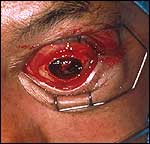
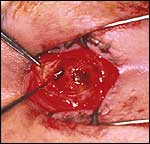
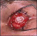
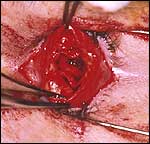
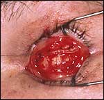
Evisceration can be performed easily under local anesthesia with sedation in less than 1 hour. A 360° peritomy is performed, adequately dissecting the conjunctiva and Tenon's capsule. The cornea is excised and the scleral contents are removed with a large chalazion curette (Slide 1, Slide 2). Half-strength peroxide is used to facilitate uveal and retinal removal, as well as coagulate small vascular channels. Once all uveal and retinal tissue has been removed, the scleral pouch may be implanted with a polymethylmethacrylate sphere, hydroxyapatite implant, or Medpor (Porex Surgical, Inc., Newnan, Ga) implant (Slide 3). Expansion sclerotomies are necessary to enhance the vascularization of the implant (hydroxyapatite or Medpor), if a peg is to be inserted later or to accommodate an 18-mm to 20-mm implant if the scleral pouch is inadequate in volume10 (Slide 4). Absolutely no tension should be on the scleral envelope over the implant to avoid late extrusion. The sclera is closed with a 5-0 Vicryl suture consisting of a few mattress sutures to take the tension off the incision followed by a running 5-0 Vicryl in a baseball fashion (Slide 5). The conjunctiva-Tenon's complex is then sutured with a 5-0 Chromic cat gut suture, taking bites of sclera to facilitate a firm closure over the scleral incision (Slide 6). No conformer is placed in the eye as the fornices have not been disturbed and avoidance of tension over the suture line is important. Temporary 6-0 silk suture tarsorrhaphies are placed medially and laterally and a firm patch is applied for 4 days to reduce postoperative edema and conjunctival chemosis (Slide 7). Additionally, systemic steroids in the form of a steroid Dosepak is given to further reduce postoperative edema that is frequently associated with evisceration. A prosthetic eye is fit at a minimum of 6 weeks when the suture line looks well-vascularized and healed (Slide 8).
References
- Meltzer MA, Schaefer DP, Della Rocca RC. Evisceration. In: Della Rocca RC, Nesi FA, Lisman RD, eds. Smith's Ophthlamic Plastic and Reconstructive Surgery. Vol. 2, St. Louis: CV Mosby. 1987:1300-1307.
- Timothy NH, Freilich DE, and Lindberg JV. Evisceration versus enucleation from the ocularist's perspective. Ophthalmic Plast Reconstr Surgery. 2003;19(9):417-420.
- Albert DM, Diaz-Rohena R. A historic review of sympathetic ophthalmia and its epidemiology. Surv Ophthalmol. 1989;34:1-14.
- Marak GE. Recent advances in sympathetic ophthalmia. Surv Ophthalmol. 1979;24:141-156.
- Bilyk JR. Enucleation, evisceration and sympathetic ophthalmia. Curr Opin Ophthalmol. 2000;11:372-385.
- Ruedemann AD Jr. Sympathetic ophthalmia after evisceration. Trans Am Ophthalmol Soc. 1963;61:274-314.
- Green WR, Maumenee AE, Sanders TE, Smith ME. Sympathetic uveitis following evisceration. Transactions of the American Academy of Ophthalmology and Otolaryngology. 1972;76:625-44.
- Levine MR, Pou CR, Lash RH. Evisceration: Is sympathetic ophthalmia a concern in the new millennium? Ophthalmic Plast Reconstr Surgery. 1999;15(1):4-8.
- Rao NA, Robin J, Hartmann D et al. The role of the penetrating wound in the development of sympathetic ophthalmia. Arch Ophthalmol. 1983;101:102-104.
- Stephenson CM. Evisceration of the eye with expansion sclerotomies. Ophthalmic Plast Reconstr Surgery. 1987;3:249-251.
Posted April 2004