Introduction
Over the years, many classifications of ptosis have been developed. However, recently with refinements in the diagnosis of ptosis, along with improved anatomic evaluation and study of the pathophysiology of certain types of ptosis, a better understanding of ptosis has developed.
Ptosis may be classified into congenital and acquired types. Congenital ptosis is a developmental dystrophy of the levator muscle of an unknown cause (Slide 1). The condition is usually sporadic, but it may be hereditary. Congenital ptosis may be simple with the defect isolated to only the levator muscle or with superior rectus muscle weakness. Congenital ptosis also includes blepharophimosis syndrome, which is hereditary and which, in addition to ptosis, may include varying degrees of telecanthus, epicanthus inverses, phimosis, and ectropion of the lower eyelids. Congenital ptosis also includes the Marcus Gunn jaw winking syndrome, which is caused by abnormal levator innervation rather than a striated muscle fiber deficiency.
Acquired ptosis is best classified by Beard1; however, other authors have added subclassifications, including aponeurotic, neurogenic, myogenic, mechanical, traumatic, and pseudoptosis.
Aponeurotic Ptosis
Aponeurotic ptosis is considered to be the most common form of acquired ptosis (Slide 2 and Slide 3). This condition may occur as the result of localized or generalized disinsertion or dehiscence of the aponeurosis from the tarsal plate. Clinical features of this aponeurotic ptosis may include a high or absent lid crease, adequate levator function, and thin eyelid tissue. Typically, aponeurotic ptosis occurs as a result of aging, including microinfarction of collagen bundles and other tissues within the aponeurosis.2 Occasionally, levator fibers may be replaced with bundles of adipose tissue. Typically, this type of ptosis is found in older patients; however, younger patients may have aponeurotic ptosis, as well. Aponeurotic ptosis may result from blunt trauma, ocular surgery, and contact lens wear (Slide 1).
Neurogenic Ptosis
Neurogenic ptosis may result from a multitude of neurologic disorders (Slide 4 and Slide 5), such as third cranial nerve palsies, Horner syndrome, ophthalmoplegic migraine, and multiple sclerosis. In third cranial nerve palsies, lesions of the superior division of the ocular motor nerve cause ptosis along with deficits of adduction, elevation, and depression of the eye. The pupil may be normal or dilated. Lesions of the superior division of the third nerve involve only the superior rectus and levator muscles with resultant ptosis and decreased levator function. Acquired third nerve palsy may be a neurosurgical emergency if the pupil is involved, especially due to a compressive lesion such as an aneurysm. Other causes of third nerve palsy include infection, cavernous sinus thrombosis, fistulas, tumors, and contiguous paranasal sinus disease.
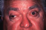
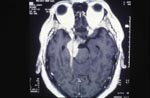
Horner syndrome consists of mild ptosis, pupillary miosis, lower lid ptosis, and at times, facial anhidrosis. This is due to decreased sympathetic tone to Müller muscle. Horner syndrome may be caused by a lesion in any of the three levels of neurons that carry sympathetic impulses to the eye.
Ophthalmoplegic migraine is a rare cause of third nerve palsy. Patients are typically young adults or children, often with a history of migraine headaches. Fifty percent of these patients have a family history of migraines. The mechanism of ophthalmoplegic migraine is unclear; however, it is believed that dilatation of the carotid artery may play a role. Compression of the third nerve usually occurs as the patient begins to complain of severe headaches, followed by ptosis and extraocular muscle paralysis. The pupil is involved in the majority of patients.
Multiple sclerosis is a disease of undetermined cause that occurs when multifocal demyelinization occurs within the white matter of the central nervous system. Multiple sclerosis occurs most often in young adults between 20 to 40 years of age. Women are slightly more often affected than men. The most common presenting symptoms are weakness, numbness, and paresthesias of the extremities.3
Myogenic Ptosis
Myogenic ptosis may sometimes overlap with ptosis of neurogenic etiology, including myasthenia gravis, chronic progressive external ophthalmoplegia, and myotonic dystrophy. Myasthenia gravis is an autoimmune disease in which antibodies to acetycholine receptor sites of neuromotor end plates impair the transfer of acetycholine from neuron to muscle. In this situation, muscle fatigue and weakness occur. Myasthenia gravis is more common in women, although it affects all races and both sexes (Slide 6 and Slide 7). Approximately 85% to 90% of patients with myasthenia gravis present with localized ocular signs, although the disease may be generalized. Ptosis is the most frequent unilateral manifestation but may be unilateral or bilateral in nature. Symptoms worsen as the day progresses or with significant fatigue.
Chronic progressive external ophthalmoplegia is a disorder resulting from a defect in the mitochondrial function in which the extraocular and periocular muscles progressively weaken. It is mostly autosomal dominant inherited, although sporadic cases may occur. Chronic progressive external ophthalmoplegia involves the extraocular and periocular muscles. The iris and ciliary body muscles are not involved. Typically, the disorder begins in childhood or adolescence and progresses slowly over 40 to 50 years. Bilateral ptosis is usually the first symptom noted and subsequently other body muscles may be affected. At an advanced stage, the eyes may be totally immobile (Slide 8). Kerns and Sayre described a form of chronic progressive external ophthalmoplegia in which retinitis pigmentosa, chronic progressive external ophthalmoplegia, and complete heart block occurred. Red ragged fibers in muscle biopsies are suggestive of mitochondrial diseases, as is the presence of spongy degeneration of the central nervous system. Patients with Kerns-Sayre syndrome require serial electrocardiograms to monitor for heart block.

Oculopharyngeal dystrophy is a myopathic dystrophy that begins after age 40 and consists of slowly progressive ptosis with dysphagia and dysarthria, which results from pharyngeal dystrophy. In some patients, the extraocular muscles, shoulder muscles, and pelvic girdle muscles may be involved. Inheritance is autosomal dominant and may have a French-Canadian ancestry.
Myotonic dystrophy is a genetic disease with an autosomal dominant inheritance and may be associated with varying degrees of bilateral ptosis, orbicularis weakness or lid closure, extraocular muscle dysfunction, poor blinking, and dry eyes (Slide 9). Additional findings may include chromatic cataracts, testicular atrophy, frontal baldness, and first degree heart block.

Traumatic Ptosis
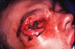
Traumatic ptosis may occur from blunt or sharp trauma to the levator muscle or aponeurosis (Slide 10 and Slide 11). Injury to the periocular tissues may cause edema, hemorrhage, or infection, which may also cause ptosis. Orbital fractures and foreign bodies may impair levator function either directly or indirectly. Ptosis may be shown to occur from injury to the levator aponeurosis as a result of ocular surgery including cataract and trabeculectomy surgery in up to 10% to 20% of cases.
Mechanical Ptosis
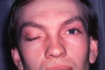
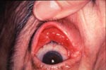
Mechanical ptosis may be caused by eyelid tumors, scarring, or blepharochalasis (Slide 12 and Slide 13). Lid tumors may cause ptosis as the result of increased weight on the upper lid or disinsertion by stretching the levator. Cicatricial ptosis may be caused as a result of trauma or tumors.
Pseudoptosis
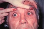
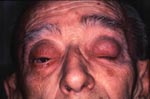
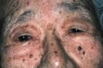
Pseudoptosis may appear with apparent ptosis when true eyelid ptosis is not actually present. Pseudoptosis may be seen in lack of posterior lid support, such as in an anophthalmic eye or posttraumatic eye, or in the presence of an orbital prosthesis. A hypotropic eye may appear to have a droopy eyelid, although this normalizes when the patient is asked to fix on an object with the hypotrophic eye. Dermatochalasis may also cause the appearance of a ptotic eyelid, although when lifting the skin off the eyelid, the margin typically returns to a normal position (Slide 14).
Evaluation of Ptosis
History
It is important to determine whether a case of ptosis is congenital. Generally, in children,
this is not a difficult task as parents who bring children for care usually know whether the
children were born with a ptosis or whether they birth trauma caused it. Older patients with
ptosis are not sure of the time of onset. Baby and childhood pictures are helpful in determining
at what stage of life the patient had the ptosis.4 A family history of other members of the
family having ptosis is important.
A history of change is important to determine whether the condition is improving, worsening, or remaining stationary. Eyelid or globe surgery, as well as contact lens wearing, is also relevant.
Physical Examination
The examination of a patient with ptosis is the most important factor in determining the type
of surgical approach. Careful examination and palpation of the eyelids and orbits help determine
the extent of asymmetry and proptosis, if present. In addition, palpation of the eyelids and
orbital rims may detect orbital masses causing a mechanical type ptosis on the lids. Everting
the upper eyelid may also determine subconjunctival masses. Exophthalmometry may be helpful to
assess globe position and possible presence of orbital masses or enophthalmos. The position of
the eyebrows, as well as head position, should be noted because the presence of ptosis may
cause a compensatory posterior head posture.
Levator function. By definition, levator function is the distance that the eyelid moves in millimeters from a maximum down gaze to a maximum up gaze without the eyebrow elevated. It is the most important factor in the correct choice of surgical procedure. Levator function may be classified as excellent (13 mm to 15 mm), very good (10 mm to 13 mm), good (8 mm to 10 mm), fair (5 mm to 7 mm), and poor (4 mm or less).
The amount of ptosis. The amount of ptosis in the unilateral case is measured as the distance between the upper and lower eyelid margins with the brow held in a relaxed position. The difference represents the amount of ptosis. The lower lid must be in normal position. In bilateral ptosis, the amount of ptosis is determined by the marginal reflex distance (MRD), that is, the light reflex from the cornea to the upper lid. The normal MRD is +4 mm to +4.5 mm with a normal eyelid position being 2 mm to 1.5 mm below the superior limbus. Ptosis is classified as mild (2 mm), moderate (3 mm), and severe (4 mm). It is important to check for ptosis in down gaze.5 The fissure in down gaze is an indication of the ability of the levator muscle to relax. In congenital ptosis, the upper lid will lag behind the normal lid. In acquired ptosis, the lid is at a lower level than the normal lid.
Prominence of lid fold is a strong indicator of the amount of levator function present.
Position of the lid fold. Accurate comparison of normal and abnormal lid folds must be determined so that the abnormal lid fold can be matched to the normal lid at the time of surgery.
Lash position. In severe ptosis, lash ptosis is also frequently present and can be improved at the time of surgery.
The amount of upper lid skin. Excess upper lid skin will frequently be present in patients undergoing large levator resection or in adult patients having ptosis correction. If this is suspected, a blepharoplasty along with ptosis surgery is appropriate.
Associated lid anomalies. Many syndromes such as blepharophimosis will have associated lid anomalies that may be corrected by surgery at the same time the ptosis is corrected or at a later date.
Synkinetic lid movement. A classic example of synkinetic lid movement is Marcus Gunn winking jaw ptosis caused by an abnormal fifth cranial nerve innervation of the lid. Other abnormal innervations such as aberrant regeneration of the third and seventh nerves must be considered.
Visual acuity must be obtained because a significant number of children with ptosis will have amblyopia secondary to anisometropia and esotropia.
Fusion. A rapid worth four dot-test for Titmus fly screening exam confirms normal function.
Extraocular muscle evaluation. Many patients with ptosis will have associated extraocular muscle imbalances or limitation, such as patients with chronic external ophthalmoplegia or myotonic dystrophy. Significant limitation of extraocular motility along with fair orbicularis tone will influence the final amount of levator surgery to avoid lid lag, lagophthalmus, and exposure keratopathy.
Bell's phenomenon is the prognostic indicator of possible exposure problems.
Tear production. A Schirmer test is important in adult-acquired ptosis to avoid complications of dry eye syndrome. A basic secretion test with topical anesthetic followed by a Schirmer strip in the inferior cul-de-sac for 5 minutes will determine basic secretion. Ten millimeters or more of moisture should be present on the filter paper strip. In addition, evaluation of tear film breakup time is also helpful (greater than 8 seconds indicates normal tear breakup time).
Corneal sensation test is usually not indicated unless a neurologic cause is suspected or a history of Herpes simplex or Herpes zoster is obtained.
Diagnostic Testing
Tensilon test. A Tensilon test is indicated in all patients when myasthenia gravis is suspected.
The test is performed by giving 2 mg (0.2 mL) intravenous as a test dose to ensure the patient
does not have a reaction, followed 30 seconds later by 8 mg (0.8 mL), and observing the patient's
lid position. If the patient has myasthenia gravis, there will be a significant elevation in lid
position within 1 to 5 minutes after giving the test dose. Cholinergic reactions are infrequent,
but injectable atropine must be available as an antidote.
Neosynepherine test. A Neosynepherine (phenylephrine HCl, Winthrop) test should be considered for all adult patients with acquired ptosis. This test will help determine if, in fact, a unilateral ptosis is a bilateral ptosis based upon Hering law. Neosynepherine 2.5% or 10% may be used and is instilled into the cul-de-sac of the abnormal eye. If the ptotic lid rises at least 2 mm to 3 mm within 5 minutes, then the patient is a candidate for a tarsal-conjunctival resection surgery or a conjunctival-Müller muscle resection surgery.
Photography
Photographs should be taken of each patient preoperatively for planning, medicolegal reasons, and insurance purposes.
Consultation
The cause of acquired ptosis can be difficult to determine. Therefore, interaction with an internist, endocrinologist, neurologist, or neurosurgeon may be helpful.
References
- Beard C. Ptosis. St. Louis, Mo: CV Mosby; 1969.
- Dortzbach RK, Satula FC. Involutional blepharoptosis: A histopathologic study. Arch Ophthalmol. 1998;98:2045.
- Siddens JD, Nesi FA. Acquired ptosis: Classification and evaluation. In: Nesi FA, Lisman RD, Levine MR, eds. Smith's Ophthalmic Plastic and Reconstructive Surgery. St. Louis, Mo: CV Mosby; 1998:379-393.
- Beard C. Examination and evaluation of the ptosis patient. In: Nesi FA, Lisman, RD, Levine MR, eds. Smith's Ophthalmic Plastic and Reconstructive Surgery. St. Louis, Mo: CV Mosby; 1998:339-344.
- Dryden RM, Kahanic DA. Worsening of blepharoptosis in down gaze. Ophthalmic Plastic Reconstruct Surg. 1992;2:126.