Introduction
Presbyopia is the gradual, age-related loss of accommodation. Unlike other refractive errors, presbyopia affects everyone at some point in his or her lifetime. Understanding the cause of this inevitable phenomenon requires an understanding of the underlying concept of accommodation.
Accommodation
Accommodation is the process that allows the eye to change its focus from far to near vision. During accommodation, the optical power of the eye increases. The crystalline lens produces this optical change, thus making it pivotal to the accommodative process. The ciliary muscle is the driving force in producing the accommodative change in the lens.
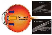
There are several theories on the mechanisms of accommodation. The most widely accepted theory was proposed by Hermann von Helmholtz in 1855. Helmholtz postulated that in the unaccommodative state when the ciliary muscle is relaxed, there is resting tension on the zonular fibers at the lens equator that keeps the lens in a flattened, unaccommodated state (Slide 1). When accommodation occurs, the ciliary muscle contracts, releasing the resting zonule tension, which allows the lens to assume a more spherical shape, thereby increasing focusing power. The human crystalline lens is ectodermal and grows throughout life with a continuous production of new cells within the lens capsule, which causes the lens to increase in size, harden, and lose its elasticity. Under Helmholtz's theory of accommodation, presbyopia may be caused by hardening of the lens that prevents the necessary spherical lens change needed during accommodation.
A competing theory of accommodation was proposed by Ronald A. Schachar, MD, in 1994.1 Schachar postulated that in the unaccommodative state when the ciliary muscle is relaxed, there is decreased tension on the zonular fibers at the lens equator (Slide 1). During accommodation, the ciliary muscle contracts, which tightens the zonules, thus increasing the diameter of the lens with a steepening of the central lens, thereby increasing focusing power.2,3 Since the lens is ectodermal and its equator grows throughout life, the working distance between the lens and ciliary body decreases as age increases. This diminishes the effective force that the ciliary muscle can apply to the lens equator during accommodation. Under Schachar's theory of accommodation, presbyopia results from the loss of ciliary body effectiveness in increasing the lens diameter as the aging equator begins to crowd the ciliary body. Adrian Glasser, PhD, refuted Schachar's theory by reporting that, in Rhesus monkeys, the lens equator and ciliary body move away from the sclera during electrically or pharmacologically stimulated accommodation as seen with ultrasound biomicroscopy.4 Schachar's theory remains controversial and is currently under study in the United States.
In addition to the lens, the ciliary muscle is necessary for accommodation. A number of studies report relatively stable strength of the ciliary muscle with increasing age.5,6 Thus, many researchers presuppose the continued functionality and strength of the ciliary muscle to respond to accommodative stimuli.
Pseudoaccommodation
It has been observed that after implantation with standard monofocal intraocular lenses (IOLs) patients seem to have functional near vision without glasses. This effect is called pseudoaccommodation and is a result of either residual postoperative myopia, small pupil size resulting in increased depth of focus, or residual postoperative against-the-rule astigmatism.7-10
Additionally, in pseudophakic patients, forward movement of the IOL during accommodative effort of ciliary muscle constriction has been reported.11,12 The ciliary muscle, upon constricting, undergoes a redistribution of its mass, bulking posteriorly and impinging on the vitreous face peripherally as demonstrated on high-resolution magnetic resolution imaging (MRI) by Strenk and colleagues.13 This impingement on the peripheral vitreous face may cause an increase in vitreous pressure. In 1986, Coleman cannulized both the anterior chamber and the vitreous cavity in primates and demonstrated increased intravitreal pressure and decreased anterior chamber pressure during electrical stimulation of the ciliary body. This pressure differential may provide a potential explanation for the axial movement of an IOL during accommodative effort (Slide 2).
The CrystaLens
Cumming reported that silicone IOLs with plate haptics demonstrated a more posterior position within the capsular bag space compared with silicone multipiece looped lenses.14 Kammann and colleagues reported a more posterior position within the capsular bag space with silicone plate lenses with a 5-mm optic.15 At the American Society of Cataract and Refractive Surgeons 2000 meeting, Findl reported the results of a comparison of the forward movement of six IOLs using partial coherence laser interferometry. Plate lenses showed the most significant forward movement of the lenses tested.
In 1991, Cumming began the development of an IOL designed to exploit and maximize the axial movement of an IOL during accommodative effort. Together with Kammann, Cumming developed an IOL with plate haptics for a more posterior position in the capsular bag and hinged haptics to facilitate a maximal forward movement of the optic. Seven IOL designs have been investigated over the past 9 years. The first six designs demonstrated evidence of axial movement, but these lenses tended to dislocate anteriorly. The seventh and current design of this axial movement IOL is the AT-45 CrystaLens (C & C Vision, Mission Viejo, Calif.) (Slide 3 and Slide 4).
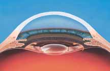
The AT-45 CrystaLens is a modified silicone, plate-haptic lens. The CrystaLens has a hinge at the junction of the haptic and optic to facilitate forward movement of the optic, and T-shaped polyimide haptics at the end of the plates. The lens is 11.5 mm from loop tip to loop tip and the length as measured from the ends of the plate haptics is 10.5 mm. The lens has a biconvex optic that is 4.5 mm in diameter.
Clinical Results
The AT-45 CrystaLens is currently
undergoing a Food and Drug Administration (FDA)-monitored, prospective,
multicenter clinical investigation at 13 investigation sites. The results of
the feasibility study and the initial phase study of the FDA clinical trial
were reported in 2001.16 The feasibility study reported on 28 eyes
in 14 patients and the initial phase study reported on 48 eyes in 48 patients
more than 3 months after implantation. Exclusion criteria included any ocular
pathology that could cause a reduction in visual acuity other than cataract.
Phacoemulsification cataract extraction was performed through a clear corneal
incision, with a capsulorrhexis of 4.0 mm to 6.0 mm, and the lens was inserted
through an enlarged 4.5-mm incision into the capsular bag. All patients
received topical atropine 1% at the end of surgery and on the first
postoperative day with the intent of paralyzing the ciliary muscle. This
facilitated centration and allowed fixation of the AT-45 CrystaLens to be
vaulted in a posterior position rather than in an anterior position in the
capsular bag during the early postoperative fibrosis period.
The main outcome measures were postoperative uncorrected distance, best-corrected distance, uncorrected near, distance-corrected near, and best-corrected near visual acuities. In the 48 clinical trial patients, 88% of patients achieved uncorrected distance visual acuity of 20/40 or better. All patients achieved best-corrected distance visual acuity of 20/40 or better; 96% of patients achieved uncorrected near visual acuity of 20/30 (J3) or better; 96% of patients achieved distance-corrected near visual acuity of 20/30 (J3) or better; and 100% of patients achieved best-corrected near visual acuity of 20/30 (J3) or better. The percentage of patients achieving 20/20 visual acuity, at distance or at near, was not reported.
In a feasibility study of 28 eyes in 14 patients, the results were reported using binocular visual acuity measurements. All patients achieved uncorrected distance visual acuity of 20/40 or better; 71% of patients achieved uncorrected distance visual acuity of 20/25 or better. All patients achieved best-corrected distance visual acuity of 20/25 or better. All patients achieved uncorrected near visual acuity of 20/30 (J3) or better; 64% of patients achieved uncorrected near visual acuity of 20/20 (J1) or better. All patients achieved distance-corrected near visual acuity of 20/30 (J3) or better; 93% of patients achieved distance-corrected near visual acuity of 20/20 (J1) or better. There have been no dislocations or complications with the AT-45 CrystaLens.
Fine, one of the principal investigators in the FDA-monitored, U.S. multicenter clinical trial of the AT-45 CrystaLens, reported his interim data on 82 patients who were at least 1 month postoperative. Ninety-two percent of patients achieved uncorrected distance visual acuity of 20/40 or better; 100% of patients achieved best-corrected distance visual acuity of 20/40 or better; 87% of patients achieved uncorrected near visual acuity of 20/30 (J3) or better; and 93% of patients achieved distance-corrected near visual acuity of 20/30 (J3) or better. In a historical comparison, Steinert and colleagues reported that 48% of patients with a monofocal IOL can read J3 or better with distance correction,17 and Lindstrom reported that 37% of patients with a monofocal IOL can read J3 or better with distance correction.18
Despite the small 4.5-mm optics size of the AT-45 CrystaLens, there have been no reports of significant glare per C & C Vision. Colvard, a principal investigator in the U.S. multicenter clinical trial, measured glare and contrast sensitivity in 10 eyes implanted with the AT-45 CrystaLens, 10 eyes implanted with a silicone multifocal IOL with a 6-mm optic, and 10 eyes implanted with a 6-mm monofocal acrylic IOL. The mean follow-up was 1 year. Test results were independently analyzed by leading contrast vision expert, Ginsburg, of the Vision Science Research Group in San Ramon, California. Colvard noted that although the small sample size precludes definitive conclusions, the results from this study indicated that neither the smaller optic nor the third-generation silicone material (Biosil) in the AT-45 CrystaLens increases glare symptoms or causes loss of contrast sensitivity.
The 1CU IOL
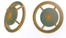
Human Optics AG (Erlangen, Germany) has developed an accommodating IOL called the Accommodative 1CU. The posterior chamber lens, which was designed by Khalil Hanna using finite-element computer-simulation models, is a one-piece, three-dimensional, foldable, acrylic IOL. The optic is 5.5 mm and the IOL has a diameter of 9.8 mm. The IOL fills the capsular bag in three dimensions which allows the capsular bag to assume its preoperative shape whereby the preoperative function of the ciliary muscle and zonular fibers are supposedly maintained. The modified haptics are intended to allow anterior movement of the lens optics upon contraction of the ciliary muscle. The 1CU IOL is a deformable three-dimensional IOL that mimics the properties of a human crystalline lens (Slide 5).
A pilot study evaluated the accommodative effect of the 1CU IOL.19 Comparisons were made among 12 eyes in 12 patients who received the 1CU IOL and 12 eyes in 12 patients who received either PMMA or acrylic posterior chamber IOLs as a control group. Phacoemulsification cataract extraction was performed though a 3.2-mm scleral tunnel incision, the IOL was injected into the capsular bag, and the haptics were unfolded manually. The results were mean distance-corrected near visual acuity, J7 vs. J13 (Birkhauser reading chart 0.34 vs. 0.15); subjective near point, 59 cm vs. 93 cm; difference between retinoscopic near and distance refraction, 1.2 D vs. 0.2 D; decrease of anterior chamber depth after 2% pilocarpine 0.63 mm vs. 0.15 mm; for the 1CU IOL subjects and the control PMMA or acrylic PCIOL subjects, respectively. The study's authors concluded that there was pseudophakic accommodation secondary to focus shift with the 1CU IOL.
A multicenter, clinical trial on the 1CU IOL is underway in Europe and Asia. Currently, researchers implant the same dimension lens in every patient, because no device can accurately measure the height, width, and depth of the capsular bag.
Summary
Two accommodating IOLs are being investigated in multicenter clinical trials. The AT-45 CrystaLens is undergoing FDA-monitored clinical trials in the United States and the 1CU IOL is undergoing clinical trials in Europe and Asia. These accommodative IOLs attempt to use the natural accommodative mechanisms in the eye and, in the future, may allow pseudophakic patients to see at near and distance without glasses.
References
-
Schachar RA, Black TD, Kash RL, Cudmore DP, Schanzlin DJ. The mechanism of accommodation and presbyopia in the primate. Ann Ophthalmol. 1995;27:58-67.
-
Schachar R, Cudmore D, Torti R, Black T, Huang T. A physical model demonstrating Schachar's hypothesis of accommodation. Ann Ophthalmol. 1994;26:4-9.
-
Schachar R, Tello C, Cudmore D, Liebmann J, Black T, Ritch R. In vivo increase of the human lens equatorial diameter during accommodation. Am J Physiol. 1996;271:670-676.
-
Glasser A, Kaufman PL. The mechanism of accommodation in primates. Ophthalmolology. 1999;106:863-872.
-
Fisher R. The force of contraction of the human ciliary muscle during accommodation. J Physiol. 1977;270:51-74.
-
Fisher R. The ciliary body in accommodation. Trans Ophthalmol Soc UK. 1986;105:208-219.
-
Huber C. Planned myopic astigmatism as a substitute for accommodation in pseudophakia. Am Intraocular Implant Soc. 1981;7:244.
-
Huber C. Myopic astigmatism as a substitute for accommodation in pseudophakia. Doc Ophthalmol. 1981;52:123.
-
Nakazawa N, Ohtsuki K. Apparent accommodation in pseudophakic eyes after implantation of posterior chamber intraocular lenses. Am J Ophthalmol. 1983;96:435-438.
-
Trindade F, Oliveira A, Frasson M. Benefit of against-the-rule astigmatism to uncorrected near acuity. J Cataract Refract Surg. 1997;23:82-85.
-
Thornton S. Lens implantation with restored accommodation. Current Canadian Ophthalmic Practice. 1986;4:60-62.
-
Lesiewska-Junk H, Kaluzny J. Intraocular lens movement and accommodation in eyes of young patients. J Cataract Refract Surg. 2000;26(4):562-5.
-
Strenk SA, Semmlow JL, Strenk LM, Munoz P, Gronlund- Jacob J, DeMarco JK. Age-related changes in human ciliary muscle and lens: A magnetic resonance imaging study. Invest Ophthalmol Vis Sci. 1999;40(6):1162-1169.
-
Coleman DJ. On the hydraulic suspension theory of accommodation. Trans Am Ophthalmol Soc. 1986;84:846-868.
-
Cumming JS, Ritter J. The measurement of vitreous cavity length and its comparison pre- and postoperatively. Eur J Implant Refract Surg. 1994;6:261-272.
-
Kammann J. Vitreous-stabilizing, single-piece, mini-loop, plate-haptic silicone intraocular lens. J Cataract Refract Surg. 1998;24:98-106.
-
Cumming JS, Slade SG, Chayet A. Clinical evaluation of the model AT-45 silicone accommodating intraocular lens: Results of feasibility and the initial phase of a Food and Drug Administration clinical trial. Ophthalmology. 2001;108(11):2005-2009; discussion 2010.
-
Steinert RF, Aker BL, Trentacost DJ, Smith PJ, Tarantino N. A prospective comparative study of the AMO ARRAY zonal-progressive multifocal silicone intraocular lens and a monofocal intraocular lens. Ophthalmology. 1999;106(7):1243-1255.
-
Lindstrom RL. Food and Drug Administration study update. One-year results from 671 patients with the 3M multifocal intraocular lens. Ophthalmology. 1993;100(1):91-97.
-
Kuchle M, Nguyen NX, Langenbucher A, Gusek-Schneider GC, Seitz B, Hanna KD. Implantation of a new accommodative posterior chamber intraocular lens. J Refract Surg. 2002;18(3):208-216.