Introduction
Acanthamoeba keratitis is an uncommon, sight-threatening condition that primarily affects contact lens wearers but may affect other individuals, as well. Men and women are equally affected and the majority of cases are unilateral. Although Acanthamoeba are ubiquitous in the environment, it is unusual to acquire Acanthamoeba keratitis without a predisposing risk factor such as:
- Contact lens wear (daily, extended-wear, rigid gas permeable [RGP], and PMMA)
- Poor sterilization and storage techniques with contact lenses
- A history of corneal trauma
- Ocular exposure to nonsterilized water or liquids
Acanthamoeba species are hardy, free-living amoebas that may be found in soil, dust, fresh water, tap water, hot tubs, swimming pools, and salt water.1 Some specimens have been cultured from the nasopharyngeal areas of healthy people, and some observers have noted that Acanthamoeba keratitis can occur following the use of saliva for wetting contact lenses. The widespread human exposure of the organism is illustrated by the fact that up to 100% of normal persons can demonstrate serum IgG antibodies against Acanthamoeba species, including Acanthamoeba castellani, Acanthamoeba culbertsoni, and Acanthamoeba astronyxis.2 Acanthamoeba can adhere to and penetrate intact human corneal epithelium.
The first cases of Acanthameba keratitis were reported in the 1970s, and the number of reported cases increased markedly through the late 1980s. It is interesting to note that thimerosal, a mercury-based preservative used in contact lens solutions, was increasingly withdrawn from use during this decade owing to reports of thimerosal-related superior limbic keratoconjunctivitis and other allergic reactions.3
Acanthamoeba keratitis is difficult to both diagnose and treat. Diagnoses are commonly delayed for weeks to months. Although both medical and surgical options exist, no consensus on the most appropriate methods of treatment has yet been reached, and the prognosis for infected eyes is generally guarded. Prevention and early diagnosis are the best current means of dealing with Acanthamoeba keratitis.
Clinical Findings
Most practitioners are aware that the classic features of Acanthamoeba keratitis include a stromal ring infiltrate in a contact lens wearer who experiences severe ocular pain out of proportion to the clinical findings. These are characteristics of advanced disease, however, and it is more useful to diagnose the keratitis at an earlier stage. In a study, the average duration of keratitis prior to diagnosis was 48 days1; in another study, the average duration was 42 days.4 Earlier diagnosis is associated with improved response to therapy and improved outcomes.
The ocular history may be one of the most important tools in the clinical assessment of patients with Acanthamoeba keratitis. It is critical to take a compete history when working up a patient with a nonspecific but nonhealing keratitis. For example, the presentation of a herpetic lesion in a contact lens wearer should raise a strong suspicion for Acanthamoeba. Suspicion should be increased with a history of ocular exposure to soil or water, or a history of trauma. It should be remembered that the disease can occur in the absence of contact lens wear; a series from India of 39 non-contact lens wearing patients with Acanthamoeba keratitis was reported in 2000.5
The stage of presentation of Acanthamoeba keratitis has been divided into three stages: early (less than 1 month), middle (1-2 months), and late (more than 2 months).6
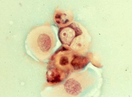
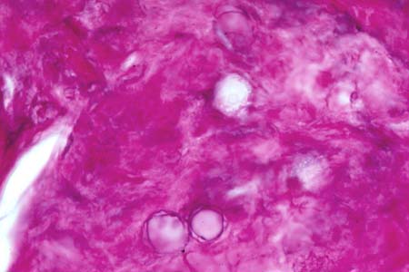
The early stages of Acanthamoeba keratitis may mimic herpes simplex keratitis in several ways. The initial features include fluctuating epithelial defects, epithelial haze, pseudodendrites, and ocular hyperemia. Severe ocular pain may accompany this presentation due to a keratoneuritis that is believed to be caused by direct invasion of Acanthamoeba trophozoites (Slide 1 and Slide 2). Despite the pain and perhaps because of the keratoneuritis, corneal sensation may be markedly diminished. In a series of six patients with Acanthamoeba keratitis, decreased corneal sensation was present in all patients.7 In another series, sensation was decreased in 29% of patients.1
Middle stages of the disease are often characterized by recurrent or persistent epithelial defects overlying nummular stromal infiltrates. This presentation often mimics herpetic disease. Frank stromal ulceration and lysis can occur during this stage (Slide 3).
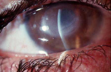
In the late or advanced stages of the disease, a ring infiltrate can appear (Slide 4), along with satellite lesions and stromal abscesses with a suppurative appearance. The presence of satellite lesions can mimic a fungal infection, but the presence of an annular keratitis and severe pain can aid in making the correct diagnosis. These features can also help to distinguish an atypical mycobacterial infection from Acanthamoeba. Progressive stromal loss can eventually lead to descemetocele formation and perforation.
Laboratory Diagnosis
Diagnostic scraping for cultures and stains should be a routine treatment for any suspicious nonhealing ulceration or keratitis; cultures are often positive. A series of 72 eyes found positive cultures in more than 70% of cases.6 HSV titers and cytology, as well as Ziehl-Neelsen stains and Lowenstein-Jensen agar, may be used to rule out other conditions. Studies have also shown that smears alone (Calcofluor White) may lead to the correct diagnosis in up to 92% of cases.
Acanthamoeba can be cultured on plain non-nutrient agar. Culture results may take 7 to 10 days if trophozoites are harvested, but may be delayed for longer periods if only cysts have been obtained, to allow time for the cysts to transform to the trophozoite stage.1 The diagnostic yield may be increased by culturing scrapings on top of a lawn of Escherichia coli plated on non-nutrient agar. Typically, the laboratory must be given prior notice to prepare these plates. E. coli are a food source for Acanthamoeba trophozoites, which typically "mow tracks" through the "lawn" of bacteria; the tracks remain empty since the non-nutrient agar does not support the regrowth of bacteria.
Although several stains may highlight Acanthamoeba trophozoites and cysts obtained from smears, Calcofluor White (M2R powder, Polysiences) and Masson trichrome stains are often preferred over Gram and Giemsa-Wright stains.
If cultures and initial smears are nondiagnostic, corneal biopsy can be considered. Even in early cases, a sample of epithelial cells can improve diagnostic yield. Later cases may require a biopsy of the stroma. The stains mentioned above can be used for such biopsies. In addition, fluorescein-labeled antibodies against Acanthamoeba, as well as electron microscopy, can aid in the diagnosis.
If available, tandem confocal microscopy can be a helpful and noninvasive method for examining corneas for the presence of both trophozoites and cysts. Various specific characteristic features of both cysts and trophozoites have been described to aid in diagnosis.8
Treatment
Antimicrobial Therapy
The main difficulty in treating Acanthamoeba keratitis is the resistance of Acanthamoeba cysts to anitmicrobials. Acanthamoeba may
persist in the encysted form for months and may reactivate after therapy has been discontinued. The topical anitmicrobials may be
toxic to the cornea, including to keratocytes, increasing the risk of long-term therapy.
Ocular medications effective against Acanthamoeba in vivo include the following (note: a 1% solution = 10 mg/mL):
- Biguanides
- Polyhexamethylene biguanide (PHMB) (e.g., Baquacil 0.02%, or a practitioner-formulated solution of 0.1%)
- Chlorhexidine biguanide (0.02% and 0.1%)
- Benzamidines
- Propamidine isethionate 0.1% (Brolene solution); also available as Dibrom-propamidine 0.15% ointment
- Pentamidine isethionate 0.05% to 0.1% (Pentam)
- Hexamidine diisethionate 0.1% (Desomedine)
- Imidazole solutions: Miconazole 1%; Clotrimazole 1%
- Neomycin (e.g., Neosporin, or Neomycin-Polymyxin B-Gramicidin)
While some antimicrobials, such as the benzamidines and biguanides, have been successful when used as monotherapy, the number of cases of organisms resistant to individual agents has led many practitioners to prefer using multiple medications when treating Acanthamoeba keratitis, both for treatment of chronic or recurrent disease as well as initial therapy. For example, initial combination therapy with a biguanide (PHMB or chlorhexidine) and a benzamidine (propamide, pentamidine, or hexamidine) is common in Europe, whereas many American practitioners prefer a combination of a benzamidine, an imidazole (miconazole or clotrimazole), and neosporin.10 Each class of medications adds another level of potential toxicity, but can also provide activity against additional and possibly coinfectious organisms (such as atypical mycobacteria, nocardia, and fungi).11 Practitioners can also consider use of oral imidazoles (e.g., systemic fluconazole, 100 mg twice a day) as adjunctive therapy in selective cases.
Frequency of drops is typically similar to frequency of use in severe bacterial keratitis (initial therapy of every hour for 3 days, then every 2 hours for 4 days, etc.). The main difference between treating bacterial and Acanthamoeba keratitis is the chronicity of therapy, since long-term maintenance therapy for Acanthamoeba can last up to a year.
Within the benzamidine family, propamidine isethionate 0.1% (Brolene solution) achieved the first documented in vivo cure of Acanthamoeba keratitis. Some researchers have found that members of the benzamidine family with more lipophilic properties, hexamidine 0.1% in particular, achieve better penetration through amebic cell walls (including cyst walls) and attain better in vivo effectiveness.12 Other researchers have found that pentamidine isethionate (Pentam) has less epithelial and endothelial toxicity than propamidine, without a compromise in efficacy,13 when delivered using the long-term therapy necessary for eradication of cyst forms.
The clinical use of PHMB was first reported in 1992. This formulation, which was found to have a lower side-effect profile than the benzamidines, is now commercially available for ophthalmic use in Europe in a 0.02% solution.13 In the United States, PHMB is available only in its formulation as a pool cleaner, but some practitioners reformulate it for ophthalmic use. The 0.02% solution is the most common formulation, but some practitioners have used higher concentrations (e.g., 0.1%). Chlorhexidine has been found to be at least as effective as PHMB in several studies, with a similarly low side-effect profile.
Adjunctive corticosteroid therapy in Acanthamoeba keratitis is somewhat controversial, but is widely used in selective cases to decrease severe corneal or anterior chamber inflammation and to reduce pain. In a series of 38 eyes in which 19 selected patients (already covered with appropriate antimicrobials) were treated with corticosteroids, the investigators found that final outcomes were not impaired with use of corticosteroids.14
Penetrating Keratoplasty
In rare cases, keratoplasty is indicated for perforation or intractable keratitis unresponsive to medical therapy and/or debridement. In
the majority of cases, however, penetrating keratoplasty is reserved for visual rehabilitation in eyes in which the infection has been
completely cleared and in which all cyst forms are believed to have been eradicated. For this reason, prolonged (e.g., 1 year or more) use
of topical antiamebic agents is indicated prior to considering penetrating keratoplasty. Because recurrence of Acanthamoeba keratitis in a
graft can be a devastating complication with a poor prognosis, use of postoperative prophylactic antiamebics, up to 1 year following surgery,
should also be a strong consideration.
Prevention
Proper contact lens disinfection and care is the most important step in preventing Acanthamoeba keratitis. A study published in 1995
suggested that the risk of developing Acanthamoeba increased markedly among daily wearers of disposable contact lenses who were less prone
to clean their lenses regularly. Those who used chlorine-based disinfectants, which are not effective against Acanthamoeba, were also at
higher risk for developing disease.15 Contact lens wearers should be specifically warned about the ubiquitous presence of this hardy
organism in soil and fresh water (including tap water).
References
- Alizadeh H, Niederkorn JY, McCulley JP. Acanthamoeba Keratitis. In: Krachmer JH, Mannis MJ, Holland EJ, eds. Cornea. St. Louis, Mo: Mosby; 1997: 1267-1273.
- Alizadeh H, Apte S, El-Agha MS, et al. Tear IgA and serum IgG antibodies against Acanthamoeba in patients with Acanthamoeba keratitis. Cornea. 2001;20(6):622-627.
- Buck SL, Rosenthal RA, Schlech BA. Methods used to evaluate the effectiveness of contact lens care solutions and other compounds against Acanthamoeba: a review of the literature. CLAO J. 2000;26(2):72-84.
- Bernauer W, Duguid GI, Dart JK. Early clinical diagnosis of acanthamoeba keratitis. A study of 70 eyes. Klin Monatsbl Augenheilkd. 1996;208(5):282-284.
- Sharma S, Garg P, Rao GN. Patient characteristics, diagnosis, and treatment of non-contact lens related Acanthamoeba keratitis. Br J Ophthalmol. 2000;84(10):1103-1108.
- Bacon AS, Frazer DG, Dart JK, et al. A review of 72 consecutive cases of Acanthamoeba keratitis, 1984-1992. Eye. 1993;7(Pt 6):719-725.
- Perry HD, Donnenfeld ED, Foulks GN, et al. Decreased corneal sensation as an initial feature of Acanthamoeba keratitis. Ophthalmology. 1995;102(10):1565-1568.
- Pfister DR, Cameron JD, Krachmer JH, Holland EJ. Confocal microscopy findings of Acanthamoeba keratitis. Am J Ophthalmol. 1996;121(2):119-128.
- Varga JH, Wolf TC, Jensen HG, et al. Combined treatment of Acanthamoeba keratitis with propamidine, neomycin, and polyhexamethylene biguanide. Am J Ophthalmol. 1993;115(4):466-470.
- Lin JC, Ward TP, Belyea DA, et al. Treatment of Nocardia asteroides keratitis with polyhexamethylene biguanide. Ophthalmology. 1997;104(8):1306-1311.
- Perrine D, Chenu JP, Georges P, et al. Amoebicidal efficiencies of various diamidines against two strains of Acanthamoeba polyphaga. Antimicrob Agents Chemother. 1995;39(2):339-342.
- Alizadeh H, Silvany RE, Meyer DR, et al. In vitro amoebicidal activity of propamidine and pentamidine isethionate against Acanthamoeba species and toxicity to corneal tissues. Cornea. 1997;16(1):94-100.
- Larkin DF, Kilvington S, Dart JK. Treatment of Acanthamoeba keratitis with polyhexamethylene biguanide. Ophthalmology. 1992;99(2):185-191.
- Park DH, Palay DA, Daya SM, et al. The role of topical corticosteroids in the management of Acanthamoeba keratitis. Cornea. 1997;16(3):277-283.
- Radford CF, Bacon AS, Dart JK, et al. Risk factors for acanthamoeba keratitis in contact lens users: A case-control study. BMJ. 1995;310:1567-1570.