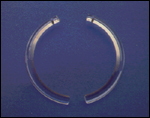
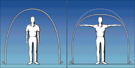
The cornea, being the principal refractive element of the eye, has been the focus of the refractive surgeon’s attention in an attempt to correct refractive errors. Contemporary refractive surgical methodologies aimed at altering the shape of the cornea are incisional or relaxation procedures (e.g., astigmatic and radial keratotomy), thermal procedures (e.g., laser thermal keratoplasty), ablative or subtraction procedures (e.g., photorefractive keratectomy, laser in situ keratomileusis [LASIK]), and additive procedures (e.g., intrastromal corneal rings [Intacs, Addition Technology, Fremont, Calif]). Barraquer first attempted to implant material in the cornea to correct refractive errors in 1949.1 Since then, several materials have been evaluated for their biocompatibility. In 1978, A. E. Reynolds conceptualized an implantable corneal 360° ring for correcting myopic and hyperopic refractive errors.2 Subsequently, this device evolved into two tiny, clear, ultra-thin, precision-engineered crescents made of polymethylmethacrylate (PMMA). Each crescent has an arc length of 150° with a fixed outer diameter of 8.1 mm and an inner diameter of 6.8 mm (Slide 1). The device serves as a spacer between arcing bundles of corneal lamellae producing predictable shortening of the central corneal arc length (Slide 2).3 This shortening is proportional to the thickness of the device, with an increased thickness resulting in greater corneal flattening. Theoretical advantages of this technology include the ability to remove the device and potentially to reverse treatment effects, to revise correction by varying the thickness of the device, and to avoid ablation of the central corneal tissue.4
Currently, the 0.25-mm, 0.30-mm, and 0.35-mm rings with predicted average corrections of -1.00 D to -1.63 D, -1.75 D to -2.25 D, and -2.38 D to -3.00 D, respectively, are approved by the U. S. Food and Drug Administration (FDA) (Table 1). Patients must have a stable refraction over the past 12 months and no evidence of corneal molding by topography. The relative contraindications of surgery include corneal diameter of less than 10 mm, central corneal thickness of less than 480 µm, peripheral thickness of less than 570 µm, and average keratometry readings greater than 46 D or less than 40 D. Other contraindications are abnormal eyelid or tear function, previous history of herpetic eye disease, and any condition that may lead to unstable corneas, such as pregnancy or systemic collagen vascular diseases. Additionally, the medications amiodarone, Imitrex (sumatriptan succinate, GlaxoSmithKline), and Accutane (isotretinoin, Roche) are listed as contraindications by the FDA.5
Preoperatively, patients are given topical antibiotic drops and an oral sedative. Several drops of nonpreserved anesthetic are administered topically prior to eye preparation with povidone-iodine. The eye is draped to fully isolate the lashes and lid margin from the surgical field. A speculum is used to hold the eyelids apart. The geometric center of the cornea is marked with a blunt Sinskey hook. A pre-inked marker is centered on the cornea to provide a visual guide for the placement of the incision at the 12 o’clock position at a 7.0-mm optical zone. Corneal ultrasonographic pachymetry is performed over the site of the peripheral incision, and a 15° diamond knife is used to make a 1.2-mm incision at a depth of 68% of the pachymetry reading along the superior corneal mark. A modified Suarez spreader is used to begin a lamellar corneal dissection. Specially designed dissecting instruments (Addition Technologies, Fremont, Calif) are inserted into the incision to create curved peripheral corneal tunnels. The intrastromal tunnels are prepared using clockwise and counterclockwise dissecting instruments. Each intrastromal corneal ring is manually rotated into a tunnel until the desired position is reached. The radial incision is gently hydrated or, alternatively, a 10-0 nylon suture may be placed. Generally, the suture is removed in 3 to 4 weeks; however, if it causes significant astigmatism, it may be removed earlier. Postoperatively, patients are given topical antibiotics and topical steroids for approximately 1 week.
The use of the intrastromal corneal ring has been shown to be safe and effective in treating low degrees of myopia (-1.00 D to -3.00 D) with an astigmatic component 1 D or less in many studies.2, 4-13 The smaller and larger ring sizes (0.21 mm, 0.40 mm, 0.45 mm) are still being evaluated in FDA clinical trials. Off-label use of intrastromal corneal rings for patients with keratoconus14, 15 and post-LASIK ectasia has also been reported with encouraging results. Intacs technology is a promising surgical option with proven safety and efficacy. It has significant advantages over other refractive surgical techniques in that the central visual axis and the normal flat to steep or prolate shape of the cornea are preserved.16, 17 This nonlaser and non-tissue-destructive procedure would be an appealing addition to a refractive surgeon’s armamentarium.
References
- Barraquer JI. Basis of refractive keratoplasty-1967. Refract Corneal Surg. 1989;5:179-193.
- Schanzlin DJ. Studies of intrastromal corneal ring segments for the correction of low to moderate myopic errors. Trans Am Ophthalmol Soc. 1999;97:815-819.
- Silvestrini T, Mathis M, Loomas B, Burris T. A geometric model to predict the change in corneal curvature from the intrastromal corneal ring (ICR). Invest Ophthalmol Vis Sci. 1994;35(suppl):2023.
- Rapuano CJ, Sugar A, Koch DD, Agapitos PJ, et al. Intrastromal corneal ring segments for low myopia: A report by the American Academy of Ophthalmology. Ophthalmology 2001;108:1922-1928.
- Linebarger E, Song D, Ruckhofer J, Schanzlin D. Intacs: The intrastromal corneal ring. International Ophthalmology Clinics. 2000;40:199-208.
- Twa MD, Karpecki PM, King BJ, Linn SH, et al. One-year results from the phase III investigation of the KeraVision Intacs. J Am Optom Assoc. 1999;70:515-524.
- Schanzlin DJ, Abbott RL, Asbell PA, Assil KK, et al. Two-year outcomes of intrastromal corneal ring segments for the correction of myopia. Ophthalmology. 2001;108:1688-1694.
- Ruckhofer J, Stoiber J, Alsner E, Grabner G. One year results of European multicenter study of intrastromal corneal ring segments. J Cataract Refract Surg. 2001;27:277-286.
- Asbell PA, Ucakhan OO. Long-term follow-up of Intacs from a single center. J Cataract Refract Surg. 2000;27:1456-1468.
- Wijdh RHJ, Van RG. Intrastromal Corneal Ring Segments (ICRS): Three and six months results. Documenta Ophthalmologica. 2000;100:27-37.
- Cochener B, Le Floch G, Colin J. Intra-corneal rings for the correction of weak myopias. J Fr Ophthalmol. 1998;21:191-208.
- Suiter B, Twa M, Ruckhofer J, Schanzlin D. A comparison of visual acuity, predictability, and visual function outcomes after intracorneal ring segments and laser in situ keratomileusis. Trans Am Ophthalmol Soc. 2000;98:51-57.
- Schanzlin DJ, Asbell PA, Burris TE, Durrie DS. The intrastromal corneal ring segments. Phase II results for the correction of myopia. Ophthalmology. 1997;104:1067-1078.
- Colin J, Cochener B, Savary G, Malet F. Correcting keratoconus with intracorneal rings. J Cataract Refract Surg. 2000;26:1117-1122.
- Colin J, Cochener B, Savary G, Malet F, Holmes-Higgin D. INTACS inserts for treating keratoconus. One-year results. Ophthalmology. 2001;108:1409-1414.
- Holmes-Higgins D, Baker P, Silvestrini, T. Characterization of the aspheric corneal surface with intrastromal corneal ring segments. J Refract Surg. 1999;15:520-528.
- Holmes-Higgins D, Burris T. Corneal surface topography and associated visual performance with Intacs for myopia: Phase III clinical trial results. Ophthalmology. 2000;107:2061-2071.