Introduction
Following are the main factors that affect accurate refractive results:
- Subjective patient response
- Objective vertex distance measurements
- Proper centering and focusing of laser
- Accuracy of actual photoablation
- Patient variation
In this tutorial, we discuss the importance of objective vertex distance measurements in achieving the most accurate refractive surgical results.
Vertex distance is the distance between the back surface of the refractive lens and the corneal surface (Slide 1). The vertex distance must be entered into the laser before every case. Historically, vertex distance has been measured by the Distometer (Haag-Streit, Waldwick, NJ), which is a device designed to obtain vertex measurements 7 mm to 23 mm from behind a spectacle or trial lens. This device works well for most spectacle readings less than 23 mm, but, because of its design and scissors-like action, it cannot take any vertex measurements behind the refractor.
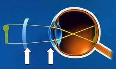
Solutions devised by refractor manufacturers involve estimation by direct sighting and through 90° mirrors located at the side of the refractor, as well as complicated correction factor tables. When used correctly, these methods can yield true vertex distance measurements, but in clinical practice the headrest is seldom correctly adjusted and correction factor tables are used infrequently. When these methods are used according to the refractor manufacturer's instructions (even with separate hand-held corneal illumination from behind the refractor), they are time-consuming and awkward, and hampered by parallax errors, dim lighting, and poor visualization.
Many refractive surgeons assume an average vertex distance of 12 mm to 14 mm for all patients without measuring vertex distance. One source of this commonly accepted convention is the Distometer operating instructions which state that "clinical experience has established that the…spectacle lenses, when fitted properly, are placed on average 8 mm to 12 mm from the cornea." Another source of this convention may be that when the forehead is adjusted so that the corneal apex is aligned with the 0 sighting line, the vertex distance from the corneal apex to the posterior lens surface is designed to be 13.75 mm (as indicated in the Ultramatic RX Master Phoroptor [Reichert, Buffalo, NY] instructions).
In this tutorial, an alternative method of directly obtaining the vertex distance is described. Using this method, it was found that the refractor vertex distance is almost always higher than commonly assumed. The clinical implications of underestimating this distance for achieving predictable results in refractive procedures are discussed, as well.
New Vertex Measuring Device
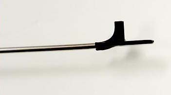
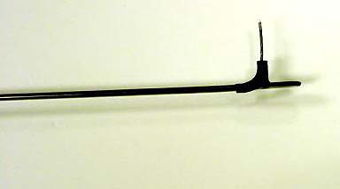
In a recent study, a new device, the Weiss Vertexometer (Oasis Medical, Glendora, Calif) (Slide 2), was used to directly measure corneal vertex distance from behind the refractor or trial lens in 189 consecutive patients. The average true vertex distance in this series was 20.4 mm. The true vertex range (eyelid thickness included) was 10 mm to 34 mm (Table 1).
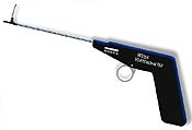
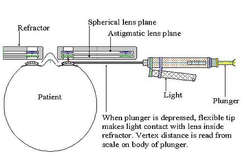
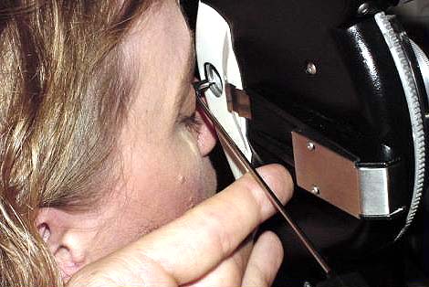
The vertex measurement was taken by having the patient close his or her eyes and by carefully positioning the Vertexometer between the patient and the refractor (Slide 3 and Slide 4). One side of the Vertexometer tip was gently placed on the apex of the closed eyelid and a flexible measuring device was advanced from the opposite side of the tip to make light contact with the lens inside the refractor (Slide 5, Slide 6). The vertex distance was indicated on a scale at the base of the Vertexometer.
Discussion
Almost all refractive surgical methods alter the refractive power of the cornea at the plane of the corneal apex (the only exception to this being refractive intraocular lenses). Spectacle trial lenses and refractors each measure the correcting refractive power of a lens in a plane other than the cornea. Therefore, the measured power at the corrective lens plane must be converted to the corrective power that is needed at the plane of the corneal apex, where the actual corneal power change takes place.
Vertex distance is defined as the distance between the back surface of the refractive lens and the corneal apex.2 This measurement is an important variable in the equation that converts the dioptric power at the refractive lens plane to the dioptric power at the corneal apex, or the corneal vertex dioptric power (Dcv).
The equation is Dcv=Dvp/(1-d*Dvp), where Dcv is the dioptric power at the corneal plane, Dvp is the power at the posterior lens plane, and d is the vertex distance in meters.3 Using this equation, we can examine the effect of vertex distance (within the range of our measured true vertex distances) on the corneal vertex dioptric power with increasing phoroptor lens powers (Table 2).
A lens power of -3.00 D corresponds to a corneal vertex power of -2.90 D with a 12-mm vertex distance and -2.71 D with a 35-mm vertex distance, which is a difference of only 0.19 D over this range, possibly clinically insignificant. However, as the lens power is increased, the differences become larger and more meaningful.
Consider what happens when we assume an average unmeasured 12-mm vertex distance that is really 25 mm. In a 6.00 D myope, we will try to correct -5.60 D at the corneal plane when we should have corrected -5.22 D, resulting in an overcorrection of 0.38 D. In this scenario, for 12.00 D and 15.00 D of myopia, we will theoretically overcorrect by 1.26 D and 1.81 D, respectively. Similarly, we can calculate that, for a 5.00 D hyperope, we will undercorrect by 0.39 D. These differences are significant.
Likewise, using an average unmeasured 12-mm vertex distance that is really 35 mm will result in larger theoretical inaccuracies; overcorrecting -6.00 D by 0.61 D, -12.00 D by 2.04 D, and -15.00 D by 2.87 D, and undercorrecting 5.00 D of hyperopia by 0.74 D. Current software versions of many excimer laser systems do not allow entry of vertex distances higher than 25 mm, a subgroup consisting of 24% or 12.4% of the quoted study.
These examples illustrate that underestimating the refractor vertex distance will result in overcorrecting a myopic patient and undercorrecting a hyperopic patient. It is clear that inaccuracy of vertex distance measurements at the refractor may account for some of the unpredictability of refractive outcomes in previously reported studies, specifically, overcorrections in higher myopes and undercorrections in higher hyperopes.
Understanding the Refractor
A crucial question is whether direct measurement from the cornea to the posterior presenting lens surface is the true vertex distance in all cases. Reichert Ophthalmic Instruments confirms that all lenses in the phoroptor are referenced to the posterior lens surface (closest to the eye) of the lenses in the strong sphere lens plane. Slide 7 is an internal schematic diagram of the lens placements within the phoroptor.
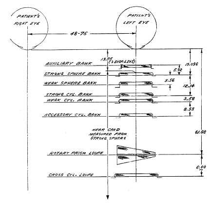
Inside the phoroptor, the lenses are divided into planes that correspond to different rotary dials that hold different power lenses; one each for an auxiliary bank, strong sphere bank, weak sphere bank, strong cylinder bank, weak cylinder bank, accessory cylinder bank, rotary prism loupe, and cross cylinder loupe, with successive planes being located farther from the eye, in the above order.
As mentioned, all lenses in the phoroptor are referenced to the back surface of the plane of the strong sphere. For all lenses in this plane (+3.00, +6.00, +9.00, +12.00, +15.00, -3.00, -6.00, -9.00, -12.00, -15.00, -18.00), the marked power is the same as the lensometer power. Thus, direct measurement of vertex distance to the back surface of lenses in the strong sphere plane is valid. The strong sphere lens plane is always in this position for marked powers of higher than -1.00 D and higher than +2.00 D, as long as the auxiliary bank is not in place at the time of measurement, as it usually is not. The only exception when direct measurement is not valid is for plano-cylindrical powers in an open tube phoroptor (without a lens aperture cover), in which case the direct measurement would be to the more distant cylinder plane, an overestimation of the vertex distance.
Weak sphere and all cylinder lenses are also referenced to the posterior surface of the strong sphere plane. For example, the actual lensometer power for a marked 5.00 D cylinder lens is 4.674 D, which corresponds to the 12.14-mm distance between lens planes.
The above confirms the validity of the fact that two lenses in combination correspond exactly to the marked phoroptor power, with a vertex distance being referenced to the back surface of the strong sphere lens plane (closest to the eye), which can then be directly measured for moderate and higher refractive errors.
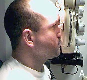
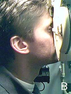
A more significant finding of the vertex study is the wide range (10 mm to 34 mm) of measured vertex distances even among patients of the same investigator, which emphasizes the need for accurate individual measurements. This relatively wide range was due in part to anatomic variations, differences in baseline phoroptor headrest position, posture, and effort. Slide 8 shows two patients with markedly different vertex distances, the farthest measuring 29 mm.
Conclusion
Accurate refractor vertex measurements in patients are essential in producing predictable refractive surgical results, especially in patients with moderate to severe refractive errors. An average vertex distance should not be applied to all patients and has no clinical usefulness except to alert clinicians to the fact that this distance varies with each patient and we should be less complacent about entering the vertex distance into the laser. The average vertex distance in one study was 20.4 mm (range, 10 mm to 34 mm). This is substantially larger than commonly used values and may be responsible for certain cases of overcorrected myopes and undercorrected hyperopes in previous studies. The issue of accurate vertex distance measurement is especially relevant with increasing demands for accuracy of refractive results.
References
- Weiss RA, Berke W, et al. Clinical importance of accurate refractor vertex distance measurements prior to refractive surgery. J Refract Surg. 2002;18:444-448.
- Benjamin WJ, ed. Borish's Clinical Refraction. Philadelphia, Pa: WB Saunders; 1998:968.
- Cogger TJ. Correction with hard contact lenses. Chapter 54. In: Duane TD, ed. Clinical Ophthalmology. Hagerstown, Md: Harper & Row; 1980;22.