Introduction
Incisions, laser ablation, and heat have been used to modify corneal curvature for the correction of refractive error and various pathological conditions. Excimer laser ablation of the corneal periphery in the form of laser in situ keratomileusis (LASIK) is currently the most popular technique for correcting myopia and hyperopia and has been reported as safe and effective for both primary surgeries and enhancements.1-3 However, complications related to the flap, such as striae, displacements, button-hole flaps, and diffuse lamellar keratitis, are not uncommon and can limit visual results.4,5
Conductive keratoplasty (CK) is a new "laserless," collagen shrinking procedure that can change corneal curvature effectively and safely. The procedure does not require a corneal flap and thus avoids flap-related complications. The mechanism of action is through the application of high frequency (radiofrequency, 350 kHz) energy that the cornea converts to heat in a controlled fashion. Treatment is applied outside of the visual axis and produces a steepening of the central cornea that corrects hyperopic refractive error.
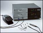
In April 2002, the U.S. Food and Drug Administration granted approval of the ViewPoint CK System (Refractec, Irvine, Calif) (Slide 1) for the treatment of mild to moderate (+0.75 D to +3.00 D), previously untreated spherical hyperopia in patients 40 years of age or older. Other applications of CK under investigation are the treatment of astigmatism, correction of hyperopia induced by previous refractive procedures, and the treatment of presbyopia through monovision surgery.
Energy Delivery
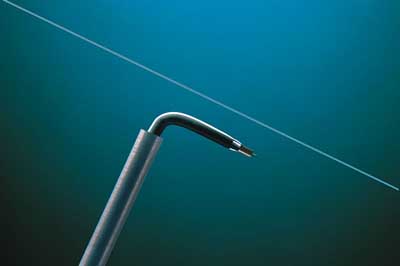
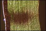
CK delivers a precise amount of radiofrequency (350 kHz) current through a fine tip (CK Keratoplast tip, Refractec) (Slide 2) inserted directly into the peripheral corneal stroma. As the current flows from the tip through the corneal tissue, resistance to the current creates localized heat. Collagen in the area surrounding the tip denatures and shrinks in a controlled fashion and forms a column of denatured collagen (Slide 3). The histology of a pig eye shows a footprint extending down to 80% of the mid-peripheral cornea after CK treatment.6,7 Since permanent contraction of collagen through thermokeratoplasty has been shown to depend on achieving a consistent, deep zone of collagen shrinkage,8 deep penetration is desirable to minimize regression. In the phase III CK clinical studies,9 endothelial damage has not resulted from CK treatment of spherical hyperopic eyes.
Treatment Effects
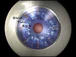
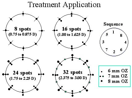
In a CK procedure, treatment is applied to eight to 32 points in a ring pattern in the corneal periphery outside of the visual axis (Slide 4). As the level of hyperopia increases, the number of treatment spots is increased up to a maximum of 32. Laser thermal keratoplasty (LTK), on the other hand, relies upon an increase in the level of pulsed laser energy (mJ) for increased effect. Small leucomas appear on the corneal surface at CK-treated spots with striae between the spots. Confocal microscopy shows that these striae are still present 12 months postoperatively9 (Slide 5) and contribute to the long-lasting effect of the procedure. The striae between the spots create a "belt tightening" effect around the mid-peripheral cornea. This increases the curvature of the central cornea and corrects hyperopia (Slide 6).
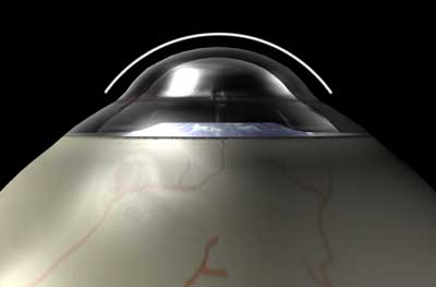
During CK energy application, the thermal effect proceeds from the point of the probe up the shaft as it finds the path of least resistance. CK-treated tissue is exposed to the same temperature at the tip of the probe (deep in the stroma) as at the top of the probe (the corneal surface). The CK collagen shrinking process is self-limiting because resistance to the flow of the current increases with the increasing dehydration of stromal collagen.
In contrast, LTK performed with the Hyperion LTK System (Sunrise Technology, Fremont, Calif) generates the greatest amount of heat at the surface of the cornea because of the high absorption of light energy in water. The holmium:YAG beam is attenuated as it passes through the cornea so that the heat energy diffuses radially and axially into the tissue. The result is corneal denaturation decreasing from top to bottom, forming a cone-shaped zone of collagen shrinkage that is wider at the surface and tapering into the stroma.10 The depth of the LTK footprint appears to be approximately 60% deep, which is more shallow when compared with the 80% depth seen in a CK-treated spot.
The System
The ViewPoint CK system consists of a portable console, corneal marker, choice of lid speculum (Lancaster, Barraquer, or Cook) that acts as the electrical return path for the radiofrequency energy delivered by the tip, a handpiece (probe) that holds the 450-µm long and 90-µm wide stainless steel tip (Keratoplast tip), and a foot pedal. The default setting for treatment is approximately 1.5 W, 60% power (0.6 W) for 0.6 seconds. An insulated stop at the base of the probe controls the depth of penetration of the Keratoplast tip. Energy is delivered into the cornea by activation of the foot pedal.
Patient Selection
Preoperatively, a complete medical and ocular history must be obtained and the requisite diagnostic examinations including determination of sphere, cylinder, and the manifest refractive spherical equivalent performed. Candidates for the procedure should demonstrate refractive stability, have visual acuity correctable to at least 20/40 in both eyes, and have pachymetry readings of at least 560 µm at the 6-mm optical zone.
Candidates should not have intractable keratoconjunctivitis sicca or unstable, progressive hyperopia. Contact lens wearers should have a stable refraction. Hard contact lens wearers should discontinue lens use 3 weeks before the procedure and soft contact lens wearers should discontinue lens use 2 weeks before the procedure.
The Procedure
CK is a simple procedure that can be performed in an office setting using topical anesthesia (proparacaine HCl, 0.5%). The eye to be treated is not dilated. Anxious patients can be given 5 mg of Valium (diazepam, Roche) preoperatively. The entire CK procedure usually takes less than 5 minutes from the time the speculum is inserted to the time the speculum is removed. The surgeon begins by consulting the nomogram and establishing a surgical plan based on the patient’s refraction. The patient is positioned under the operating microscope, the lid speculum is inserted, and the patient is asked to fixate on the fixation light. A circular mark with eight intersections is then made with a special CK marker dampened in gentian violet or rose bengal stain.
The Keratoplast tip is inserted into the stroma at defined spots around the peripheral cornea, according to the supplied nomogram (Slide 7). An increasing number of spots is applied for higher levels of hyperopia at the default level of power (60%). If mild myopia is desired for monovision, an overcorrection can be designed by the surgeon. The coagulated epithelium overlying the treatment site is removed to promote epithelial healing. Postoperative medications include a broad-spectrum topical antibiotic, a topical nonsteroidal anti-inflammatory, a mild topical steroid (e.g., fluorometholone), and an oral analgesic, if needed.
When CK is used to correct astigmatism, the surgeon must customize the procedure based on the corneal topography and apply selected spots to steepen the appropriate axis. Treatment patterns and a nomogram for treatment of primary astigmatism are currently being studied. CK has the potential for replacing astigmatic keratotomy for the correction of astigmatism; however, such use is not approved by the U.S. Food and Drug Administration.
Another ongoing study for treating presbyopia with CK by inducing monovision suggests that the procedure may be effective and safe when performed in one eye to induce low myopia.
Advantages of CK
Current treatment options for patients with hyperopia include the Sunrise Hyperion LTK system, hyperopic LASIK, hyperopic photorefractive keratectomy (PRK), and hyperopic phakic intraocular lenses for hyperopia higher than 3.00 D. Regression of effect has been associated with the LTK procedure, although published, well-controlled studies are few. Alio and associates reported complete regression in 31% after 6 months,11 whereas Tutton reported 50% regression after 2 years.12 Clinical trial results of CK with up to 2 years of follow-up, on the other hand, have not demonstrated significant regression. This may be due to the deeper stromal penetration and cylindrical footprint of CK (80% corneal depth) compared with the more shallow, cone-shaped penetration of the LTK treatment.
There is no evidence of recurrent erosions, infections, visually significant corneal scarring, or corneal haze following the CK procedure. Irregular astigmatism may result from asymmetric application of the high frequency energy, but such occurrences have been infrequent. Compared with hyperopic LASIK treatment, CK does not invade the central cornea, is technically easier to perform, has a shorter surgical learning curve, has no flap complications, and does not compromise the integrity or structure of the cornea. CK results in a larger effective optical zone than can be achieved with either LASIK or PRK. In addition, CK may be rapidly performed, requires only topical anesthesia, and the small portable CK unit is less expensive than other refractive technologies.
Summary
Recent advances in refractive surgery have expanded the treatment options for patients with hyperopia. Although LASIK is currently the most popular treatment for patients with low to moderate hyperopia, a simple nonincisional, nonablative technique such as CK offers several advantages in safety, ease of treatment, and cost. Noncontact LTK treatment with the holmium:YAG laser has shown inherent deficiencies in predictability and stability. CK may offer the advantages of a nonablative technique without the associated concern regarding refractive stability. The FDA has recently approved the ViewPoint CK System for the temporary reduction of mild to moderate (+0.75 D to +3.00 D), previously untreated spherical hyperopia. Treatment of astigmatism, correction of hyperopia induced by previous refractive procedures, and the treatment of presbyopia through monovision surgery are being explored as potential applications internationally and in the United States.
References
- Sher NA. Hyperopic refractive surgery. Curr Opin Ophthalmol. 2001;12:304-308.
- Choi RY, Wilson SE. Hyperopic laser in situ keratomileusis: primary and secondary treatments are safe and effective. Cornea. 2001;20:388-393.
- Rojas MC, Haw WW, Manche EE. Laser in situ keratomileusis for consecutive hyperopia after myopic overcorrection. J Cataract Refract Surg. 2002;28:37-43.
- Melki SA, Azar DT. LASIK complications: etiology, management, and prevention. Surv Ophthalmol. 2001;46:95-116.
- Stulting RD, Carr JD, Thompson KP, et al. Complications of laser in situ keratomileusis for the correction of myopia. Ophthalmology 1999;106:13-20.
- Goth P, Stern R. Conductive Keratoplasty, Principles and Technology. Presented at the American Society for Cataract and Refractive Surgery. Boston, Mass; April 2000.
- Pearce J. Corneal reshaping by radio frequency current: numerical model studies. Proc SPIE. 2001:4247.
- Brinkmann R, Radt B, Flamm C, Kampmeier J, Koop N, Birngruber R. Influence of temperature and time on thermally induced forces in corneal collagen and the effect on laser thermokeratoplasty. J Cataract Refract Surg. 2000;26;744-754.
- McDonald MB, Hersh PS, Manche EE, Maloney RK, Davidorf J. Conductive Keratoplasty for the Correction of Low to Moderate Hyperopia: U.S. Clinical Trial One-Year Results on 355 Eyes. Ophthalmology. Accepted for publication.
- Koch DD, Kohnen T, Anderson JA, et al. Histologic changes and wound healing response following 10-pulse noncontact holmium:YAG laser thermal keratoplasty. J Refract Surg. 1996;12:621-634.
- Alio JL, Ismail MM, Sanchez Pego JL. Correction of hyperopia with non-contact Ho:YAG laser thermal keratoplasty. J Refract Surg. 1997;13:17-22.
- Tutton MK, Cherry PM. Holmium:YAG laser thermokeratoplasty to correct hyperopia: two years follow-up. Ophthalmic Surg Lasers. 1996;27(5 Suppl):S521-S524.