Introduction
The standard conjunctival incision used in extraocular muscle surgery is a peritomy at the limbus. The conjunctiva and Tenon's capsule are incised as a single tissue plane in the limbal incision. This approach is useful when there is conjunctival scarring as is frequently encountered in re-operations or in patients with long-standing restrictive strabismus. Using a limbal incision, the conjunctiva can be recessed upon completion of surgery. The limbal incision provides excellent visualization of the operative field. Therefore, it is particularly useful when there is no assistant surgeon1 or when an adjustable suture technique is needed.
In the standard limbal approach, three incisions of the conjunctival Tenon's layer are made. One incision is made parallel to the limbus, and two relaxing radial or oblique incisions are made at the peripheral edges of the limbal incision.2 One of the authors (A.R.C.) has devised a limbal approach that uses a single incision of conjunctival Tenon's layer instead of the three required in the standard limbal approach.3
Surgical Technique
The simplified limbal approach can be used for all rectus muscle surgery but is best suited to surgery performed on the horizontal rectus muscles, as is described here. A 6-0 traction suture is placed through the conjunctiva and episclera at the 6 o'clock and 12 o'clock limbal positions and held by a hemostat to allow exposure of the surgical aspect of the eye. The conjunctiva and Tenon's capsule are lifted (with 0.3-mm toothed forceps) 4 mm up from the eyeball and 3 mm away from the limbus at the 9 o'clock or 3 o'clock position for horizontal rectus muscles, or at the 6 o'clock or 12 o'clock position for vertical rectus muscles (Slide 1). Wescott scissors (Norfolk, VA) are brought across the cornea and parallel to the lid margin to engage the conjunctiva 1 mm to 2 mm away from the limbus. The tips of the Wescott scissors should extend from the point of the conjunctival engagement (1 mm to 2 mm away from limbus) towards the muscle insertion, approximately 7 mm away from the limbus (Slide 2). The one-snip technique results in a horseshoe-shaped conjunctival incision that is approximately 18 mm to 20 mm long (Slide 3).
Then, the combined conjunctival and Tenon's layer is reflected with a forceps, and the extraocular muscle insertion is easily exposed (Slide 4). In many cases, no dissection of Tenon's capsule is required as the muscle is completely exposed after the original incision. The muscle is hooked (Slide 5).
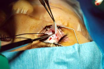
Upon completion of the muscle surgery, the conjunctival Tenon's flap can be either returned to the original anatomical position or recessed a few millimeters away from the limbus with two interrupted 6-0 plain sutures.
We have used this one-snip technique on more than 300 patients. There were no intraoperative surgical complications; however, care must be taken not to extend the incision too far posterior over the muscle. The conjunctiva protects the underlying muscle from being cut. As with any conjunctival incision, if Tenon's capsule is left exposed, then a granuloma can result.
Discussion
The three most widely used conjunctival approaches are the Swan incision, the limbal incision, and the cul-de-sac or fornix incision.3 The Swan incision, placed anterior to the rectus muscle insertion, risks copious hemorrhage from an accidental cutting of the muscle. The cul-de-sac incision, also known as the fornix approach, provides superior patient comfort immediately after surgery and superior postoperative cosmetic appearance. However, the surgical technique is difficult and requires an assistant who is well-trained in strabismus surgery. The exposure is limited and the conjunctiva cannot be recessed with this incision. An adjustable suture procedure may also be more difficult to perform.
The limbal or peritomy incision for strabismus surgery is the easiest of the three conjunctival approaches to perform. Use of this incision allows the conjunctiva to be recessed for an adjustable suture procedure or to relieve a restriction to ocular rotation resulting from conjunctival scarring or shortening.
The one-snip limbal approach we have described is simpler and faster to perform than the standard limbal approach because the two radial relaxing conjunctival incisions required in the standard limbal approach are not needed. We timed a number of the two different limbal approaches and found that it took, on the average, four to five times longer to perform a standard limbal approach than the one-snip conjunctival incision.
Also, we believe there is less bleeding and tissue damage with the one-snip approach because it involves fewer incisions, causing less conjunctival vessel damage. However, we have no accurate means of measuring tissue damage. The one-snip incision leads directly to the muscle insertion, providing good surgical exposure for both muscle recession and resection procedures as well as for horizontal and vertical rectus muscle procedures. However, results obtained in original extraocular muscle procedures were better than those achieved from repeat surgeries.
Because no significant data are reported regarding the complication rates of different types of conjunctival incisions, we cannot explicitly compare the results of the one-snip procedure in terms of complications with those achieved with different incisions. We have not had any serious complications related to either the limbal or the cul-de-sac conjunctival approaches in more than 20 years of practice in pediatric ophthalmology. The simplified limbal technique reduces the duration of surgery and is associated with both excellent healing and superior surgical results.
References
- Calhoun JH, Nelson LE, Harley RD. Conjunctival incisions. In: Calhoun JH, et al, eds. Atlas of Pediatric Ophthalmic Surgery. 1st ed. Philadelphia, Pa: WB Saunders; 1987:23-31.
- Parks MM. Limbus incision. In: Parks MM, ed. Atlas of Strabismus Surgery. 2nd ed. Philadelphia, Pa: Harper & Row; 1983:78-84.
- Caputo AR, Guo S, DeRespinis P, Wagner RS. Simplified limbal incision for rectus muscle surgery. Ophthalmic Surgery. 1991;22:406-408.
- Parks MM. Fornix incision for horizontal rectus muscle surgery. Am J Ophthalmol. 1968;65:907-915.
- VonNoorden GK. The limbal approach to surgery of the rectus muscles. Arch Ophthalmol. 1968;80:94-97.