Introduction
Cutaneous carcinoma is the most common malignancy in the United States. Approximately 500,000 people are treated annually for basal or squamous cell carcinoma of the skin. Approximately 5% to 9% of all skin cancers arise in the eyelids. The most common malignancies of the periocular region are basal, squamous, and sebaceous cell carcinoma and malignant melanoma. Among immunosuppressed patients, Kaposi sarcoma is being recognized with increasing frequency. Management of any malignant eyelid lesion requires an early and accurate diagnosis followed by total eradication of the tumor and reconstruction of the eyelid for restoration of both function and cosmesis.
Basal Cell Carcinoma
Incidence
Basal cell carcinoma is the
most common malignant tumor of the eyelids. It accounts for 90% of all eyelid
malignancies. It most frequently occurs in the lower eyelid (50% to 66%) and
medial canthus (25% to 30%). The upper eyelid (15%) and lateral canthus (5%) are
less frequently involved (Slide 1).
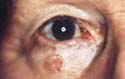
Sun exposure is an important factor in the development of basal cell carcinoma. Fair skinned people are particularly susceptible to the effects of ultraviolet radiation. Basal cell carcinoma is rare among black individuals. Most patients with basal cell carcinoma are older than 50 years of age, but approximately 5% to 15% of cases occur in patients between 20 and 40 years of age. There is no sex predilection. Patients with acquired immunodeficiency syndrome (AIDS) experience a greater risk of basal cell carcinoma.1
Presentation
Basal
cell carcinoma of the eyelid has a variety of clinical presentations.
These presentations are most often dependent on the histologic
characteristics. The most common histologic types of basal cell
carcinoma include the nodular, noduloulcerative, pigmented, cystic, and
morphea form varieties.
The most common, nodular basal cell carcinoma presents as a firm, pearly, dome-shaped nodule, with multiple telangiectatic vessels. The overlying epithelial surface is smooth in appearance. A few lesions may bleed spontaneously after minimal trauma. The tumor is generally painless. The interior of the tumor may outgrow its blood supply, leading to a central ulceration. Eventually, this may appear as a slowly enlarging ulcer, leading to the common noduloulcerative basal cell carcinoma variant with characteristic ulcer crater within a raised, pearly margin. This lesion is often referred as a “rodent ulcer.” The nodular type of tumor tends to be the least aggressive, and rarely has subcutaneous extensions that are not clinically apparent.
Pigmented basal cell carcinomas are infrequent and account for less than 10% of basal cell cancers excised. These lesions are occasionally misdiagnosed as pigmented nevi or malignant melanomas. Cystic basal cell carcinoma may develop with mucin accumulation or degenerative necrosis within a solid tumor. They may be hard to distinguish from a benign epithelial inclusion cyst or apocrine hidrocystoma. Morphea form basal cell carcinoma appears as a flat, indurated plaque with telangiectasia. These tumors are most often asymptomatic. If situated along the lid margin, it often simulates a localized blepharitis. The borders may be fairly indistinct. In some cases, there is widespread subcutaneous involvement that is not visible clinically. Incompletely resected morphea form of basal cell carcinoma is 10 times more likely to recur than the nodular type. This type of tumor is best resected with a Mohs resection. Superficial basal cell carcinoma is the least common variety of basal cell carcinoma. These flat superficial lesions appear as erythematous, scaly patches with well-defined pearly borders. They grow slowly. These lesions often are mistaken for psoriatic plaques or Bowen disease.
Treatment
Any suspicious eyelid lesion
should undergo a biopsy to establish a definitive histologic diagnosis prior to
complete excision and repair. Several treatment modalities have been used in
the management of eyelid basal cell carcinomas. Surgical excision, Mohs
micrographic surgery, cryosurgery, and radiotherapy have all been reported to
achieve 5-year cure rates of 90% or higher. Surgical excision with microscopic
evaluation of the margins is the best technique for eliminating basal cell
carcinomas. Frozen section control of tumor margins is performed by noting the
clinical boundaries of the tumor edges and excising an additional 3-mm cuff of
normal appearing tissue. A pathologist then examines each of the margins, and
if no tumor cells are noted, immediate repair is undertaken.
Prognosis
Surgical excision with
standard frozen section control or Mohs micrographic surgery are the best
available treatment modalities with a 5-year cure rate of 95%. Treatment cure
rate is dependent on several variables, including location, size, histologic
characteristics of the basal cell carcinoma, immune status of the patient, and
previous treatment failure. Basal cell carcinomas located in the medial canthus
are more prone to be deeply infiltrative than those arising from the central or
lateral eyelid margin. With regard to tumor histology, incompletely excised
nodular basal cell carcinoma have been found to recur in only 8% of cases,
whereas ulcerative and morphea form basal cell carcinomas have recurrence rates
of 60% and 75%, respectively. Recurrent lesions have a propensity to be more
aggressive and have a higher recurrence rate than primary tumors. Tumors that
have previously been radiated recur at a higher rate than lesions treated by
other means. The majority of recurrent basal cell carcinomas occur within 5
years of primary excision.
Patients with one basal cell carcinoma are at risk for developing other new lesions. A new basal cell carcinoma will develop in 20% to 30% of patients within 1 year after having the initial tumor excised. After 5 years, the incidence of developing a new lesion increases to 36%.
Basal Cell Nevus Syndrome
Presentation
Basal cell nevus syndrome is
an uncommon autosomal dominant dermatosis. It is associated with numerous organ
system abnormalities including jaw cysts and musculoskeletal anomalies. The
most common and serious ophthalmic manifestation in basal cell nevus syndrome
is basal cell carcinomas of the eyelids. Patients may present with a few basal
cell carcinomas to hundreds of tumors. Histologically, these tumors are
indistinguishable from actinically induced basal cell carcinomas. These tumors
can be locally invasive and result in disfigurement. In young patients, the
presence of basal cell carcinoma should suggest the possibility of basal cell
nevus syndrome.
Treatment
Although surgical excision
with microscopic evaluation of the margins is the best technique for removing
basal cell carcinoma, the surgery itself may cause disfigurement and become
difficult because of lack of tissue available for multiple reconstructions.
Nonsurgical techniques, such as cryotherapy, curettage, and electrocoagulation
can be beneficial for these lesions. Mohs micrographic excision technique is
recommended for large invasive lesions or for recurrent lesions. Radiation
therapy is contraindicated because of its increased mutagenic potential in
these patients.
Squamous Cell Carcinoma
Incidence
Squamous cell carcinoma is
the second most common eyelid malignancy. Patients with squamous cell carcinoma
average 70 years of age. It typically occurs in patients with a fair complexion
who have a history of chronic sun exposure. Patients with albinism and
xeroderma pigmentosa have an increased incidence of squamous cell carcinomas at
a young age. Patients with lymphoma, leukemia, and patients with
immunosuppression from organ transplantation have an increased risk for
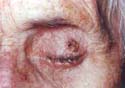
developing squamous cell carcinoma (Slide 2).
Presentation
Squamous cell carcinoma has
a predilection for developing at the mucocutaneous junction of the lower
eyelid. It characteristically develops as a painless, slightly raised, plaque
or nodule with scaling, crusting, or ulceration. It may also appear as a
papillomatous growth along the eyelid margin. This tumor is locally invasive.
It can invade the orbit and sinuses by contiguous extension. Squamous cell
carcinoma can also spread via perineural invasion and lymphatic spread, and
metastasize to distant sites. Actinic keratosis is considered a premalignant
precursor to squamous cell carcinoma.2
Treatment
Any suspicious eyelid lesion
should be biopsied to establish a definitive histologic diagnosis prior to
complete excision and repair. Several treatment modalities have been used in
the management of eyelid squamous cell carcinomas. Surgical excision with
microscopic evaluation of the margins is the best technique for eliminating
squamous cell carcinomas. Frozen section control of tumor margins is performed
by noting the clinical boundaries of the tumor edges and excising an additional
3 mm cuff of normal appearing tissue. A pathologist then examines each of the
margins, and if no tumor cells are noted, immediate repair is undertaken. For
patients with orbital squamous cell carcinoma with distant metastasis,
radiation therapy can be used for palliative treatment.
Sebaceous Cell Carcinoma
Incidence
Sebaceous cell carcinoma is
the third most common eyelid malignancy. It tends to affect patients between
the fifth and ninth decades of life, with an average at diagnosis of 61 years
of age. Women are affected at a rate of 2.5 times greater than men
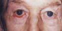
(Slide 3).
Presentation
Sebaceous cell carcinoma has
a predilection for the upper eyelid. It can originate from meibomian glands and
sebaceous glands of the eyelid, eyebrow, and caruncle. The typical presentation
of a sebaceous cell carcinoma is that of a thickened eyelid margin with
associated inflammation. A slowly growing, firm lesion of the eyelid margin
with associated loss of eyelashes may be present. Sebaceous cell carcinoma will
frequently have a yellow hue to the tumor.3 Patients are often times
misdiagnosed with blepharoconjunctivitis, blepharitis, and chalazions prior to
having the lesions biopsied. Any patient with a chronic eyelid inflammation or
recurrent chalazion that does not respond to medical and or surgical treatment
should have the eyelid biopsied.
Treatment
Any suspicious eyelid lesion
should be biopsied to establish a definitive histologic diagnosis, prior to
complete excision and repair. If sebaceous cell carcinoma is suspected, the pathologist
should be made aware so that he or she stains the tissue with oil-red-0 stain
for fat. Surgical excision with microscopic evaluation of the margins is the
best technique for eliminating sebaceous cell carcinomas. Frozen section
control of tumor margins is performed by noting the clinical boundaries of the
tumor edges and excising an additional 5-mm cuff of normal appearing tissue. A
pathologist then examines each margin, and if no tumor cells are noted,
immediate repair is undertaken.
Prognosis
Sebaceous cell carcinoma
recurs in approximately 9% to 36% of patients. It may recur with orbital
involvement in 6% to 17% of patients. Regional lymph nodes are the most common
site of metastases. Distant metastatic site may include the lungs, liver, skull,
and brain. The incidence of metastases is approximately 17% to 28%. The 10-year
tumor death rate is approximately 28%.
Malignant Melanoma
Incidence
Cutaneous malignant
melanomas of the eyelid are rare, representing less than 1% of all eyelid
tumors. However, the incidence has been slowly increasing over the last few
decades.4 The majority of cutaneous melanomas seem to develop de
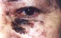
novo without antecedent melanocytic nevi (Slide 4).
Presentation
The principal types of
cutaneous melanoma involving the eyelid are lentigo maligna melanoma (10%),
superficial spreading melanoma (80%), and nodular melanoma (10%).
Lentigo Maligna Melanoma
Lentigo maligna (Hutchinson freckle) is considered the premalignant lesion of lentigo maligna melanoma. While lentigo maligna melanoma is responsible for 90% of head and neck melanomas, it represents only 10% of eyelid melanomas.
Lentigo maligna typically presents as a flat, tan to brown macule with irregular borders in sun-exposed areas. The lesion may have a long radial or horizontal growth phase superficially, before vertical invasion into the dermis occurs, resulting in the development of lentigo malignant melanoma. At this point, the lesion becomes elevated. Approximately 25% to 30% of lentigo maligna undergo malignant transformation, and metastasis is rare. Wide surgical excision with careful histologic examination of the margins is the most efficacious treatment.
Superficial Spreading Melanoma
Superficial spreading melanoma is the most common eyelid melanoma. It typically presents as an elevated, multinodular lesion with variegated colors. It occurs on nonexposed skin surfaces and usually develops over 2 to 5 y ears.
Nodular Melanoma
Nodular melanoma constitutes 10% of eyelid melanomas. It arises in sun-exposed areas and exhibits early vertical invasion.
Treatment
A biopsy should be performed
on all pigmented eyelid lesions suspected of being a melanoma. Mohs resection
is recommended to assure total resection followed by immediate reconstruction
of the defect. A metastatic work up should also be done because these tumors
often spread to the lymph nodes, liver, and other organs.
Sweat Gland Carcinomas
Sweat gland carcinomas are rare skin tumors derived from eccrine or apocrine glands. Mucin-secreting adenocarcinoma is the most common type and typically occurs in the sixth and seventh decades. It usually presents as a firm, cystic nodule with either reddish induration or normal-appearing overlying skin. A less common sweat gland carcinoma is the malignant syringoma, which usually appears on the upper lip. While sweat gland carcinomas have low potential for metastasis, they may spread to adjacent structures, the orbit, and regional lymph nodes. The treatment of choice is complete surgical excision and evaluation for possible metastasis.5
Kaposi Sarcoma
Kaposi’s sarcoma is a malignant vascular tumor that most often develops on the skin. It may also involve the mucous membranes, lymph nodes, and visceral organs. This previously rare tumor is a frequent manifestation of AIDS occurring in approximately 24% to 35% of AIDS patients (Slide 5).
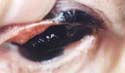
Ophthalmic Kaposi sarcoma typically occurs on the eyelids, conjunctiva, caruncle, and lacrimal sac. The eyelid lesion presents as a chronic, nontender reddish nodule, which may cause ocular discomfort or visual obstruction. The differential diagnosis includes pyogenic granuloma, hemangioma, lymphoma, and malignant melanoma.6
Because AIDS-related Kaposi sarcoma is a multifocal disease, excision of isolated lesions does not result in a cure. However, the lesions are slow growing and seldom invasive, so observation may be appropriate for some patients. Lesions that cause functional or significant cosmetic problems may be treated with excision, radiation, cryotherapy, or intralesional chemotherapeutic agents. Surgical excision is the most effective treatment for localized, well-delineated lesions. Large or diffuse lesions are better treated with one of the other modalities.
References
- Shields CL. Basal cell carcinoma of the eyelids. Int Ophthalmol Clin. 1993:33:1-4.
- Reifler DM, Hornblass A. Squamous cell carcinoma of the eyelid. Surv Ophthalmol. 1986;30:349-365.
- De Potter P, Shields CL, Shields JA. Sebaceous gland carcinoma of the eyelids. Int Ophthalmol Clin. 1993;33:5-9.
- Grossniklaus HE, McLean IW. Cutaneous melanoma of the eyelid. Ophthalmology. 1991;98:1867-1873.
- Cohen KL, Peiffer RL, Lipper S. Mucinous sweat gland adenocarcinoma of the eyelid. Am J Ophthalmol. 1981;92:183-188.
- Soll DB, Redovan EG. Kaposi’s sarcoma of the eyelid as the initial manifestation of AIDS. Ophthalmic Plast Reconstr Surg. 1989;5:49-51.