Introduction
Normal eyelids protect the globe from desiccation and trauma, exhibit specialized movement, and allow for vision and movement of fluid bathing the cornea. The upper eyelid margin is normally situated 1.5 mm to 2 mm below the superior limbus and 4.5 mm to 4 mm above the center of the cornea. The lower eyelid margin is normally situated at the inferior limbus. Eyelid retraction is a condition in which the eyelid margin is displaced superiorly or the lower eyelid margin is displaced inferiorly. Although eyelid retraction is the most common ocular clinical feature of Graves disease, this sign is encountered relatively infrequently in general ophthalmology. Published case reports subclassify eyelid retraction into three categories: neurogenic, myogenic, and mechanical. Other categories may be listed as follows1:
- Inflammatory
- Thyroid eye disease (Slide 1)
- Orbital pseudotumor
- Cicatricial conjunctival disease
- Chronic dermatitis
- Traumatic
- Eyelid laceration
- Orbital floor fracture
- Inferior rectus disinsertion
- Postoperative
- Blepharoplasty (Slide 2 and Slide 3)
- Blepharoptosis surgery
- Eyelid tumor resection and reconstruction
- Orbital floor fracture repair
- Inferior rectus recession
- Glaucoma filtering surgery
- Retinal detachment surgery
- Involutional, congenital, neurogenic
- Aberrant regeneration of oculomotor nerve(Slide 4)
- Marcus Gunn jaw winking
- Sympathetic overactivity (Claude Bernard syndrome)
- Midbrain lesion (Collier sign)
- Facial nerve palsy
- Pseudoretraction from contralateral ptosis (Hering law)
- Metabolic
- Cirrhosis
- Hypokalemic periodic paralysis
- Uremia
- Cushing syndrome
- Pharmacologic
- Sympathomimetic agents
- Corticosteroids
- Proptotic, myopic
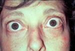
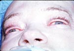
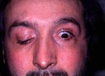
Eyelid retraction can be present at birth (Slide 5). Preterm infants may have a benign transient conjugate downward gaze with upper eyelid retraction. This condition has been ascribed to immature myelinization of vertical eye movement systems and immaturity of the extra-geniculate visual pathways. An "eye-popping" reflex in normal infants can occur when ambient lighting levels suddenly are reduced. This reflex can be elicited in 80% of children in the first 5 months of life.2
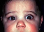
The cause of eyelid retraction in Graves disease is not clearly understood, but several factors may be contributory. In the upper lid, contributory factors include3:
- Müller muscle overaction from sympathetic stimulation
- levator muscle contraction from inflammation and fibrosis of the levator muscle
- levator adhesions to the orbicularis muscle and orbital septum
- overaction of the levator superior rectus complex in response to a hypophoria produced by fibrosis and retraction of the inferior rectus
In the lower eyelid, adrenergic stimulation of the Müller muscle plays a smaller role, but fibrosis of the inferior rectus exerting a retraction action on the lower eyelid through its capsulopalpebral head appears to be more influential.
Clinical Findings
Lid retraction may be mild, moderate, or severe and may involve upper lids, lower lids, or both. The patient's history and the cause of lid retraction will lead to pertinent clinical observations. High axial myopia with an enlarged globe can result in apparent eyelid retraction and, therefore, simple observation of the patient's spectacles or refraction will determine the cause. In Graves disease, diffuse eyelid inflammation can result in shortening of the extraocular muscles and the levator muscle. Therefore, extraocular motility with identification of a hypotropia from a tight and retracted inferior rectus muscle may lead to increased tone in the superior rectus/levator complex resulting in an accentuated overshoot of the upper eyelid due to Hering law.
After trauma, the eyelid tissue may become fibrotic and shortened, resulting in eyelid retraction. Eyelid retraction appears most commonly if the insult is perpendicular to the eyelid margin because scar bands fold the yield margin toward the orbital margin. In addition, chronic dermatitis or burns can result in ocular exposure problems with blurred vision, discomfort, infection, corneal erosions, and an undesirable cosmetic result.
Diagnostic Features
Unilateral/bilateral eyelid retraction is the most common orbital finding in Graves disease, occurring in approximately 50% of the cases. This may be combined with lid lag. Therefore, a patient with unexplained lid retraction should be evaluated for thyroid disease with laboratory diagnostic tests. Serum T4, T3, and third-generation, thyroid-stimulating hormone (TSH) assays should be ordered. Approximately 90% of patients with thyroid disease have elevated T4 or T3 levels. The sensitive TSH assay measures depressed levels of this hormone, which reflects the suppressive state induced by a diseased hyperproductive thyroid gland. An independently functioning thyroid gland associated with eyelid and orbital changes characteristic of Graves disease virtually confirms the diagnosis. Although rarely, Hashimoto thyroiditis may mimic Graves disease. Other supporting tests including antithyroid receptor antibodies, antimicrosomal antibodies, and computed tomography looking for enlarged extraocular muscles (inferior rectus, medial rectus more common) may be helpful.
Treatment
Prior to correction of lid retraction, the immediate treatment goal is to protect the cornea by establishing the best precorneal tear film for as long a duration as possible. Treatment includes preservative-free tears supplemented with bland lubricating ointments or gels, punctal plugs, moist chamber shields, and taping the eyelids shut during sleep.
It is critical to perform eyelid retraction surgery in Graves disease when the disease is in a quiescent state. Failure to do so may compromise surgical results. The systemic thyroid status and eyelid position on serial exams should be stable for 6 to 12 months before surgery. Severe exposure keratitis unresponsive to medical management may necessitate earlier intervention in rare instances. An easily reversible tarsorrhaphy may also be considered in these circumstances.
Sympathetic ganglion blockers, such as guanethidine monosulfate, have an upper eyelid lowering effect, but toxicity with both topical and systemic modalities preclude their use.
As therapies for other manifestations of Graves disease can affect eyelid position, eyelid retraction surgery should be delayed until after these treatments can be completed and the effects have stabilized. These treatments include treatment and stabilization of hyperthyroidism, systemic corticosteroids, orbital irradiation, orbital decompression, and extraocular muscle surgery diplopia. Either two may have a significant effect on eyelid position.
There are many approaches for surgical correction of lid retraction secondary to Graves disease, including the anterior/posterior approach with or without spacers. In all of these procedures, alterations are made in one or both of the upper eyelid retractors, that is, Müller muscle and the levator palpebrae superioris muscle. Müller muscle may be excised or recessed. The levator aponeurosis is sometimes implanted between the recessed upper eyelid retractors and the tarsus. Although such spacers remain helpful in the correction of lower eyelid retraction, they are seldom used in the upper eyelids.5 A graded recession of a levator/Müller muscle complex, more temporal and central than nasal leaving the orbital septum intact, provides satisfactory outcome (Slide 6 and Slide 7).6
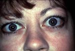
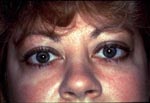
If traumatic scarring is the cause of eyelid retraction and a vertical discrete cicatrix is present, the treatment involves excision of the scar and closure of the healthy tissues. Tissue transfer procedures, such as the Z plasty or VY plasty, are commonly used.
Anterior lamellar shortening involving the skin resulting from dermatitis, burns, or excessive skin removal following upper eyelid blepharoplasty is best remedied by releasing the scar and placement of full thickness skin graft.
References
- Glatt HJ, Putterman AM. Management of eyelid retraction. In: Stewart WB, ed. Surgery of the Eyelid Orbit and Lacrimal System, 2nd ed. American Academy of Ophthalmology Monographs; 1994:134-161.
- Bartley GB. The differential diagnosis and classification of eyelid retraction. Ophthalmology. 1996;103:168-176.
- Tse DT. Management of thyroid-related eyelid retraction. In: Tse DT, ed.Color Atlas of Ophthalmic Surgery. Philadelphia, Pa: JB Lippincott; 1992:387-412.
- Lemke BN. Lid retraction. In: Stephenson CM, ed. Ophthalmic Plastic Reconstructive and Orbital Surgery. Boston, Mass: Butterworth Heinemann; 1997:539-555.
- Lisman RD, Milite JP. Management of eyelid malposition in thyroid ophthalmopathy. In: Nesi FA, Lisman RD, Levine MR, eds. Smith's Ophthalmic Plastic and Reconstructive Surgery, 2nd ed. St. Louis, Mo: CV Mosby; 1998:1133-1163.
- Levine MR, Chu A. Surgical treatment of thyroid lid retraction: A new variation. Ophthal Plast Reconstr Surg. 1991;22:90-94.