Introduction
The surgical procedure to remove an eye should be performed with efforts to minimize cosmetic deformity. The anophthalmic socket must be able to hold a prosthesis and allow movement of the prosthesis that matches the movements of the contralateral eye. The eyelids also must be positioned so their movement matches the contralateral eyelids. To achieve this, a socket with the following characteristics is required:
- A centrally placed, buried, inert implant of adequate size attached to the extraocular muscles
- Deep fornices
- A lower eyelid that can support a prosthesis
- A superior eyelid with position and movement symmetric to the normal eyelid
- Prosthesis and implant volume equal to that of the natural eye
- An implant peg that couples with the prosthesis
These characteristics apply to both patients undergoing enucleation and patients undergoing evisceration. Many different implants and techniques can be used to meet these criteria.
Orbital Implants in Anophthalmos
Implants can be integrated or nonintegrated. Nonintegrated implants have no direct attachments to the extraocular muscles and are usually inert spheres of silicone or methylmethacrylate, placed in Tenon's capsule within the muscle cone, deep to anterior Tenon's fascia and conjunctiva. The rectus muscles may be sutured together anterior to the implant.
With an integrated implant, the rectus muscles are attached directly to it. Allen implants have tunnels into which the muscles are attached. With spherical integrated implants made of silicone or hydroxyapatite, the muscles are sutured to a wrap consisting of sclera, fascia, or an artificial substance. Polyethylene implants can be sutured directly and, therefore, do not require a wrap. They are significantly less expensive than hydroxyapatite implants.
Before 1985, glass, acrylic, or silicone implants were used. Since then, coralline hydroxyapatite spheres have become available. These spheres are popular because they are porous and, therefore, allow fibrovascular ingrowth. This reduces the incidence of extrusion, migration, and infection; however, the pores may trap infectious agents during placement. These implants are not recommended for use in immunocompromised patients or patients with panophthalmitis.
Implant porosity and fibrovascular ingrowth allow drilling and placement of a peg for direct coupling to the prosthesis. The peg is fitted into an indentation into the back of the prosthesis so movement of the prosthesis is equal to that of the implant and the other eye.1 The peg also supports the weight of the prosthesis, preventing lower eyelid sag if the prosthesis is large. Many patients with porous implants do not require peg insertion; insertion rates range from 11% to 76% in published series.2 Porous spheres made from polyethylene, ceramic,3 and bovine4 and manufactured hydroxyapatite5 have later become available.
Sclera has been the most commonly used wrap for implants. However, the occurrence of transmissible diseases related to sclera wraps have resulted in a rise in popularity of other implant wraps. Autogenous tissue, such as fascia lata and posterior auricular muscle complex, and synthetic materials, such as high porosity expanded polytetrafluoroethylene, have been used effectively.6
Implant Size
The socket tissue, implant, and prosthesis must have a total volume equal to the volume of the natural eye to achieve symmetry (Slide 1). The size of the prosthesis selected is limited by the weight that can be supported by the lower eyelid. Implants typically are less than 3 cc in volume. In patients requiring a larger prosthesis, a peg can be used for additional support. The thickness of the prosthesis is limited by the depth of the anterior chamber. Increased thickness is required to allow for an indentation for a peg. The ideal volume of a prosthesis is 2 cc to 2.5 cc.7
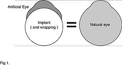
The volume of implant used should be equal to the volume of the enucleated eye (pre disease if phthisical) minus the volume of the prosthesis. If the prosthesis size is 2.5 cc and the volume of an eye is 7.2 cc with an axial length (AL) of 24 mm, then the implant volume should be 4.7 cc. If spherical, the diameter is 21mm (Table 1).8
Globe volume - prosthesis volume = implant volume7.2 cc (24 mm AL) - 2.5 cc = 4.7 cc (21 mm diameter)
A scleral wrap adds approximately 1.5 mm to the diameter of the implant. See Table 1 to calculate implant size for different sized eyes. An intermediate implant size can be created by shaving a larger implant.
Enucleation
Enucleation is the removal of the entire eyeball, which is usually replaced with a spherical orbital implant.
Indications for enucleation include:
- Intraocular malignancy untreatable by other means
- Ruptured globe, resulting in a blind eye, with risk of sympathetic ophthalmia
- A blind eye producing sympathetic ophthalmia
- A blind, painful eye, usually secondary to end-stage glaucoma, retinal detachment, chronic inflammation, or trauma
In enucleation surgery, when removing the diseased tissue, a socket must be created that is large enough to hold an adequate prosthesis and preserves the most conjunctiva possible. The implant must be of adequate volume with good movement.9,10
Technique
General anesthesia is preferred during an enucleation procedure, although local anesthesia with sedation may also be used. A retrobulbar
injection of 0.75% bupivicaine and 1:100,000 epinephrine is administered even with general anesthesia to achieve hemostasis and prevent
postoperative pain.
A 360° peritomy, with lateral and medial relaxing incisions, is made. The four rectus muscles are dissected out, double-end 6-0 polyglactin sutures are attached, and the muscles are divided at their insertions. The oblique muscles are divided. The optic nerve is divided using a snare, which is slowly tightened, or by clamping for several minutes then cutting with scissors. These methods promote hemostasis by crushing the central retinal vessels. Further hemostasis is achieved with bipolar cautery, thrombin, and Gelfoam.
The surgeon should confirm that the predetermined implant will fit with sizing spheres placed in the muscle cone. If it does not fit, then the largest fit is used.
The sclera is prepared by turning it inside out and removing any residual choroid. Relaxing incisions are made on either side of the hole where the cornea was located to allow placement of the implant within. The corneal hole becomes the posterior pole and the optic disc becomes the anterior pole when placed in the socket. Holes are cut for each of the rectus muscles adjacent to the optic disc. The holes can be cut using a 2-mm circular punch biopsy blade and making two or three adjoining cuts. The implant is placed within the sclera and secured by partially closing the sclera with 5-0 polyester sutures. The rectus muscle holes and the posterior hole where the cornea was removed allow fibrovascular ingrowth.
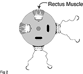
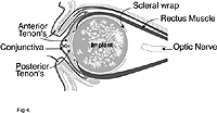
The implant is placed through posterior Tenon's fascia into the muscle cone. The rectus muscles are attached to the anterior lips of the scleral windows with 6-0 polyglactin sutures (Slide 2). Posterior Tenon's fascia is pulled forward in quadrants between the rectus muscles and a 6-0 polyglactin purse-string suture used to close this layer in front of the implant (Slide 3). Anterior Tenon's fascia is closed horizontally with interrupted 6-0 polyglactin sutures and conjunctiva closed horizontally with continuous 6-0 polyglactin sutures (Slide 4).
Antibiotic ointment is placed in the socket. A conformer that places no tension on the wound with the lids closed should be placed. A pressure dressing is applied and left on for 48 hours. A prosthesis is fitted 4 to 8 weeks after enucleation when all swelling has settled.
When the implant is vascularized, usually after 6 to 12 months, the implant can be drilled and a peg placed. Vascularization can be confirmed with computed tomography (CT) scan with contrast sensitivity, magnetic resonance imaging (MRI) with gadolinium, or a Technetium-99 bone scan. A number of small drill holes made into the implant at the time of surgery will increase the rate of vascularization. Initially, single-piece plastic pegs were used. These have been associated with complications such as loosening, extrusion, coral exposure, pyogenic granuloma, and infection. Newer titanium, screw in, sleeve and peg systems may decrease these complications.11
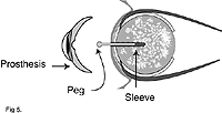
The peg is inserted using local anesthetic under sterile conditions. To help correctly place the peg, a template can be made by the prosthetist with a hole at the anterior pole of the implant. The conjunctiva is cut and cauterized, then, with the template in position, a hole is drilled. The sleeve is screwed in and a flat peg placed in the sleeve, over which the prosthesis is worn. After 4 weeks, the prosthetist replaces the flat peg with a round peg and makes an impression in the back surface of the prosthesis into which the peg fits. This produces prosthesis movement equal to that of the implant (Slide 5 and Slide 6).
Complications
Potential complications of enucleation include the following:
- Removal of the wrong eye can be avoided by marking the forehead on the side for enucleation while the patient is awake. In a normal looking eye, dilate the pupil and confirm the presence of tumor. This reassures the patient.12
- Incomplete enucleation is important to avoid in patients with intraocular tumors and infection to prevent seeding the orbit. With retinoblastoma, as much optic nerve as possible, perhaps 10 mm, should be removed because this tumor spreads down the nerve. Traction sutures at the horizontal rectus muscle insertions can be used to pull the globe forward.
- Hemorrhage can be minimized by achieving hemostasis and ceasing anticoagulants an adequate time preoperatively.
- Swelling and hematoma formation can be minimized by padding the eye postoperatively for 48 hours.
- Infection of the implant may be treated with systemic antibiotics if it is vascularized. If the implant is not vascularized, then removal is usually necessary with replacement later. Systemic antibiotics perioperatively and soaking the implant in antibiotics will help prevention.
- Wound breakdown with implant exposure is the most common complication with hydroxyapatite implants, more so than with silicone implants, with reports of 3% to 20% versus 1% to 5%.13,14 This may occur early due to tension on the wound or later due to pressure of a poorly fitting prosthesis. Discharge, pyogenic granuloma formation, and infection can occur.
- Tension on the wound can be avoided by careful hemostasis, posterior placement of the implant by anterior attachment of the rectus muscles, closure of posterior Tenon's fascia anterior to the implant, ensuring the conformer does not pull on the wound with the eye closed, a pressure bandage for 48 hours, and ensuring the implant is not too large. Wrapping the hydroxyapatite implant is important because the coarse nature of its surface can cause breakdown of the conjunctiva and Tenon's capsule over it. 15 One study suggested that autologous wrapping may decrease exposure. 16
- A poorly fitting prosthesis may cause erosion and should be modified.
- Implant exposure of less than 3 mm may heal spontaneously. A defect with exposure of a vascularized implant may also heal spontaneously. A persistent defect should be repaired with a patch graft of sclera or fascia covered by a flap of conjunctiva. The patch should be twice the size of the exposed area and placed in a pocket between the conjunctiva and Tenon's capsule. A bucket handle flap of conjunctiva should be mobilized above and brought down to cover the patch. The defect above is left to epithelialize.17
- Alternatively, free autogenous tissue grafts of hard palate mucosa or thin dermis-fat graft may be used after burring of the hydroxyapatite and freshening of the conjunctival edges.18
- Implant extrusion rarely occurs with porous implants. It occurs with wound breakdown and epithelial ingrowth around a nonporous implant.
- Migration outside the muscle cone may occur with a nonwrapped, nonporous implant. Closing the defect in posterior Tenon's capsule through which the optic nerve passed prior to implantation may prevent this. A migrated implant is removed and replaced by an integrated implant.
- Anophthalmic enophthalmos, or deep superior sulcus syndrome, or post enucleation socket syndrome occurs when the implant is too small or has migrated inferiorly. There is enophthalmos and a deep upper sulcus. Ptosis and lower lid sagging may also be present. If the implant is too small, ptosis and lower lid sagging may be corrected by enlarging the prosthesis. However, the larger prosthesis often causes discomfort and the extra weight may not be adequately supported by the lower eyelid. Replacement with a larger implant or an orbital floor implant may be required.
- A socket with a short inferior fornix cannot hold an implant. Deepening the fornix is difficult and requires mucous membrane grafting.
- Inadequate movement of the prosthesis may be improved by placing a peg into the implant, if it is porous and vascularized.
- Ptosis should be corrected using an anterior approach so that the conjunctiva is preserved.
- Conjunctival cyst formation can present as an enlarging mass. A CT scan can help in diagnosis; then the cyst is excised.
- Discharge and irritation caused by the prosthesis is common and giant papillary conjunctivitis may occur. Treatment involves ensuring that the prosthesis fits properly, eliminating protein deposits on the prosthesis and treating with steroid drops, long-term if necessary.
- Retardation of orbital growth may occur with enucleation in childhood. A dermis-fat graft may grow with the patient and, therefore, may help prevent this retardation.
- Peg problems including discharge, pyogenic granuloma, peg falling out, poor transfer of movement, clicking, the conjunctiva overgrowing the peg, a peg drilled at an angle or off center, hydroxyapatite exposure around the peg hole, excess movement of peg, and implant infection occurred in 37% of pegged implants in one study.19
Evisceration
Evisceration is the removal of the contents of the eyeball, leaving the sclera (and sometimes the cornea) intact and preserving the extraocular muscle attachments.
The advantages of evisceration over enucleation are that evisceration is easier to perform, quicker, and less disruptive of the orbital contents than enucleation. Also, it provides better movement20 and a better cosmetic result than enucleation. Extrusion is less frequent. Psychologically, evisceration may be better accepted by patients because the whole eye is not removed. With intraocular infection, the subarachnoid space is not opened so the theoretical risk of infection spread into the brain is avoided.
Disadvantages of evisceration include the risk of sympathetic ophthalmia21,22 and difficulty in placing an implant in severe phthisis. It is contraindicated in patients with intraocular tumor and if there is a need for histological examination of the entire globe.
Indications for evisceration include:
- A blind, painful eye (if intraocular malignancy can be eliminated)
- Corneal perforation
- Endophthalmitis (evisceration is preferred in this situation to prevent intracranial spread via incised dura)
Aims of evisceration are to eliminate the disease process and produce a comfortable socket with a cosmetically acceptable prosthesis. The requirements for this have been described above in the Introduction. 23
Technique
Many different techniques both with and without keratectomy have been described.24-26 Described below is one method,
with keratectomy, which is applicable to most situations in which an evisceration is indicated.
Local anesthesia with intravenous sedation may be used, although general anesthetic is often preferred to decrease patient anxiety.
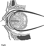
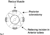
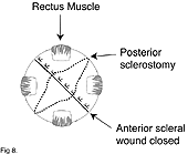
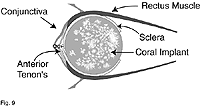
A 360° peritomy with horizontal relaxing incisions is performed. The cornea is excised and relaxing incisions made at 45° (i.e., between the rectus muscles) (Slide 7). The ocular contents are removed carefully ensuring no choroidal tissue is left. Hemostasis is achieved with cautery. The size of the cavity should be checked with sizing spheres. If, as is usual, the cavity is too small for the required implant, then an expansion sclerostomy may be required. One technique involves making a posterior scleral incision at 90° to the anterior scleral relaxing incisions (Slide 7). This should extend around the optic nerve and forward to the equator at both ends so the implant can protrude posteriorly through it. This also helps with fibrovascular ingrowth into a porous implant. The implant is placed into the sceral shell and the sclera is closed anteriorly with interrupted vertical mattress sutures of 5-0 braided polyester so the edges overlap (Slide 8). The anterior Tenon's fascia is closed with interrupted 6-0 polyglactin sutures and the conjunctiva closed with continuous 6-0 polyglactin sutures (Slide 9).
Implants can consist of glass, silicone, methylmethacrylate, hydroxyapatite, or polyethylene. The porous implants, hydroxyapatite and polyethylene, allow fibrovascular ingrowth and, therefore, can be drilled for peg placement later to improve prosthesis motility.
Complications
Potential complications of evisceration include the following:
- Hemorrhage within the sclera may cause expulsion of the implant if there is no posterior opening.
- Infection of the implant may be treated with systemic antibiotics if it is fully vascularized. If the implant is not vascularized, then removal and later replacement are required. If the implant must be removed, it can be replaced with a prosthesis implanted behind the posterior sclera, which provides an extra layer of cover.
- Wound breakdown with implant exposure may be due to tension on the wound or pressure of a poorly fitting prosthesis. Wound tension can be relieved by removing the implant and performing a posterior scleral relaxing incision, and replacing the prosthesis with a smaller implant or burring down of the implant. The defect can also be covered with a flap or patch graft.
- Implant extrusion rarely occurs with porous implants; it occurs with wound breakdown and epithelial ingrowth around a nonporous implant. In these cases, the implant should be replaced.
- Inadequate movement of the prosthesis may be improved by placing a peg into the implant, if it is porous and vascularized. This fits into an indentation in the prosthesis to produce movement equal to that of the implant.
- Discharge and irritation caused by the prosthesis is common and giant papillary conjunctivitis may occur. Treatment involves ensuring a proper fit of the prosthesis, eliminating protein deposits on the prosthesis, and treating with steroid drops, long-term if necessary.
Dermis-fat graft
The dermis-fat graft is an alternative to an implant and is useful when there is a contracted socket and inadequate socket volume. It can be used after enucleation or evisceration and has been most often used to replace an extruded implant.
During the procedure, general anesthesia is used. The donor site of the graft may be the non-weight-bearing lower abdomen or thigh. The dermis-fat graft should be in contact with orbital fat to enhance graft viability. Thus, Tenon's fascia, sclera, or pseudocapsule left after implant extrusion should be incised or excised to facilitate this.
The graft is harvested by first marking a 25-mm circle. The epidermis and superficial dermis are excised using a scalpel. An incision is made into subcutaneous tissue to a depth of 15 mm and the graft is placed into the socket.
The edge of the dermis is sutured to the rectus muscles, in enucleation, or to the anterior scleral ring, in evisceration. Tenon's capsule and conjunctiva are approximated to the dermis circumferentially. Excess fat may be removed; however, over correction of 20% to 30% is necessary to allow for graft atrophy. If there is enough conjunctiva, this can be sutured to cover some or all of the dermis.
Full epithelialization of the dermis occurs after 4 weeks, so the prosthesis may be fitted after this time.
Complications
Complications of dermis-fat grafting include the following.
- Fat atrophy and volume loss are variable and may require further dermis-fat grafting.
- Graft failure usually is associated with a compromised orbital vascular supply after radiation treatment or chemical injury. It may be more likely to occur in patients with advanced age, infection, vascular disease, or diabetes, patients who experienced graft trauma from manipulation, excessive cautery, or inadequate hemostasis, and patients who smoke.
- Minor complications include conjunctival cysts, granulomas, graft ulcers, socket keratinization and cilia retention.17
Exenteration
Exenteration is the removal of the entire contents of the orbit, which is usually performed to remove life-threatening orbital malignancy or infection, such as mucor mycosis. It may also be done for severely disfiguring or painful conditions such as orbital lymphangioma or sclerosing pseudotumor. Exenteration may be total, subtotal, or extended. Subtotal exenteration spares the eyelids and can be performed when the disease is behind the globe. Extended exenteration includes removing surrounding bone. Histopathological diagnosis with permanent sections along with patient evaluation for metastatic disease should be made before exenteration.27
Technique
Total exenteration. General anesthesia is required during total exenteration. The procedure is begun by marking the position of the orbital rim on the skin. The surgeon should infiltrate along the incision with local anesthetic, and 1:100,000 epinephrine is administered for hemostasis. Next, an incision is made down to the orbital rim, through the periosteum, to expose bone (Slide 10). The periosteum is elevated from the orbital bones. Tissue is divided at the inferior and superior orbital fissures and the nasolacrimal duct. Care must be taken not to perforate the thin bone of the medial orbital wall as this will cause sinus-orbit fistularization. The soft tissue orbital contents are transsected as far posteriorly as possible and removed (Slide 11). Hemostasis is achieved with bipolar cautery to avoid damage to adjacent cavernous sinus or middle cranial fossa contents.
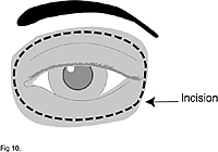
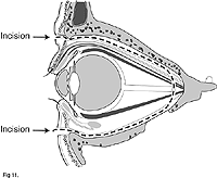
The cavity is then packed with antibiotic- and saline-soaked gauze and allowed to granulate over the next 8 to 12 weeks. During this period, the gauze should be removed, the cavity cleaned and repacked every 3 days. Alternatively, and for faster rehabilitation, a split skin graft can be used to line the cavity. This should be sutured to the surrounding skin and either meshed or punctured to allow drainage of blood and exudate. It is packed and changed regularly.
When healed, a prosthesis is fitted to an eyeglass frame or to titanium pegs inserted into the orbital rim. Alternatively, a black patch may be worn.28
Subtotal exenteration. When performing a subtotal exenteration, an incision is made around the palpable fissure, outside the lashes (Slide 12). The dissection plane is between the orbicularis muscle and orbital septum to the orbital rim (Slide 13). The periosteum is incised and the orbital contents removed as described above for the total exenteration procedure. A drain tube is placed in the orbit and the lids sutured together.
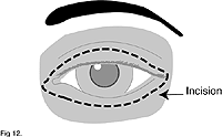
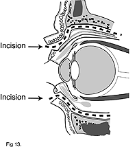
Complications
Complications of subtotal exenteration include the following:
- Sensation loss in the first division of the trigeminal nerve occurs due to removal of that nerve.
- A leak of cerebral spinal fluid may occur if there is damage to the roof of the orbit. This is life threatening and is treated initially with antibiotics. The leak is then repaired with temporalis muscle flap through the lateral orbital wall, or a mid forehead skin flap, onto the defect.
- Fistularization to a sinus may occur if there is damage to the medial wall or floor of the orbit. This may cause discharge into the socket or difficulty wearing the prosthesis. It may be repaired by rotation flap from adjacent orbital wall or with a temporalis or forehead flap.
- Skin graft necrosis is treated by debriding then regrafting or allowing granulation and epithelialization.
- After identification of the organism, infection is managed with frequent cleaning and dressing of the orbit and topical and systemic antibiotics.
- Cluster headaches on the side of the exenteration have been reported. These headaches are treated with oral analgesia.
- Tumor recurrence requires further excision with or without adjuvant chemotherapy or radiotherapy.
References
- Ashworth JL, et al. The hydroxyapatite orbital implant: A prospective study. Eye. 1996;10:29-37.
- Custer PL. Enucleation: Past, present, and future. Ophthal Plast Reconstr Surg. 2000;16:316-321.
- Jordan DR, et al. The bioceramic orbital implant: A new generation of porous implants. Ophthal Plast Reconstr Surg. 2000;16:347-355.
- Jordan DR, et al. The Molteno m-Sphere. Ophthal Plast Reconstr Surg. 2000;16:356-362.
- Jordan DR, et al. Brazilian hydroxyapatite implant. Ophthal Plast Reconstr Surg. 2000;16:363-369.
- Klapper SR, et al. Hydroxyapatite implant wrapping materials: Analysis of fibrovascular ingrowth in an animal model. Ophthal Plast Reconstr Surg. 2000;16:278-285.
- Kaltreider SA. The ideal ocular prosthesis. Analysis of prosthetic volume. Ophthal Plast Reconstr Surg. 2000;16:388-392.
- Custer PL, Trinkaus KM. Volumetric determination of enucleation implant size. Am J Ophthalmol. 1999;128:489-494.
- Older JJ. Enucleation. In: Levine MR, ed. Manual of Oculoplastic Surgery, 2nd edition. Boston, Mass: Butterworth-Heinemann; 1996:245-254.
- Custer PL, Reistad CE. Enucleation of blind, painful eyes. Ophthal Plast Reconstr Surg. 2000;16:326-329.
- Jordan DR, Klapper SR. A new titanium peg for hydroxyapatite orbital implants. Ophthal Plast Reconstr Surg. 2000;16:380-387.
- Woog JJ, Angrist RC, White WL, Dortzbach RK. Enucleation, evisceration and exenteration. In: Dortzbach RK, ed. Ophthalmic Plastic Surgery: Prevention and Management of Complications. New York: Raven Press Ltd; 1994:251-268.
- Downes R. Orbital implants: Food for thought. Eye. 1996;10:1-4.
- Oestreicher JH, Liu E, Berkowitz M. Complications of hydroxyapatite orbital implants. A review of 100 consecutive cases and a comparison of dexon mesh (polyglycolic acid) with scleral wrapping. Ophthalmology. 1997;104:324-329.
- Rubin PAD, Popham JK, Bilyk JR and Shore JW. Comparison of fibrovascular ingrowth into hydroxyapatite and porous polyethylene orbital implants. Ophthal Plast Reconstr Surg. 1994;10:96-103.
- Remulla HD, et al. Complications of porous spherical orbital implants. Ophthalmology. 1995;102:586-593.
- Levine MR, Fagien S. Enucleation and Evisceration. In: Stewart WB, ed. Surgery of the Eyelid, Orbit & Lacrimal System, Vol. 3. San Francisco, Calif: American Academy of Ophthalmology; 1995:84-111.
- Kim YD, et al. Management of exposed hydroxyapatite orbital implants. Ophthalmology. 1994;101:1709-1715.
- Jordan DR, et al. Complications associated with pegging hydroxyapatite orbital implants. Ophthalmology. 1999;106:505-512.
- Ghabrial R, et al. Assessment of the anophthalmic socket with dynamic cine-MRI. Orbit. 1997;16:207-216.
- Levine MR, Pou CR, Lash HL. Evisceration: Is sympathetic ophthalmia a concern in the new millennium? Ophthal Plast Reconstr Surg. 1999;15:4-8.
- Bellan L. Sympathetic ophthalmia: A case report and review of the need for prophylactic enucleation. Can J Ophthalmol. 1999;34:95-98.
- Shore JW. Evisceration. In: Levine MR, ed. Manual of Oculoplastic Surgery, 2nd edition. Boston, Mass: Butterworth-Heinemann; 1996:245-254.
- Long JA, Tann TM, Girkin CA. Evisceration: A new technique of trans-scleral placement. Ophthal Plast Reconstr Surg. 2000;16:322-325.
- Massry GG, Holds JB. Evisceration with scleral modification. Ophthalmic Plast Reconstruct Surg. 2001;17:42-47.
- Yang JG, et al. Hydroxyapatite implantation with scleral quadrisection after evisceration. Ophthalmic Surg Lasers. 1997;28:915-919.
- Slonim CB. Exenteration. In: Levine MR, ed. Manual of Oculoplastic Surgery, 2nd edition. Boston, Mass: Butterworth-Heinemann; 1996:245-254.
- McNab AA. Orbital exenteration. In: Manual of Orbital and Lacrimal Surgery, 2nd ed. Boston, Mass: Butterworth-Heinemann; 1998:49-57.