Introduction
Congenital ptosis, or dysmyogenic ptosis, is the most common ptosis seen in childhood. It is caused by primary dystrophy of the levator muscle. In congenital ptosis, Müller superior tarsal muscle is normal.1 A decrease in size and number of fibers results in an inability of the muscles to contract sufficiently to elevate the eyelid to its normal position in primary gaze. It also causes an inability to relax, resulting in lid lag on down gaze. Congenital ptosis is usually sporadic; however, occasionally, there is a positive family history. Congenital ptosis is 75% unilateral and is generally a pure ptosis. However, approximately 5% of patients may be associated with superior rectus weakness because the levator and superior rectus arise from the same mesodermal bud (Slide 1). Other conditions associated with congenital ptosis include Marcus Gunn synkinetic jaw winking ptosis and blepharophimosis syndrome. In patients with Marcus Gunn jaw winking ptosis, the involved eyelid retracts when the mouth opens and the mandible moves to the side opposite the ptotic lid from contraction of the external pterygoid muscle. Less commonly, the eyelid elevates following teeth clenching and use of the internal pterygoid muscle. The underlying cause is not well defined, but the levator may be partially innervated by the fifth cranial nerve. Blepharophimosis syndrome is a bilateral condition characterized by severe ptosis, epicanthus inverses, telecanthus, phimosis, and varying degrees of ectropion (Slide 2).2
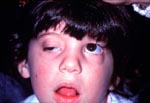
A strong family history is associated with congenital ptosis. In congenital ptosis, amblyopia is detected in approximately 20% of patients, usually secondary to convergent strabismus, high astigmatism, and anisometropia.3,4 Anderson and Baumgartner report rare instances of amblyopia due solely to the ptotic occlusion of the pupil.5
Clinical Features
Congenital ptosis may be unilateral or bilateral. If the ptosis is mild, the patient will present with ptosis alone without eyebrow and head posturing. Conversely, with severe ptosis and poor levator function, the brow or brows may be elevated with a chin up position since the patient may, in effect, look under the eyelids to see. With poor levator function, the eyelid crease is poorly formed and there is lid lag or down gaze indicative of a dystrophic muscle. The patient may have normal vision or amblyopia secondary to a convergent strabismus or anisometropia. Variability of ptosis by history or physical exam should alert the physician to the possibility of myasthenia gravis, which can occur in the younger age group.
Diagnostic Features
The amount of ptosis in the unilateral case is measured as the distance between the upper and lower eyelid margins with the brow held in a relaxed position. The difference represents the amount of ptosis. In bilateral ptosis, the amount of ptosis is determined by the marginal reflex distance (MRD), that is, the light reflects from the cornea to the upper lid. The normal MRD is +4 mm to +4.5 mm with the normal eyelid position being 2 mm to 1.5 mm from the superior limbus. The levator muscle function is the measurement of the upper lid excursion from down gaze to far up gaze with the eyebrow held in a fixed position to eliminate frontalis muscle function. The levator muscle function is approximately 15 mm. The height and contour of the upper eyelid creases are observed and measured to determine the point of action and the point of insertion of the levator aponeurosis. Marcus Gunn synkinetic jaw winking ptosis must be ruled out because the condition is treated differently from the standard congenital ptosis. This is based upon the amount of ptosis and the severity of the jaw winking component. In the blepharophimosis syndrome, levator function is generally poor and it is a bilateral condition. Myasthenia gravis is considered when levator function and the amount of ptosis are variable. Myasthenia gravis is a disorder of muscular weakness and fatigue caused by the presence of antibodies to the acetyl choline receptors of the muscle motor end plate. Acetyl choline receptor antibodies are detected in one third to one half of the patients with ocular myasthenia and 80% to 90% of the patients with generalized myasthenia.6 Although an edrophonium chloride (Tensilon) test is the first-line test to establish a diagnosis of myasthenia gravis, it is not always practical to perform the test on young children. There also may be difficulty in performing a single-fiber electromyelogram on children. Therefore, myasthenia gravis may be diagnosed based upon a detailed patient history, an up gaze fatigue test, an acetyl choline receptor antibody test, or an oral Mestinon (pyridostigmine, ICN Pharmaceuticals) or intramuscular Prostigmin (neostigmine, ICN Pharmaceuticals) trial and, most recently, an ice test.
Treatment
Traditionally, ptosis has been classified as mild (2 mm), moderate (3 mm), and severe (4 mm). Levator function may be classified as excellent (13 mm to 15 mm), very good (10 mm to 13 mm), good (8 mm to 10 mm), fair (5 mm to 7 mm), and poor (4 mm or less). The amount of ptosis and levator function determines the amount of aponeurotic and levator muscle resection with the levator function being the most important.
The earliest age to repair a congenital ptosis is 4 to 5 years old. At this age, the eyelid tissues are larger and the patients are cooperative to ensure accurate preoperative measurements. The only exception occurs when the pupillary axis is completely covered by the eyelid, which should be elevated on an urgent basis to prevent deprivational amblyopia. Patients are seen multiple times to obtain consistent measurements before surgery.
Congenital ptosis can be corrected by three operative procedures: levator resection by the skin; levator resection by the conjunctiva; and eyebrow suspension of the eyelids.7
Surgeons’ preferences are based on experience. It is important to choose a procedure where the anatomy is familiar and used often to achieve the desired consistent results. Levator function is a more important measurement than the amount of ptosis. Therefore, the best surgical approach in congenital ptosis is levator aponeurotic or muscle resection via the skin approach. If the levator function is 4 mm or less with severe ptosis, an autogenous frontalis sling is the appropriate procedure. The Table shows the amount of levator resection for a given amount of ptosis and levator function with emphasis on the levator function. The amount of resection of the levator aponeurosis or muscle is measured from the disinserted end proximal with the muscle on little to no stretch. Following are the steps of a levator resection:
(1)
Under general anesthesia, intubation and sterile skin preparation, the upper eyelid
crease is marked at the desired height so it is symmetrical with the opposite
upper eyelid crease.
(2)
Xylocaine 1% (lidocaine HCl, Astra) is injected subconjunctivally
and subcutaneously along the eyelid crease.
(3)
The skin is incised along the methylene blue marks with a #15 blade. A Stevens scissors
is used to button-hole the orbicularis muscle at the pretarsal border and
extend medial and laterally the length of the tarsal plate.
(4)
The pretarsal skin and orbicularis are dissected off the upper half of the tarsal
plate.
(5)
With a double-pronged skin hook pulling inferiorly on the skin-muscle flap, a
Stevens scissors is used to sharply dissect underneath preseptal skin and
orbicularis superiorly in a plane underneath the orbicularis muscle and
anterior to levator muscle.
(6)
With gentle pressure on the inferior lid, the orbital septum and preaponeurotic fat
are prolapsed forward.
(7)
The orbital septum is opened, some of the preaponeurotic fat is prolapsed, excised,
and cauterized.
(8)
A double battery hand-held cautery is used to dissect up levator aponeurosis
along with Müller muscle from conjunctiva as superior as necessary to
accomplish the predetermined levator/Müller muscle resection based upon the
Table (Slide 3). The medial and lateral horns of the levator are cut only as
necessary to allow resection of the amount of levator desired.8
(9) Three double-armed 6-0 silk sutures are placed through the anterior half of the tarsal plate, central nasal and temporal, 4 mm to 5 mm from the superior tarsal border and back through the levator/Müller muscle with the levator muscle being placed inferiorly with little stretch and tied over a 4-0 silk suture for release if necessary (Slide 4).9
(10) The amount of levator resection is measured from the inferior edge of levator aponeurosis, which is held in the forceps. The eyelid height and contour are evaluated, and adjustments are made.
(11) The three 6-0 silk sutures are tied down firmly.
(12) The excess levator and Müller muscle are excised. Two millimeters to 3 mm of skin are then excised superiorly to the incision before closure.
(13) Four 6-0 chromic cat gut sutures form the eyelid crease by suturing pretarsal orbicularis to levator aponeurosis. The skin is closed with running 6-0 fast absorbing sutures as well as some interrupted sutures.
(14) A 4-0 silk frost suture is placed in the lower eyelid margin and taped to the forehead for ocular protection for 24 hours and the eye is double patched. Postoperatively, the patch is removed the next day and the eye is lubricated with lubricating ointment for several days followed by artificial tears for approximately 1 week as needed (Slide 5 and Slide 6).
References
- Berke RN, Wadsworth JAC. Histology of levator muscle in congenital and acquired ptosis. Arch Ophthalmol. 1955;53:413-417.
- Mustarde JC. Congenital soft tissue deformities. In: Nesi FA, Lisman RD, Levine MR, eds. Smith’s Ophthalmic Plastic and Reconstructive Surgery. St. Louis, Mo: CV Mosby; 1998:977-999.
- Anderson RL, Baumgartner SA. Amblyopia in ptosis. Arch Ophthalmol. 1980;98:1068-1072.
- Merriam WW, Ellis FD, Helveston EM. Congenital blepharoptosis, anisometropia and amblyopia. Am J Ophthalmol. 1980:89:401-407. p>
- Anderson RL, Baumgartner SA. Strabismus in ptosis. Arch Ophthalmol. 1980;98(6):1062-1067.
- Savino PJ, Schanzer B, Moster ML. Ptosis in neurologic disease. In: Nesi FA, Lisman RD, Levine MR, eds. Smith’s Ophthalmic Plastic and Reconstructive Surgery. St. Louis, Mo: CV Mosby; 1998:345-354.
- Callahan MA. Congenital ptosis. In: Nesi FA, Lisman RD, Levine MR, eds. Smith’s Ophthalmic Plastic and Reconstructive Surgery. St. Louis, Mo: CV Mosby; 1998:355-378.
- Anderson RL. Age of aponeurotic awareness. Ophthal Plast Reconstr Surg. 1985;1:77-79.
- Beard C. Ptosis surgery: Past, present and future. Ophthal Plast Reconstr Surg. 1985;1:69-72.