Blepharoplasty
Introduction
The term blepharoplasty comes from the Greek words blepharon (eyelid) and plasso (surgical procedure
for repair of a defect or restoration of form or function). The conditions requiring blepharoplasty can most
easily be divided into the restoration of functional visual loss and cosmetic rejuvenation. Although visual
loss is the primary reason a patient will seek medical attention for blepharoplasty, this does not preclude
them from seeking an optimally aesthetic result.
Functional visual loss is most often attributed to the involutional changes in the skin and subcutaneous fascia, characterized by a redundancy in the skin of the eyelids and compromise of the levator aponeurosis. It is frequently associated with protrusion of orbital fat.1 These problems contribute to true blepharoptosis and lead to reduction of the superior visual field, as well as complaints of decreased vision at near from a diminished palpebral fissure on downgaze. Also, upper eyelid dermatochalasis is often associated with eyelash ptosis, brow ache, and complaints of a sleepy appearance and heaviness around the eyes.2 Involutional changes of the lower eyelid are most often associated with entropion and ectropion, and complaints of epiphora and conjunctival injection attributable to exposure keratopathy. As in the upper eyelids, age-related changes in the lower eyelids are often associated with orbital fat protrusion. This fatty appearance has been shown to occur as a true hernia, due to separation of the orbital septum from the levator aponeurosis in the upper eyelid and from the capsulopalpebral fascia in the lower lid.3
Preoperative Evaluation
A thorough medical evaluation should be performed before blepharoplasty. Specific attention should be drawn
to any medical problems that may pose a risk to the patient with use of general anesthesia. The patient must
stop taking anticoagulants before surgery. Nonsteroidal anti-inflammatory drugs and aspirin-containing products
traditionally are stopped 2 weeks before surgery, whereas warfarin sodium (Coumadin, DuPont) is metabolized
in 3 to 4 days. It is important to clear all medication changes with the prescribing doctor in the event that a
patient poses a high risk for a thromboembolic event. In these instances, special arrangements can be made
for intraoperative titration of heparin. The surgical team can also reduce the risk of abnormal bleeding in these
situations by using a CO2 laser for the incision and dissection, and by induction of hypotensive anesthesia.4
Patients must also be aware of vitamins, homeopathic remedies, and food items that may have anticoagulant effects.
Proper documentation of ocular function, deficits, and associated eye conditions should be made before blepharoplasty. This documentation includes guidelines that must be fulfilled for insurance reimbursement.
The preoperative ocular evaluation should include:
- Routine ophthalmic examination
- Assessment of dry eye by evaluation of tear film integrity, basal secretion rate, reflex tearing, and signs and symptoms of exposure keratopathy
- Visual field testing to document superior field deficits
- Evaluation of Bell phenomenon
- Preoperative photographs of both eyelids in the frontal and oblique positions, as well as in downgaze
- Measurements of palpebral fissure (PF) height centrally, and at the temporal and medial limbus in primary gaze, margin to increase distance (MCD); the distance from lid margin to lid crease in downgaze, margin to lid fold distance (MFD); the distance from lid margin to upper lid fold in primary gaze, margin reflex distance-1 (MRD1); the light reflex to upper lid margin in primary gaze with brow fixation, and PF in downgaze
- Lower eyelid snap test (pull lower eyelid down and quickly release) to assess laxity of the lower lid
- Documentation of lagophthalmos
- Measurement of scleral show, due to eyelid retraction
- Evaluation of fat pads, excess skin, and periorbital rhytids
- Evaluation of cheek bags
Surgical Technique
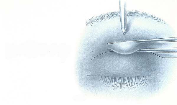
Before any sedation, a sterile surgical marking pen is used to delineate an incision in the eyelid crease. In
women, this delineation is placed approximately 11 mm centrally, as opposed to a lower 10-mm crease in
men. Excess skin is delineated using smooth forceps (Slide 1). Sedation can then be administered, and
local anesthetic (2% lidocaine with epinephrine, 1:100,000) is infiltrated diffusely in the upper eyelid. Then
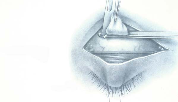
a traction suture is placed in the upper lid. The skin incision is performed with a #15 blade. While traction is
placed inferiorly on the upper lid, the orbicularis is tented up centrally and a Westcott scissors is used to
penetrate the suborbicularis plane (Slide 2). The skin muscle flap is excised with a Colorado needle
(Colorado Biomedical). The septum is released in a similar fashion, and the fat pads are removed using a
combination of a straight hemostat to grasp the fat, a #15 blade to excise it, and a Bovie cautery for
hemostasis (Slide 3). Before release of the fat stump, it is grasped with forceps to inspect for any residual
bleeding before allowing it to retract back into the orbit. Then a lid crease is formed with the use of a 5-0
Mersilene (polyester fiber) suture, which is tied from levator to orbicularis muscle. Then a 6-0 Vicryl
(polyglactin) suture is used to tie the skin to levator, and the wound is closed with a running 6-0 silk suture.
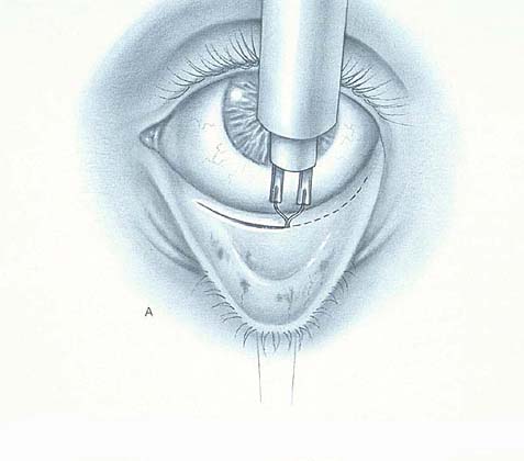
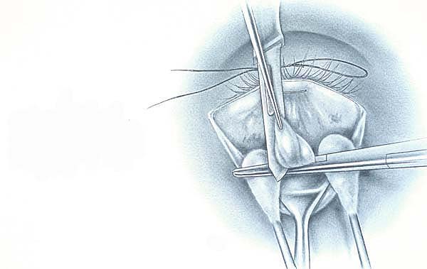
In the lower eyelid, the fat pads are injected with local anesthetic. A similar traction suture is placed, and the lid is everted over a Desmarres retractor. A Colorado needle is used to dissect through the conjunctiva and lower eyelid retractor (Slide 4). The lower lid retractors are then placed on traction suture superiorly, while blunt dissection is carried out to expose the fat pads (Slide 5). Special care is taken not to damage the inferior oblique muscle, which can be observed in a course between the medial and central fat pads. The fat is removed as was done in the upper eyelids. Finally, the conjunctiva can be closed with a 6-0 fast absorbable plain gut suture.
Postoperative Considerations
At the conclusion of surgery, topical ophthalmic antibiotic ointment is applied to the surgical wounds. Ice-cold
compresses are placed over the affected areas and visual assessments are made every 15 minutes until the
surgeon has cleared the patient for discharge. Although a relatively rare occurrence, blindness has been
reported after blepharoplasty, and central retinal artery occlusion from a retrobulbar hemorrhage has been
implicated as the cause.5 For this reason, any dressing that may conceal the signs and symptoms
of retrobulbar bleeding should not be used. Patients are instructed to intermittently continue applying cold
compresses and checking their vision for the first 24 hours.
Before surgery, a patient must be informed of the possible complications of surgery. Complications of blepharoplasty range form innocuous to severe.
Upper Eyelid Complications
- Blindness
- Lid asymmetry
- Ptosis
- Lagophthalmos and lid retraction
- Exposure keratopathy
- Diplopia
- Infection
- Canthal webbing
Lower Eyelid Complications
- Blindness
- Entropion
- Ectropion
- Diplopia
- Canalicular laceration
- Chemosis
Browplasty
Introduction
As the field of ophthalmic plastic surgery has evolved, surgeons have changed their focus. Traditional views
of the periorbital region as a separate entity have been supplanted in favor of a global approach toward facial
rejuvenation of an aesthetic unit. One such example is the relationship between the brow and upper eyelid.
As a result, oculoplastic surgeons have included brow lifting into their surgical armamentarium.
Eyebrow ptosis is an important problem in cosmetic oculoplastic surgery. Frequently, ptosis of the brow significantly contributes to a large upper eyelid fold that may be only partially caused by excessive upper eyelid skin.6 In an attempt to reduce excess upper eyelid skin, patients with brow ptosis often find it necessary to maintain their brows in a constantly elevated state through contraction of the frontalis muscle. Surgical fixation of the brow helps to alleviate this problem and avoids unhappy blepharoplasty patients who notice the same postoperative fullness in the upper eyelids after the brow returns to its ptotic state.
Preoperative Evaluation
A preoperative evaluation is necessary for determining the presence of brow ptosis and for selecting the
proper surgical procedure. It is essential to fixate the brow when evaluating the upper eyelid and brow. In
conjunction with the aforementioned preoperative evaluation for blepharoplasty, the following measurements
should be included in a brow workup:
- The distance between the first row of brow lashes and the upper eyelid in primary gaze (BUL)
- The distance from the inferior to the first row of brow lashes in primary gaze (ILB)
- Brow symmetry
- Forehead wrinkling
Anatomical Considerations
An intimate knowledge of the eyelid and facial anatomy is essential to avoid the dreaded complications of
sensory and motor loss. The supraorbital nerve provides sensation to the forehead and anterior scalp. Care
must be taken during any dissection around its eminence from the supraorbital notch and as this nerve courses
vertically on the frontalis muscle. Special consideration must also be given to avoid damage to the nerve in the
small subset of the population that possesses a separate foramen located centrally and approximately 1 cm
superior to the orbital rim. The temporal branch of the facial nerve provides motor innervation to the forehead.
It runs just superficial to the deep temporal fascia in the area of the lateral orbital rim. It is important for a
surgeon to maintain a deep plane on the temporal fascia to avoid injury to the nerve, which could produce a
permanent eyebrow paralysis.7
Choice of Procedure
The following procedures are used in browplasty:
- Internal brow lift: useful for mild brow ptosis confined to the central and temporal aspects of the brow8
- Corneal forehead lift: for total forehead and brow elevation
- Pretrichal forehead lift: accomplished by placing the coronal incision at the junction of the hairline and cephalic forehead9
- Midforehead lift: incisions are placed in prominent forehead creases
- Temporal lift: for isolated elevation of the lateral aspect of the brow
- Direct brow lift: excision of skin directly at the superior aspect of the brow
Endoscopy-Assisted Small-Incision Forehead and Brow Lift
The endoscopic approach achieves mild lifting of the forehead and brow while avoiding the morbidity associated
with large ear-to-ear incisions as well as the numbness, bruising, hair loss, and recuperation time often
associated with coronal lifting.7 This approach also provides options to men with male pattern baldness, in
whom incisions can be either placed in the hairline or hidden due to small vertical incisions. The primary
advantage of endoscopic lifting is that smaller vertical incisions are placed in the frontal plane.
Before endoscopic brow surgery, patients are told they can expect mild hair loss around the incision sites. Therefore, we instruct patients to apply topical minoxidil for 2 weeks before surgery.
Prior to surgical preparation, incisions should be delineated with a surgical marking pen. We then place two circumferential incisions on the temporal fossa, approximately 4 cm to 5 cm from the hairline along a plane that lines up the lateral canthus and the side of the nose. Vertical incisions are placed in line with the medial canthus and lateral canthus, approximately 1 cm in the hairline. Once the hair is thoroughly scrubbed and tied in a manner that will best avoid hair in the wounds, a frontal block is performed and local anesthetic is injected into the future planes of dissection. Although there are many variations, we prefer to use a mixture of 1L lactated Ringer's solution mixed with 1,500 units of hyaluronidase (Wydase, Wyeth-Ayerst), 1 mL of epinephrine (1:100,000), 50 mL of 2% lidocaine (Xylocaine, Astra), and 4 mL 0.5% bupivacaine hydrochloride (Marcaine, Sanofi Winthrop) with epinephrine. This mixture is administered with a hydrodissection pump.
The vertical incisions are placed through the periosteum, and the dissection plane is connected with the circumferential incisions, which are placed down to the shiny white, deep temporalis fascia (Slide 6). Direct visualization can be used to ensure that the conjoined tendon has been completely resected before the endoscope is introduced. Under endoscopic control, the dissections are carried about the supraorbital nerve. The periosteum is then released. Any attenuation of the corrugator, frontalis, supraciliary orbicularis, or procerus muscles can be performed at this time (Slide 7). Although variations exist, we prefer to dissect the periosteum at the supraorbital notch under direct visualization from an upper blepharoplasty incision. We can also dissect the corrugator with the same approach.
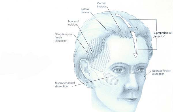
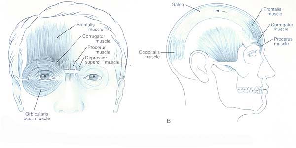
Once the periosteum has been adequately released, the forehead and brow can be lifted and tacked down using a combination of screws and staples for fixation and wound closure (Slide 8). A pressure bandage is placed over the wounds and the patient is taken to the recovery area.
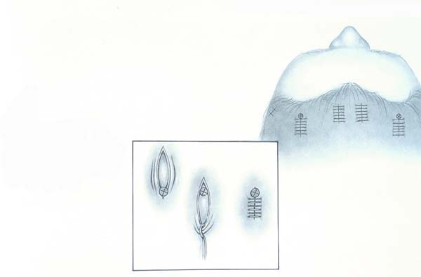
In the immediate postoperative period, the patient must be carefully observed for the presence of a hematoma, the most dreaded postoperative complication. The patients are also given a headband to aid in tissue adhesion once they have removed their bandage on the first postoperative day. Staples and screws are left in for 2 weeks, at which time they are removed in the surgeon's office.
References
- Putterman AM, McCord CD Jr. Cosmetic Blepharoplasty. In: Continuing Ophthalmic Video Education. San Francisco, CA: American Academy of Ophthalmology; 1985.
- Putterman A. Upper eyelid ptosis surgery improves the ability to read. Chicago Magazine. 1995:98:10-13.
- Putterman A, Urist M. Baggy eyelids-a true hernia. Ann Ophthalmol. 1973;5:1029-1032.
- Nesi FA, Waltz KL, Vega JF. Basic principles of ophthalmic plastic surgery. In: Nesi FA, Lisman RD, Levine MR, eds. Smith's Ophthalmic Plastic and Reconstructive Surgery, 2nd ed. St. Louis, MO: Mosby; 1998:81.
- Putterman A. Temporary blindness after cosmetic blepharoplasty. Am J Ophthalmol. 1975;80:1081-1083.
- Tardy ME, Alex J, Hendrick D. Rejuvenation of the aging brow and forehead. In: Putterman AM, ed. Cosmetic Oculoplastic Surgery, 3rd ed. Philadelphia, PA: WB Saunders; 1999:267.
- Goldberg RA. Browlift - an oculoplastic surgeon's perspective. In: Millman AL, Romo III T, ed. Aesthetic Facial Plastic Surgery: A Multidisciplinary Approach. New York: Thieme; 2000:213.
- Bowman CB, Tanenbaum M, McCord CD Jr. Internal brow lift: Browplasty and browpexy. In: Putterman AM, ed. Cosmetic Oculoplastic Surgery, 3rd ed. Philadelphia, PA: WB Saunders; 1999:113.
- Tardy ME, Alex J, Hendrick D. Rejuvenation of the aging brow and forehead. In: Putterman AM, ed. Cosmetic Oculoplastic Surgery, 3rd ed. Philadelphia, PA: WB Saunders; 1999:280.