Introduction
Currently, lasers are used in the periorbital region to treat a wide spectrum of disorders. The following discussion will focus on cutaneous skin resurfacing, the most common laser application.
Cutaneous laser resurfacing or laser skin resurfacing is a procedure in which a laser is used to modify the skin for the purpose of reducing the signs of aging and actinic damage. Usually, this treatment involves the removal of unwanted superficial lines, wrinkles, and pigmentary changes, which generally require stimulation of collagen remodeling and/or production at the level of the dermis. The goal of improving the skin’s surface was first realized with the carbon dioxide (CO2) laser; but subsequently has been accomplished with the Er:YAG laser, intense pulsed light devices (flashlamps producing light energy with a wide range of wavelengths), and a variety of other laser wavelengths. Until recently, skin resurfacing was accomplished by removing the epidermis. Ablative skin resurfacing creates a wound that takes 3 to 10 days to heal and is associated with a prolonged period of postoperative erythema. Newer techniques such as nonablative skin resurfacing produce a less dramatic degree of rejuvenation changes via an intact epidermis. Nonablative skin resurfacing produces erythema that lasts for only a few hours. Currently, there is a role for both ablative and nonablative resurfacing techniques.
The CO2 laser has been used to perform soft tissue surgery for almost 30 years. Its first application to the periorbital region in the late 1970s was for the removal of ocular adnexal tumors.1,2 In 1980, the CO2 laser was first used as an incisional device for aesthetic surgery by Baker, who used a continuous wave laser to perform upper blepharoplasty.3 Laser resurfacing was first performed by Remington in the mid to late 1980s using a defocused continuous wave laser to ablate the outer layers of a lip in a patient with actinic cheilitis, a premalignant condition. In 1993, a breakthrough in laser technology permitted CO2 lasers to be used with a greater degree of precision and safety, even allowing treatment of delicate areas such as the periorbital region. However, an understanding of the basic principles underlying laser skin resurfacing is required if the patient, the surgical team, and the surgeon are to be protected from injury.
Laser Physics
The CO2 laser has been in clinical use for more than 30 years. Detailed information about the theory behind its
development and application can be found elsewhere in literature.4,5 Lasers with wavelengths of 532 nm, 595 nm, 980 nm,
1064 nm, 1320 nm, and intense pulsed light devices have been used to successfully remodel the dermis through an intact epidermis.
The Er:YAG laser emits at a wavelength of 2940 nm. The absorption by water of Er:YAG energy exceeds that of CO2
energy by a factor of approximately 13.
Laser Tissue Interaction
When a laser beam interacts with tissue, the laser beam may be reflected, scattered, transmitted, or absorbed. The clinical effects
of any laser are realized only when laser energy is absorbed by a target tissue known as a chromophore.6 Water, the major
component of soft tissue, is the chromophore for both the CO2 and Er:YAG lasers (Slide 1). When laser energy of sufficient
concentration is applied to soft tissue, intracellular water is rapidly heated to its boiling point and converted to steam, effectively ablating the
surrounding tissue. Thus, the laser plume produced when CO2 or Er:YAG lasers interact with soft tissue contains vaporized
intracellular water and accompanying cellular debris. In addition to producing ablation, a portion of the laser energy will heat the surrounding
tissue, denaturing proteins and causing thermal damage. To achieve tissue ablation, a threshold energy density (fluence) must be delivered.
The threshold fluence for soft tissue ablation is approximately 4 J/cm2 to 5 J/cm2 for CO2
radiation and 2 J/cm2 to 3 J/cm2 with the Er:YAG laser. When considering nonablative resurfacing, the
energy required to induce dermal changes seems to be unique to each device. The mechanism by which dermal changes occur is not
well understood and may be related to generalized dermal heating, transmission of an unknown signal from epidermal keratinocytes to
the underlying dermis, or other factors.
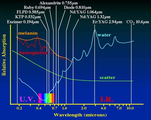
Due to its lower absorption coefficient, the CO2 laser produces less tissue ablation and a 5- to 10-fold greater amount of residual thermal effect in the remaining dermis than the Er:YAG laser. This thermal effect appears to be required to induce neocollagenesis, the development of new collagen. When used in purely ablative mode, the Er:YAG laser more effectively removes tissue but produces less dermal remodeling and, therefore, less smoothing of rhytids than the CO2 laser. In an effort to produce a more versatile device, two Er:YAG lasers are available that can produce the same tissue and clinical effects as a CO2 laser.
Laser Safety
The successful use of lasers requires close attention to laser safety requirements and guidelines. Failure to observe these rules
can endanger the patient, surgeon, operating room personnel, and other staff members. Lasers must be operated within office, hospital,
state, and federal guidelines. The American National Standards for the Safe Use of Lasers in Health Care Facilities (Z316.3) are safety
standards that have been established by the American National Standards Institute (ANSI), a nongovernmental agency that administers the
U.S. voluntary standardization and conformity assessment system.7 A copy of these guidelines should be present in every
facility where a medical laser is used.
Detailed information on laser safety is available elsewhere in the literature.8-10 Particular importance should be assigned to ocular protection when performing skin resurfacing. The cornea may be burned when performing laser skin resurfacing, especially when using the CO2 laser. The CO2 laser can have a serious thermal effect on the cornea. All personnel within the operating room must use appropriate eye protection. Surgical loupes utilize a double lens system that is protective of CO2 and Er:YAG radiation, but laser-safe side shields are also recommended.
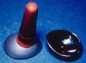
A laser-safe device must protect the globe of patients receiving periocular skin resurfacing procedures. The safest option is an internal metal shield that is placed behind the eyelid. Internal metal shields such as the Cox II and III shields (Oculo-Plastik, Montreal, Canada) are also effective when hair-removal lasers are used to treat trichiasis on the eyelids (Slide 2). Plastic cornea protectors can melt when exposed to energy produced by the CO2 and Er:YAG lasers.
Indications
Facial rhytids are the most common indication for laser skin resurfacing. The periorbital region, including the "crow’s feet" lateral to the lateral canthal angle and the glabella, are areas of concern to many patients. Ablative resurfacing techniques may also be used to treat a variety of cutaneous disorders characterized by pathologic changes found in the epidermis or superficial dermis. Examples of these conditions include benign dermal tumors including syringomas and xanthelasma,11 sebaceous hyperplasia, adenoma sebaceum,12 superficial basal cell carcinoma, actinic keratosis,13 seborrheic keratosis, and epidermal nevi.14 Skin resurfacing may also play a role in the management of actinic cheilitis and rhinophyma.15-20 Laser resurfacing is not advantageous for the treatment of vascular lesions such as hemangiomas or telangiectasia, or pigmented lesions such as lentigines, cafe-au-lait spots, ephelides, and some decorative tattoos. Although some of these conditions may be improved or eliminated with the CO2 and/or Er:YAG lasers, other more specific treatments are available for each.21 Ablative techniques are used when there is significant actinic injury (e.g., pigmentary changes, bronzing, or hyperkeratosis) to the epidermis as well as changes within the dermis. When epidermal and dermal changes are mild, nonablative resurfacing may be indicated. In these cases, epidermal modification with retinoids, superficial chemical peels, or microdermabrasion (crystal skin resurfacing) is often required to achieve the best results.
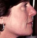
When selecting patients for skin resurfacing, patients with significant dyschromia due to excessive sun exposure should be identified and counseled before undergoing treatment of the entire face (Slide 3). Patients meeting these criteria always have severe photodamage on their neck and chest and even when "feathering" techniques are employed to treat these patients, it is difficult to avoid creating an easily apparent demarcation line. A series of preoperative superficial to medium depth chemical peels of the neck and chest may help soften the contrast between treated and untreated areas. While the neck and chest can be resurfaced, the thinner, adnexal-poor skin of these areas is prone to a greater risk of scarring and a prolonged recovery period.
Static vs. Dynamic Rhytids
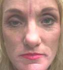
The glabella and periorbital regions often demonstrate a combination of static lines resulting from dermal changes and dynamic
rhytids produced by repetitive contraction of underlying facial musculature (Slide 4). The static component of these rhytids responds
best to skin resurfacing while the dynamic component requires inactivation of the musculature via botulinum toxin injection and/or surgery.
For optimal results, botulinum toxin injections should be administered 2 to 3 weeks before laser resurfacing, whereas surgical modification
of the muscles via endoscopic forehead and brow lifting may be performed simultaneously with resurfacing.
Contraindications
There are few absolute contraindications to laser skin resurfacing. Due to the risk of postoperative infection, resurfacing should not be performed in the presence of any active bacterial, fungal, or viral infections. Patients with pilosebaceous units that are impaired and unable to generate an epithelial response after resurfacing should not be treated. Examples of such patients include those who have taken isotretinoin within the past 2 years or those whose skin has been exposed to injurious doses of ionizing radiation (usually from an external beam source). Relative contraindications to skin resurfacing include a history of hypertrophic scarring or keloids, and hypersensitivity to sun exposure that may occur in patients with discoid lupus or polymorphous light eruption. Patients with Type IV to VI skin are at high risk for postinflammatory hyperpigmentation and should be counseled accordingly.22,23 Perhaps the most important contraindication to laser resurfacing is unrealistic expectations of the results. Patients who are undergoing resurfacing with the expectation of achieving a secondary gain such as improvement in their marriage, sex life, or status at work should be approached with extreme caution.
Treatment
Preoperative Regimen
There is no conclusive evidence to suggest that pretreatment of the skin is either necessary or beneficial. Patients who are at particular
risk for postinflammatory hyperpigmentation or those undergoing resurfacing for the treatment of a disorder of pigmentation such as melasma
should be treated preoperatively with a bleaching agent. Hydroquinone is the agent used most often to bleach the skin, although kojic acid in
an alphahydroxy acid base may be used as well. Hydroquinone prevents the conversion of tyrosine to L-dopa and, therefore, prevents the
synthesis of melanin by melanocytes. It does not remove or destroy melanin that has already been produced. Hydroquinone is tolerated best
when used in a cream base as the gel and solution forms tend to be more irritating to the majority of patients. The cream base hydroquinone
is available for use with or without sunblock. The ideal duration of therapy prior to resurfacing is not known, but a duration of at least 2 weeks
is recommended. If a patient reports significant irritation from the hydroquinone preparation, treatment should be discontinued or modified by
diluting the agent or mixing it with moisturizer or hydrocortisone cream prior to its application.24 Laser treatment of inflamed skin
is not recommended.
Anesthesia Requirements for Laser Resurfacing
Nonablative laser resurfacing is performed in an office setting without supplemental anesthesia. Ablative resurfacing is usually performed
in a procedure room or operating room setting. For ablative resurfacing, most surgeons prefer supplementing local anesthesia with intravenous
sedation. A small percentage of resurfacing procedures are performed under general anesthesia. Lidocaine 1% or 2% with epinephrine added
at a concentration of 1:100,000 or 1:200,000 is the most commonly used agent. Hyaluronidase may be added if desired. When treating the
periorbital area, local infiltration (as opposed to regional nerve blocks) is required. A small, 27-gauge or 30-gauge needle should be used to
minimize patient discomfort.
Ablative Laser Resurfacing
The skin does not require special preparation prior to laser skin resurfacing. Eye protection specific for the wavelength in question is
required for the patient, surgeon, and all personnel in the room. When performing full face resurfacing, the teeth should be protected with a wet
sponge or laser-safe mouth guard to prevent damage to the enamel. When using a focused delivery system, the handpiece must remain
equidistant from the skin throughout the treatment to produce a uniform result. In contrast, a collimated delivery system allows variability in
handpiece position of up to 18 cm without altering the laser’s tissue effects.
The skin must be dry when treatment is administered because moisture on the skin’s surface will absorb the incident laser energy and prevent it from producing the desired cutaneous effects. The laser spots should be placed in a confluent fashion so that the entire aesthetic unit receives uniform treatment. When using the CO2 laser, it is important to avoid treating the same area twice without first removing the desiccated skin created by the previous pass. Transfer of the laser energy to this carbonized, desiccated skin causes it to heat rapidly to a temperature of several hundred degrees Celsius. As cooling occurs, this heat is transferred to the surrounding tissue resulting in thermal damage that can impair wound healing and lead to the development of scarring. In contrast, ablation with the Er:YAG laser is a more explosive process, due to the greater coefficient of absorption by water. Less debris is left on the skin’s surface, therefore, some surgeons do not find it necessary to remove desiccated debris between passes with the Er:YAG laser.
The laser energy settings may require adjustment to reflect differences in skin thickness in various portions of the face. The eyelid skin is the thinnest in the body, and extreme care must be taken in treating this area. No more than two passes should be made with the CO2 laser on the eyelids. The pretarsal skin of the lower lids should be treated gently, especially in the medial portion of the lid as cicatricial ectropion may be produced with overzealous treatment of the eyelid skin. The skin of the upper lip is also relatively thin, although it is thicker than the skin of the eyelids. Therefore, the eyelids and upper lip are the areas most prone to the development of scarring following laser resurfacing.
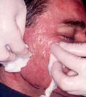
Once an entire area has been treated, the desiccated skin is debrided using saline soaked sterile gauze (Slide 5). Debridement should be gentle, not vigorous, to avoid the creation of mechanical effects in addition to the changes caused by the laser. Additional passes are made as required to achieve the desired clinical result or until a depth of treatment has been reached beyond which the surgeon feels the risk of complications such as delayed healing and scarring outweigh the benefits. Recent evidence suggests that after three passes have been completed with the CO2 laser, additional passes do not produce more tissue ablation. Generally, hemostasis is absolute during CO2 or long-pulsed Er:YAG skin resurfacing due to the associated thermal effect.
When the Er:YAG laser is used, treatment is carried to a depth necessary to achieve the clinical goals. If most of the pathology is in the epidermis and the dermis is healthy (Glogau I), 60 m m to 80 m m of ablation may be sufficient to achieve the desired result. In contrast, if the dermis must be treated more aggressively, a greater depth of ablation is required. The depth of ablation may be approximated by multiplying the fluence by 4 m m. For example, 25 J/cm2 fluence with the Er:YAG laser will ablate approximately 100 m m of tissue. In these cases, treatment is carried to the level of the papillary or superficial reticular dermis if bleeding does not limit the depth of treatment. If bleeding occurs, pressure is applied and attention is shifted to other areas until the bleeding stops. Application of cautery, silver nitrate, and other potentially injurious agents intended to secure hemostasis should be avoided.
If less than a full-face treatment has been performed, many surgeons elect to "feather" the edges of the treated area to help create a smooth transition to the untreated skin. Strategies for feathering include application of the laser in a tangential orientation, increasing the spacing between spots, or gradually decreasing the energy per pulse at the borders of the treated skin. Due to the frequent appearance of demarcation zones despite feathering, full-face resurfacing is recommended if more than one aesthetic unit is to be treated.
Nonablative Laser Resurfacing
Nonablative laser resurfacing is much simpler than ablative laser resurfacing from a procedural standpoint. Energy is applied in
a nonoverlapping fashion at an energy setting that produces mild erythema after several minutes. The energy settings will vary with the
device used. Approximately 4 to 6 treatments must be administered to accomplish the desired clinical effect, and these treatments are
usually performed over 3 to 6 weeks.
Postoperative Regimen
The goal of any postoperative treatment regimen is to accomplish reepithelialization of the wound while maintaining patient
comfort. Numerous wound care regimens have been used following laser resurfacing, each with a unique set of advantages and
disadvantages. However, there is no single regimen that should be recommended universally. The two most commonly used regimens
are occlusive dressings and an "open" technique consisting of only topical preparations.
Occlusive wound dressings are often used in the care of patients who have received burns of any nature. These dressings promote wound healing while maintaining patient comfort but are not popular with many patients. Occlusive dressings are usually opaque and should be changed regularly to permit wound assessment.
Topical preparations alone are the dressing of choice for many surgeons. A bland, preservative-free ointment that does not contain antibiotics is sufficient. The ointment is applied continuously until the wound is healed, usually 7 to 10 days after surgery. Some physicians also recommend the use of cold compresses made from a diluted vinegar solution to help retard bacterial and fungal wound infection.
A study by Dover and colleagues demonstrated an advantage to using occlusive dressings for the first 48 hours and then switching to an open technique. Once epithelialization is complete, patients may return to the use of commercial makeup or moisturizing products. All products must contain sunblock offering a sun protection factor (SPF) of least 15. Failure to keep the wounds moist and well lubricated can lead to adherent crusts, delayed wound healing, infection, and dermal scarring.25
There are no specific wound care requirements after nonablative skin resurfacing. The skin should remain moist and sunblock must be used regularly.
General Postoperative Instructions
Following ablative skin resurfacing, patients may resume low level activity immediately after treatment. Physical exertion is
discouraged postoperatively for approximately 2 weeks and sun exposure should be discouraged for at least 3 to 6 months. Usually,
postoperative discomfort is minimal to mild and is usually managed with acetaminophen. A minority of patients will request narcotics
for pain relief.
Results
Patient satisfaction following laser resurfacing is generally high. Clinical results will vary from patient to patient, depending on t
he degree of preoperative actinic damage, the depth of wrinkles present, and the amount of treatment given. Appropriate preoperative
evaluation and counseling is critical in patient satisfaction. This is especially true of nonablative resurfacing where the clinical results
tend to be more subtle.
Complications
Complications of laser resurfacing are uncommon when suggested guidelines for patient selection and treatment parameters
are followed. Complications that do occur are usually related to infection, abnormal pigmentation, or scarring. Complications following
nonablative resurfacing are less common than after an ablative procedure.
With appropriate antibiotic and antiviral prophylaxis, infections following laser resurfacing are most uncommon. Fungal infections have been reported and are usually caused by Candida species. As is true of bacterial and viral infections, Candidiasis usually presents during the initial recovery period prior to completion of epithelial healing. Patients may present with significantly increased erythema, crusting, and even bleeding in the treated area. The diagnosis may be confirmed by Gram stain, potassium hydroxide preparation, or culture of the exudate found on the skin. Candida infections may be treated with a combination of oral ketoconazole and topical soaks with diluted vinegar solution to create a local environment undesirable to fungus.
Bacterial infections are also most likely to occur prior to wound reepithelialization. Bacterial infections are most often caused by Staphylococcus or Streptococcus species and are characterized by increasing erythema and exudate. The diagnosis is made by culture and Gram stain of the involved area. Treatment with systemic antibiotics is required but topical antibiotic ointment may be used as well. Infection with more uncommon pathogens such as Pseudomonas and Enterobacter species may occur, underscoring the need for culture and Gram stain.
Viral infections after laser resurfacing are usually caused by herpes simplex. However, patients may not present with the vesicles typical of herpes infection due to the fact that the epidermis is missing. Patient may have erosions, erythema, and pain. Systemic antiviral therapy is indicated immediately to treat herpetic infection. If the herpetic infection is identified late or left untreated, significant dermal scarring will result.
Postinflammatory hyperpigmentation is a condition characterized by the development of increased pigment in response to cutaneous inflammation. The pigment may be located in the epidermis or dermis and generally appears several days to several months after treatment. It may be blotchy or uniform in appearance. The pigment generally fades with time and sun avoidance, but may take months to years to disappear completely. Treatment can be initiated to help decrease the amount of pigment produced and the length of time it remains. At the first sign of postinflammatory hyperpigmentation, usually 3 weeks after surgery, treatment should begin with the use of a bleaching agent such as hydroquinone, kojic acid, or azelaic acid. Once the skin is able to tolerate retinoic acid, Retin-A (tretinoin, Ortho Dermatological) or Renova (tretinoin emollient cream, Ortho Dermatological) is usually added. Alpha hydroxy acids may be used instead of retinoic acid. If the pigmentation fails to respond to topical therapy and is located in the epidermis as indicated by a prominent appearance under a Wood’s lamp, gentle laser therapy, or chemical peels may be useful. These treatments must be gentle so as not to create too much inflammation that can aggravate the problem. Understandably, some patients who have developed postinflammatory hyperpigmentation are reluctant to undergo any further treatments.
Hypopigmentation is a well-documented phenomenon following laser skin resurfacing. The incidence of this problem may be as high as 50% following CO2 resurfacing. The risk of hypopigmentation may increase with the depth of treatment and/or the amount of residual thermal injury left in the dermis. Hypopigmentation may represent either true pigment wipe-out or the return of skin to its natural color and appearance. When contrasted against adjacent damaged skin, the treated skin appears relatively hypopigmented. Both conditions may be remedied by treating the remainder of the face.
Erythema after skin resurfacing generally resolves in 3 to 16 weeks, depending on the type of laser used and the aggressiveness of treatment. Rarely, erythema may persist for many months. This is usually a sign of infection, scarring, hypersensitivity, or chemical irritation and the etiology must be carefully investigated.
Hypertrophic scarring usually occurs following overly aggressive treatment, inadequately treated infection, or inappropriate wound care. This problem is rare in the periorbital region, but overtreatment of the lower eyelids can result in a full thickness burn and cicatricial ectropion.26
Freshly resurfaced skin is extremely sensitive and, therefore, easily irritated. Antibiotic ointments and fragranced soap, makeup, or perfumes should be avoided. If a local allergic reaction develops, the responsible agent should be discontinued and appropriate therapy should be initiated.27
References
- Hornblass A, Herschorn BJ. Carbon dioxide laser surgery in hemophilia. Am J Ophthalmol. 1980;96(5):689-690.
- Beckman H, Fuller TA, Boyman R. Carbon dioxide laser surgery of the eye and adnexa. Ophthalmology. 1980;87:990-1000.
- Baker SS, Muenzler WS, Small RG, Leonard JE. Carbon dioxide laser blepharoplasty. Ophthalmology. 1984;91(3):238-244.
- Patel CKN. Continuous-wave laser action on vibrational-rotational transitions of carbon dioxide. Phys Rev. 1964;136:A1187-A1193.
- Trost D, Smith MFW, Zacherl A. Surgical lasers and their tissue interaction. In: Smith MFW, ed. Neurological Surgery of the Ear. St. Louis, Mo: Mosby Yearbook; 1992.
- Anderson RR. Laser Tissue Interactions. In: Goldman M. Cutaneous Laser Surgery: The Art and Science of Selective Photothermolysis. St. Louis, Mo: Mosby Yearbook; 1994.
- American National Standards Institute. American National Standards for the Safe Use of Lasers in Health Care Facilities. Standard Z136.3-1996. New York: ANSI Publications; 1996
- Gloster HM Jr, Roenigk RK. Risk of acquiring human papillomavirus from the plume produced by the carbon dioxide laser in the treatment of warts. J Am Acad Dermatol. 1995;32(3):436-441
- Bergbrant IM, Samuelsson L, Olofsson S, Jonassen F, Ricksten A. Polymerase chain reaction for monitoring human papillomavirus contamination of medical personnel during treatment of genital warts with CO2 laser and electrocoagulation. Acta Derm Venereol. 1994;74(5):393-395.
- Riley JR. Safety considerations in the use of the CO2 laser in facial skin resurfacing. Lasers Surg Med. 1997;(suppl):61.
- Wheeland RG, Bailin PL, Reynolds OD, Ratz JL. Carbon dioxide laser vaporization of multiple facial syringomas. J Dermatol Surg Oncol. 1986;12:225-228.
- Bellack GS, Shapshay SM. Management of facial angiofibromas in tuberous sclerosis: Use of the carbon dioxide laser. Otolaryngol Head Neck Surg. 1986;94:37-40.
- Wheeland RG, Bailin PL, Ratz JL, Roenigk RK. Carbon dioxide laser vaporization and curettage in the treatment of large or multiple superficial basal cell carcinomas. J Dermatol Surg Oncol. 1987;13:119-125.
- Ratz JL, Bailin PL, Lakeland RF. Carbon dioxide laser treatment of epidermal nevi. J Dermatol Surg Oncol. 1986;12:567-570.
- Defresne RG, Garrett AB, Bailin PL, Ratz JL. Carbon dioxide laser treatment of chronic actinic cheilitis. J Am Acad Dermatol. 1988;19:876-878.
- David LM. Laser vermilion ablation for actinic cheilitis. J Dermatol Surg Oncol. 1985;11:605-608.
- Zelvekson BD, Roenigk RK. Actinic cheilitis. Treatment with the carbon dioxide laser. Cancer. 1990;65:1307-1311.
- Ali KM, Callari RH, Mobley DL. Resection of rhinophyma with CO2 laser. Laryngoscope. 1989;99:453-455.
- Shapshay SM, Strong MS Anastasi GW, Vaughan CW. Removal of rhinophyma with the carbon dioxide laser. Arch Otolaryngol. 1980;106:257-259.
- Wheeland RG, Bailin PL, Ratz JL. Combined carbon dioxide laser excision and vaporization in the treatment of rhinophyma. J Dermatol Surg Oncol. 1987;13:172-177.
- Fitzpatrick RE, Goldman MP. Advances in carbon dioxide laser surgery. Clin Dermatol. 1995;13:35-47.
- Ho C, et al. Laser resurfacing in pigmented skin. Dermatol Surg. 1995;21:1035-1037.
- Lowe NJ, et al. Laser skin resurfacing: Pre- and posttreatment guidelines. Dermatol Surg. 1995;21:1017-1019.
- Rubin MG. Basic concepts in skin peeling. In: Manual of Chemical Peels: Superficial and Medium Depth. Philadelphia, Pa: JB Lippincott Co; 1995:44-50.
- Dover R, Otto WR, Nanchahal J, Riches DJ. Toxicity testing of wound dressings materials in vitro. Br J Plast Surg. 1995;48(4):230-235.
- Weinstein C. UltraPulse carbon dioxide laser removal of periocular wrinkles in association with laser blepharoplasty. J Clin Laser Med Surg. 1994;12:205-209.
- Waldorf HA, Kauvar ANB, Geronemus RG. Skin resurfacing of fine to deep rhytids using a char-free carbon dioxide laser in 47 patients. Dermatol Surg. 1995;21:940-946.