Introduction
Involutional entropion and ectropion are malpositions of the lower eyelid, commonly encountered in clinical practice. This tutorial will discuss the etiology, symptoms, and differential diagnosis of these disorders, as well as my management strategies that have evolved during 11 years of experience in subspecialty oculoplastic surgery.
Etiology
Involutional Entropion
Involutional entropion is caused by any combination of horizontal lower eyelid laxity,
attenuation, and dehiscence of the lower eyelid retractors, and the preseptal orbicularis
muscle’s overriding the pretarsal orbicularis muscle.1-3 Although in the past, involutional
atrophy of the orbital fat has been cited as a cause of involutional entropion, the role of
this factor has been recently questioned.4
Involutional Ectropion
Involutional ectropion is primarily caused by horizontal lower eyelid laxity. Attenuation
and/or dehiscence of the lower eyelid retractors is an additional cause in so-called tarsal
ectropion, a variant of involutional ectropion characterized by severe eversion of the lower
eyelid.5-7
Symptoms and Signs
Involutional Entropion
Symptoms of involutional entropion of the lower eyelid may include foreign body sensation, pain,
itching, tearing, and mucoid discharge. Signs may include constant or intermittent entropion,
superficial punctate keratopathy, and mucoid discharge. Tests such as “squeeze tests” and “supine
tests” may allow the physician to document cases of latent entropion that may not be obvious upon
initial examination. A squeeze test requires patients to forcibly close their lids multiple times.
In a supine test, the physician conducts an ocular examination while patients are in a supine
position.8 Performing these two tests together may reveal the subtlest cases of involutional
entropion.
Involutional Ectropion
Symptoms of involutional ectropion of the lower eyelid may include tearing, redness, foreign body
sensation, itching, pain, and mucoid discharge. Signs may include redness and hypertrophy of the
tarsal conjunctiva, eversion of the lower punctum, superficial punctate keratopathy, and mucoid
discharge.
Differential Diagnosis
Effective management of involutional entropion and ectropion is contingent upon accurate diagnosis, because different diagnostic entities are treated with different surgical procedures.
Involutional Entropion
Lower eyelid lashes and skin may come in contact with the cornea and conjunctiva in cases of
trichiasis, cicatricial entropion, and involutional entropion. Trichiasis is characterized by
misdirected lashes that emerge from the lid margin in an aberrant direction and point toward the
globe. Entropion is characterized by inward rotation of the lid margin. Trichiasis and entropion
may coexist. Cicatricial entropion is characterized by shortening of the posterior lamella of
the eyelid, that is, scarring of the tarsus and/or conjunctiva. Cicatricial entropion may occur
as a result of cicatricial pemphigoid, Stevens-Johnson syndrome, trachoma, trauma, or chemical
or thermal burns.
Involutional Ectropion
Eversion of the lower lid margin may be a result of cicatricial ectropion, tarsal ectropion,
or involutional ectropion. Cicatricial ectropion is characterized by shortening of the anterior
lamella of the eyelid, or a skin deficiency. Cicatricial ectropion may occur as a result of
facial skin surgery, trauma, chemical or thermal burns, chronic sun damage, chronic dermatitides,
or facial sagging. Tarsal ectropion is a variant of involutional ectropion characterized by severe
eversion of the lower eyelid.
The distinction among the three types of lower lid ectropion can be blurred in some patients. For example, scarring of the lower lid skin will often cause secondary dehiscence of the lateral and medial canthal tendons. Conversely, cases of predominantly involutional ectropion with severe lower lid laxity may be associated with mild degrees of downward skin traction in an elderly patient population prone to facial sagging and chronic sun damage. Ultimately, surgeons must decide which of the distinct procedures appropriate for cicatricial ectropion, tarsal ectropion, or involutional ectropion will best serve their patients.
Treatment
Involutional Ectropion
Myriad procedures for the treatment of involutional lower lid entropion have been described.8,9
Surgical procedures for involutional lower lid entropion generally address some or all of the
causative factors of the condition, that is, horizontal lower eyelid laxity, attenuation, and/or
dehiscence of the lower eyelid retractors, and orbicularis override. Correction of horizontal
lower eyelid laxity is a particularly important component of the repair, as it tends to prevent
both overcorrection and undercorrection.10-12 A surgeon’s choice of procedure should be based on
his or her training level, comfort level, and success rate.
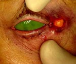
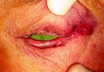
Transconjunctival Entropion Repair and Tarsal Strip
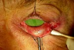
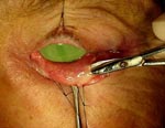
The following is a description of the procedure for involutional lower eyelid entropion that
I use in my practice. Similar to procedures described by Dresner and Karesh,13 and Migliori,14
my technique combines a tarsal strip procedure 15 and a transconjunctival approach to lower lid
retractor repair, partial excision of the preseptal orbicularis, and placement of rotational
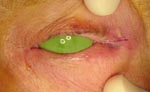
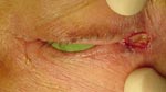
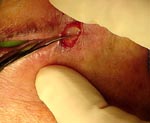
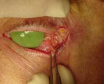
sutures (Slide 1A).
- Preoperatively, the surgeon administers topical proparacaine hydrochloride in both eyes and an anesthetist or anesthesiologist administers intravenous anesthetic. The choice of anesthetic agents varies according to the patient’s age, weight, and medical condition. Typical anesthetic consists of 0.5 mg to 1.0 mg of Versed (midazolam HCl, Roche), 0.5 cc to 1.0 cc of fentanyl citrate, and 15 mg to 30 mg of Brevital (methohexital sodium, Lilly). Subcutaneous 2% lidocaine with 1:100,000 epinephrine is injected into the subcutaneous tissues of the lower eyelid, lateral canthus, and central upper lid. The face is draped in the typical sterile manner and a protective contact lens is inserted.
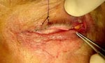
- A line is drawn, beginning at the lateral canthus and extending laterally
approximately 7 mm (Slide 1B).
- The skin is incised with a 15 Bard-Parker blade. The orbicularis is transected with a
thermal cautery (Slide 1C).
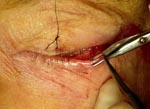
A lateral canthotomy is performed with Westcott scissors (Slide 1D).
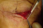
Inferior cantholysis is performed with a thermal cautery
(Slide 1E and Slide 1F).
- The upper lid is retracted superiorly with a 5-0 silk suture. Two 5-0 silk sutures are placed in the conjunctiva and tarsus just inferior to the lower margin in two places across the lid.
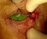
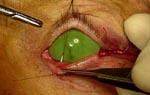
- The lower eyelid is everted over the Desmarres retractor (Slide 1G). Blunt dissection with
Westcott scissors is performed to separate the lower lid retractors from the orbicularis
(Slide 1H).
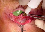
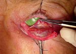
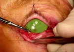
- Thermal cautery transects the conjunctiva and lower lid retractors at the level of the
inferior tarsal border. A plane is developed inferiorly below the lower lid retractors and
the orbicularis (Slide 1I and Slide 1J).
Thermal cautery is used to remove a strip of preseptal
orbicularis approximately 4 mm in vertical dimension across the entire lid. Care is taken
not to penetrate the skin during this step.
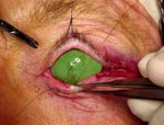
- The 5-0 silk sutures are removed from the lower lid. The lower eyelid is pulled laterally
until the appropriate tension is reached (Slide 1K). With the lid in this slightly tightened
configuration, the intersection of the lower lid with the cut edge of the upper lid at the
lateral canthus is marked with a marking pen. In this marked off area, an 11 blade dissects
between anterior and posterior lamellae (Slide 1L).
- The anterior lamella, including lashes and follicles, is removed. A cut is made below
the inferior tarsal border. The conjunctival epithelium of the posterior lamella of the
lid margin is removed with the Westcott scissors (Slide 1M). Conjunctival epithelium on
the posterior surface of the tarsal strip is scraped off with a 15 Bard Parker blade
(Slide 1N).
- A 5-0 vicryl suture is placed through the edge of the tarsal strip. The tarsal strip
is grasped with forceps, and a site for attachment in the lateral canthal area is selected.
This site of attachment may be the periosteum of the lateral rim, the superior crus of the
lateral canthal tendon, or other adjacent soft tissue. The vicryl suture is tied in a temporary
manner (Slide 1O).
- With the patient sitting in an upright position, the surgeon assesses the lower lid and lateral canthus. The lower lid attachment site to the lateral canthal area is adjusted until acceptable lateral canthal height and lower lid positions are achieved. The 5-0 vicryl suture is temporarily loosened.
- Next, 5-0 chromic sutures are used to attach the lower lid retractors to the inferior
tarsal border and through the lower lid skin to the anterior surface of the lid to serve as
rotational sutures. To accomplish this, each 5-0 chromic suture enters the eyelid skin
approximately 2 mm below the eyelashes and exits the posterior surface of the lid through
the wound. The needle is reversed and passed through the inferior tarsal border
(Slide 1P).
The needle then passes through the conjunctiva and lower lid retractors inferiorly
(Slide 1Q).
- Finally, the needle enters the wound in the back of the lid and exits the anterior surface
of the lid, 2 mm from the lashes and approximately 3 mm away from the initial entry site of the
chromic suture (Slide 1R).
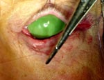
When these sutures are tied, the lower lid retractors are reattached
to the inferior tarsal border and rotational sutures are placed
(Slide 1S and Slide 1T). A combination
of the previous excision of the preseptal orbicularis and the placement of these rotational
sutures prevents orbicularis override. The 5-0 vicryl suture connecting the tarsal strip to the
lateral canthal tissues is then tied in a permanent manner. The lateral canthal angle is formed
with a 5-0 chromic suture with a buried knot. The wound is closed with subcutaneous 5-0 chromic
sutures and a cutaneous running 6-0 plain suture (Slide 1U). The contact lens is removed,
erythromycin topical ointment is applied, and a pressure patch is placed.
Involutional Ectropion
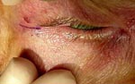
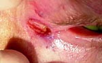
Numerous procedures for the treatment of involutional ectropion have been described.16 Surgical
procedures for involutional ectropion generally involve correction of lower lid laxity and
inward rotation of the punctum. Correction of lower lid laxity is usually performed at the
lateral canthus in the form of a tarsal strip procedure.15 Occasionally, lower lid laxity is
corrected by repair of the inferior crus of the medial canthal tendon 17 or by a wedge resection
of the lower lid. The medial repair has the obvious disadvantage of potential transection or
kinking of the inferior canaliculus, but is occasionally necessary in cases with massive medial
dehiscence. Medial repair is less problematic in cases with previous traumatic canalicular
obstruction and/or lacrimal insufficiency. Wedge resection is occasionally indicated in cases
with focal trichiasis or lid notching. Inward rotation of the punctum is accomplished by
resection of an ellipse of posterior lamella combined with rotational sutures.18,19 In cases of
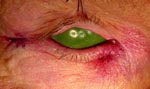
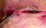
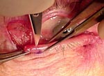
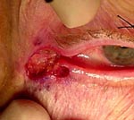
tarsal ectropion, the tarsal strip procedure is combined with a transconjunctival approach to
lower lid retractor repair and placement of rotational sutures.7 Some cases of apparent
involutional lower lid ectropion have downward skin traction secondary to facial sagging.
The surgeon must decide whether the degree of traction is sufficient to mandate placement of a
skin graft, that is, a cicatricial ectropion repair.
Omission of horizontal lower lid tightening is occasionally appropriate in patients with prominent globes, shallow orbits, and/or maxillary recession. In such cases, horizontal tightening will pull the lower lid under the globe with resultant lid retraction. In mild cases of medial ectropion with punctal eversion and unfavorable circumstances for lower lid tightening, inward rotation of the punctum alone may be sufficient to address patients’ complaints
Inward Rotation of the Punctum and Tarsal Strip Procedure
- Preoperatively, the surgeon administers topical proparacaine hydrochloride in both eyes and an anesthetist or anesthesiologist administers intravenous anesthetic. The choice of anesthetic agents varies according to the patient’s age, weight, and medical condition. Typical anesthetic consists of 0.5 mg to 1.0 mg of Versed (midazolam HCl, Roche), 0.5 cc to 1.0 cc of fentanyl citrate, and 15 mg to 30 mg of Brevital (methohexital sodium, Lilly). Subcutaneous 2% lidocaine with 1:100,000 epinephrine is injected into the subcutaneous tissues of the lower eyelid, lateral canthus, and central upper lid. The face is draped in the typical sterile manner and a protective contact lens is inserted.
- A line is drawn, beginning at the lateral canthus and extending laterally approximately 7
mm (Slide 2A).
The skin is incised with a 15 Bard-Parker blade. The orbicularis is transected
with a thermal cautery (Slide 2B).
A lateral canthotomy is performed with Westcott scissors (Slide 2C).
Inferior cantholysis is performed with a thermal cautery
(Slide 2D). The upper lid is retracted superiorly with a 5-0 silk suture.
- The lower punctum is dilated with a punctal dilator. A 00 Bowman probe is placed through the lower punctum and is passed through the lower canaliculus into the lacrimal sac until bone is felt. The Bowman probe is then used to elevate and evert the lower lid in the region of the punctum.
- Forceps grasp the conjunctiva, and a thermal cautery is used to penetrate conjunctival
and subconjunctival tissue (Slide 2E).
The superior incision of the ellipse is made approximately
3 mm below the punctum beginning approximately 2 mm medial to the punctum and ending 2 mm lateral
to the punctum. Dissection is then carried in the plane posterior to the subconjunctival tissue
for a short distance. Thermal cautery then makes the second incision of the ellipse 3 mm to 4 mm
below the superior incision (Slide 2F).
- Two 6-0 chromic sutures are then used to close the ellipse and also to serve as rotational
sutures. The sutures are placed as follows: Each 6-0 chromic suture enters the skin approximately
3 mm below the lid margin. It exits the posterior surface of the lid through the wound
(Slide 2G).
- The needle is reversed and passed through the superior edge of the wound
(Slide 2H).
The needle then passes through the inferior edge of the wound
(Slide 2I). Finally, the
needle enters the wound in the back of the lid and exits the anterior surface of the lid 3
mm to 4 mm from the lid margin and approximately 3 mm from the original entry point
(Slide 2J). When tied, these sutures close the ellipse and rotate the punctum inward
(Slides 2K and Slide 2L).
Two of these sutures are placed, one medial to the punctum and one
lateral to the punctum. Care must be taken when passing the medial suture to avoid penetration
of the canaliculus. The amount of rotation can be modulated by altering the entry site on the
skin surface. For example, if the punctum is not sufficiently inverted, the suture can be placed
more inferiorly on the skin surface. Conversely, if there is excessive rotation of the punctum,
then the sutures can be placed more superiorly.
- The lower eyelid is pulled laterally until the appropriate tension is reached. With the
lid in this slightly tightened configuration, the intersection of the lower lid with the cut
edge of the upper lid at the lateral canthus is marked with a marking pen
(Slide 2M). In this
marked area, an 11 blade dissects between anterior and posterior lamellae
(Slide 2N). The
anterior lamella, including lashes and follicles, is removed (Slide 2O).
- The epithelium of the posterior lamella of the lid margin is removed with the Westcott
scissors (Slide 2P).
A cut is made below the inferior tarsal border (Slide 2Q). Conjunctival
epithelium on the posterior surface of the tarsal strip is scraped off with a 15 Bard Parker
blade (Slides 2R and Slide 2S).
A 5-0 vicryl suture is placed through the edge of the tarsal strip.
The tarsal strip is grasped with forceps, and a site for attachment in the lateral canthal area
is selected. This site of attachment may be the periosteum of the lateral rim, the superior crus
of the lateral canthal tendon, or other adjacent soft tissue. The vicryl is tied in a temporary
manner (Slides 2T and Slide 2U).
- With the patient sitting upright, the position of the lower lid and lateral canthus can
be assessed. The lower lid attachment site to the lateral canthal area is adjusted until
adequate lateral canthal height and lower lid position are achieved. The 5-0 vicryl suture
connecting the tarsal strip to the lateral canthal tissues is then tied in a permanent manner.
The lateral canthal angle is formed with a 5-0 chromic suture with a buried knot
(Slide 2V, Slide 2W, and Slide 2X).
The wound is closed with subcutaneous 5-0 chromic sutures and a
cutaneous running 6-0 plain suture (Slide 2Y).
The contact lens is removed, erythromycin eye-ointment is applied, and a pressure patch is placed.
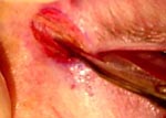
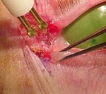
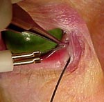
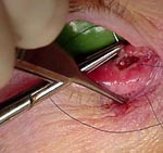
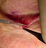
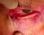
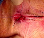
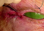
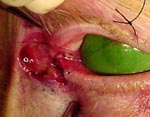
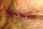
Transconjunctival Ectropion Repair and Tarsal Strip for Tarsal Ectropion
- Preoperatively, the surgeon administers topical proparacaine hydrochloride in both eyes and an anesthetist or anesthesiologist administers intravenous anesthetic. The choice of anesthetic agents varies according to the patient’s age, weight, and medical condition. Typical anesthetic consists of 0.5 mg to 1.0 mg of Versed (midazolam HCl, Roche), 0.5 cc to 1.0 cc of fentanyl citrate, and 15 mg to 30 mg of Brevital (methohexital sodium, Lilly). Subcutaneous 2% lidocaine with 1:100,000 epinephrine is injected into the subcutaneous tissues of the lower eyelid, lateral canthus, and central upper lid. The face is draped in the typical sterile manner and a protective contact lens is inserted.
- A line is drawn, beginning at the lateral canthus and extending laterally approximately 7 mm. The skin is incised with a 15 Bard-Parker blade. The orbicularis is transected with a thermal cautery. A lateral canthotomy is performed with Westcott scissors. Inferior cantholysis is performed with a thermal cautery.
- The upper lid is retracted superiorly with a 5-0 silk suture. Two 5-0 silk sutures are placed in the conjunctiva and tarsus just inferior to the lower margin in two places across the lid. The lower eyelid is everted over the Desmarres retractor. Thermal cautery transects the conjunctiva and lower lid retractors at the level of the inferior tarsal border. A plane is developed inferiorly below the lower lid retractors and the orbicularis.
- The 5-0 silk sutures are removed from the lower lid. The lower eyelid is pulled laterally until the appropriate tension is reached. With the lid in this slightly tightened configuration, the intersection of the lower lid with the cut edge of the upper lid at the lateral canthus is marked with a marking pen. In this marked area, an 11 blade dissects between anterior and posterior lamellae. The anterior lamella, including lashes and follicles, is removed. A cut is made below the inferior tarsal border. The conjunctival epithelium of the posterior lamella of the lid margin is removed with the Westcott scissors. Conjunctival epithelium on the posterior surface of the tarsal strip is scraped off with a 15 Bard Parker blade.
- A 5-0 vicryl suture is placed through the edge of the tarsal strip. The tarsal strip is grasped with forceps and a site for attachment in the lateral canthal area is selected. This site of attachment may be the periosteum of the lateral rim, the superior crus of the lateral canthal tendon, or other adjacent soft tissue. The vicryl suture is tied temporarily.
- With the patient sitting in an upright position, the lower lid and lateral canthus are assessed. The lower lid attachment site to the lateral canthal area is adjusted until good lateral canthal height and lower lid position are achieved. The 5-0 vicryl is temporarily loosened. 5-0 chromic sutures are used to attach the lower lid retractors to the inferior tarsal border and to come through the lower lid skin to the anterior surface of the lid to serve as rotational sutures. To accomplish this, each 5-0 chromic enters the eyelid skin approximately five millimeters below the eyelashes. It exits the posterior surface of the lid through the wound. The needle is reversed and passed through the inferior tarsal border. The needle then passes through the conjunctiva and lower lid retractors inferiorly.
- Finally, the needle enters the wound in the back of the lid and exits the anterior surface of the lid five millimeters from the lashes approximately three millimeters away from the initial entry site of the chromic. When these sutures are tied, the lower lid retractors have been reattached to the inferior tarsal border, and rotational sutures have been placed. The 5-0 vicryl connecting the tarsal strip to the lateral canthal tissues is then tied in a permanent manner. The lateral canthal angle is formed with a 5-0 chromic suture with a buried knot. The wound is closed with subcutaneous 5-0 chromic sutures and a cutaneous running 6-0 plain suture. The contact lens is removed, erythromycin eye ointment is applied, and a pressure patch is placed.
References
- Dalgleish R, Smith J. Mechanics and histology of senile entropion. Br J Ophthalmol. 1966;50:79-91.
- Schaefer A. Variation in the pathophysiology of involutional entropion and its treatment. Ophthalmic Surg. 1983;14:653-655.
- Benger R, Musch D. A comparative study of eyelid parameters in involutional entropion. Ophthalmic Plast Reconstr Surg. 1989;5:281-287.
- Kersten R, Hammer B, Kulwin D. The role of enophthalmos in involutional entropion. Ophthalmic Plast Reconstr Surg. 1997;13:195-198.
- Putterman A. Ectropion of the lower eyelid secondary to Müller’s muscle-capsulopalpebral fascia detachment. Am J Ophthalmol. 1978;85:814-817.
- Wesley R. Tarsal ectropion from detachment of the lower eyelid retractors. Am J Ophthalmol. 1982;93:491-495.
- Tse D, Kronish J, Buus D. Surgical correction of lower eyelid tarsal ectropion by reinsertion of the retractors. Arch Ophthalmol. 1991;109:427-431.
- Glatt H. Follow-up methods and the apparent success of entropion surgery. Ophthalmic Plast Reconstr Surg. 1999;15:396-400.
- Benger R, Frueh B. Involutional entropion: a review of the management. Ophthalmic Surg. 1987;18:140-142.
- Lance S, Wilkins R. Involutional entropion: a retrospective analysis of the Wies procedure alone or combined with a horizontal shortening procedure. Ophthalmic Plast Reconstr Surg. 1991;7:273-277.
- Van den Bosch W, Rosman M, Stijnen T. Involutional lower eyelid entropion: results of a combined approach. Ophthalmic Surg Lasers. 1998;29:581-586.
- Danks J, Rose G. Involutional lower lid entropion: to shorten or not to shorten? Ophthalmology. 1998;105:2065-2067.
- Dresner S, Karesh J. Transconjunctival entropion repair. Arch Ophthalmol. 1993;111:1144-1148.
- Migliori M. Internal repair of entropion and tarsal ectropion. Presented at the annual meeting of the American Society of Ophthalmic Plastic and Reconstructive Surgery. Atlanta, Ga; 1995.
- Anderson R, Gordy D. The tarsal strip procedure. Arch Ophthalmol. 1979;97:2192-2196.
- Benger R, Frueh B. Involutional ectropion: A review of the management. Ophthalmic Surg. 1987;18:136-139.
- Jordan D, Anderson R, Thiese S. The medial tarsal strip. Arch Ophthalmol. 1990;108:120-124.
- Tse D. Surgical correction of punctal malposition. Am J Ophthalmol. 1985;100:339-341.
- Lowry J. How to treat punctal eversion. Review of Ophthalmology. 1998(Feb);100-101.