Introduction
Superior limbic keratoconjunctivitis (SLK) is a disease characterized by inflammation of the upper palpebral and superior bulbar conjunctiva.1 Secondary epidermalization of the superior limbal conjunctiva with keratin formation may follow, often accompanied by a superior filamentary keratitis (one-third of cases).2 This idiopathic inflammation of the upper tarsus, superior limbus and corneal epithelium is associated with a fine, papillary upper tarsal conjunctival reaction in all cases (Slide 1). The superior bulbar conjunctiva characteristically shows rose bengal staining (Slide 2).
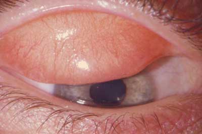
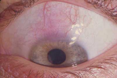
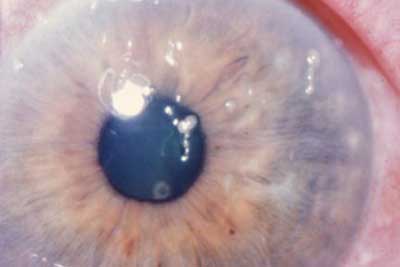
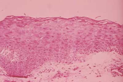
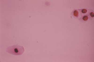
The corneal alterations are secondary to irritation by the superior tarsal papillary reaction, which results in the disturbance of the superior bulbar conjunctiva, leading to the development of tenacious mucus, which in turn leads to filamentary keratitis (Slide 3). If not treated or in severe cases, the superior bulbar conjunctiva may undergo epidermalization (transformation of mucous membrane to a tissue with the characteristics of skin, primarily keratinization) (Slide 4, Slide 5). Histologic changes of the conjunctiva consist of hyperplasia of the squamous layer and induction of a granular layer that indicates keratin production. The presence of keratinized epithelium either by superficial scraping or full-thickness biopsy supports the clinical diagnosis but is not specific.3 Diseases commonly associated with SLK include keratoconjunctivitis sicca (KCS), hyperthyroidism and hyperparathyroidism.1-3 These associations suggest an autoimmune etiology for SLK.
Treatment
Several currently used treatment modalities for SLK such as topical corticosteroids, cromolyn sodium and lodoxamide tromethamine have modulating effects on the local immune environment. Others have proposed a mechanical hypothesis.1-3 Numerous treatment modalities, such as 0.5% silver nitrate application, topical vitamin A, punctal occlusion, pressure patching, bandage contact lens, conjunctival cauterization, and conjunctival resection that change the mechanical surface of the conjunctiva have been reported as being successful.1-10 This implies a multifactorial causation and points to the need to be aware of several treatment options for long-term control.
Cyclosporine
Due to its effectiveness as an immunomodulator and lack of chronic ocular sequelae such as cataracts and glaucoma, topical cyclosporine A (tCSA) has been used in place of steroids in the treatment of graft rejection, dry eye syndrome and vernal conjunctivitis.11-14 In dry eye disease, tCSA has been shown to decrease T cell infiltration of the lacrimal gland.15-17 In the postoperative management of penetrating keratoplasty, tCSA has been shown to be effective in avoiding corticosteroid-induced ocular hypertension and in preventing and treating graft rejections, which is a T cell mediated mechanism.18-19 The conjunctival epithelium in vernal keratoconjunctivitis has been shown to possess a unique profile of T cell lymphocytes.13 Recently, conjunctival specimens from patients with SLK have also been shown to have an alteration in T cell functioning.20 Given the success of tCSA in several ocular pathologies with a common etiology of T cell dysfunction, it follows that tCSA may have utility in the treatment of SLK. The purpose of this tutorial is to review SLK and alert our colleagues to the potential benefit of tCSA 0.5% in the treatment of SLK.24
TCSA is beneficial to patients with SLK for many reasons. First, tCSA has recently been shown to be of value in treating patients with KCS.15-17 KCS occurs in high frequency in patients with SLK.1,3,5 The benefit of treatment of the KCS component of SLK may be more than increasing basal tear secretion and improving dry eye symptoms. Since the mode of action of tCSA in treating patients with KCS is mainly through T cell modulation, this action may not only help decrease lacrimal gland tissue infiltration, but also may positively affect the other signs and symptoms of SLK. This would be especially likely if, in some cases, SLK represents a localized form of myxedema which is in part a T cell mediated disease.18-20,22-24 Conjunctival specimens from patients with SLK have also been shown to have an upregulation of transforming growth factor beta-2 (TGF b -2).20 T cell functioning and proliferation have been shown to be intricately related to TGF b -2 secretion. In our series of patients, the response to topical CSA was not rapid but occurred over 4 to 6 weeks.24 This most likely reflects the time necessary to dissipate the circulating lymphocyte pool.16-20
Second, both cromolyn and lodoxamide tromethamine are mast cell stabilizers and have been shown to be effective in treating SLK.5-6 The effectiveness of tCSA in the treatment of experimental allergic conjunctivitis caused by mast cell–mediated hypersensitivity reaction to immunoglobulin E-bound antigens has been reported.11 The success reported in this experimental study, coupled with previous reports of success with tCSA in recalcitrant cases of vernal catarrh and our experience in treating corneal grafts, suggests that this agent may be beneficial in treating patients with SLK.12-14
References
- Theodore FH. Further observations on superior limbic keratoconjunctivitis. Trans Am Acad Ophthalmol Otolaryngol. 1967;71:341-351.
- Theodore FH, Ferry AP. Superior limbic keratoconjunctivitis. Clinical and pathological correlations. Arch Ophthalmol. 1970;84:481-484.
- Udell IJ, Kenyon KR, Sawa M, Dohlman CH. Treatment of superior limbic keratoconjunctivitis by thermocauterization of the superior bulbar conjunctiva. Ophthalmology. 1986;93:162-166.
- Yang HY, Fujishima H, Toda I, et al. Lacrimal punctal occlusion for the treatment of superior limbic keratoconjunctivitis. Am J Ophthalmol. 1997;124:80-87.
- Confino J, Brown SI. Treatment of superior limbic keratoconjunctivitis with topical Cromolyn sodium. Ann Ophthalmol. 1987;19:129-131.
- Grutzmacher RD, Foster RS, Feiler LS. Lodoxamide tromethamine treatment for superior limbic keratoconjunctivitis. Am J Ophthalmol. 1995;120:400-402.
- Mondino BJ, Zaidman GW, Salamon SW. Use of pressure patching and soft contact lenses in superior limbic keratoconjunctivitis. Arch Ophthalmol. 1982;100:1932-1934.
- Passons GA, Wood TO. Conjunctival resection for superior limbic keratoconjunctivitis. Ophthalmology. 1984;91:966-968.
- Bleik JH, Tabbara KF. Topical cyclosporine in vernal keratoconjunctivitis. Ophthalmology. 1991;98:1679-1684.
- Ohashi Y, Watanabe H, Kinoshita S, et al. Vitamin A eyedrops for superior limbic keratoconjunctivitis. Am J Ophthalmol. 1988;15:105:523-527.
- Donshik PC, Collin HB, Foster CS, et al. Conjunctival resection treatment and ultrastructural histopathology of superior limbic keratoconjunctivitis. Am J Ophthalmol. 1978;85:101-110.
- Whitcup SM, Chan CC, Luyo DA, et al. Topical cyclosporine inhibits mast cell-mediated conjunctivitis. Invest Ophthalmol Vis Sci. 1996;37:2686-2693.
- Mendicute J, Aranzasti C, Eder F, et al. Topical cyclosporin A 2% in the treatment of vernal keratoconjunctivitis. Eye (England). 1997;11(pt 1):75-78.
- Secchi AG, Tognon MS, Leonardi A. Topical use of cyclosporine in the treatment of vernal keratoconjunctivitis. Am J Ophthalmol. 1990;10:641-645.
- Stern ME, Beuerman R, Fox RL, Gao J, et al. The ocular surface in dry eye: A therapeutic target. Societas Ophthalmologica Europaea. 1997;11:911-919.
- Sall K, Stevenson OD, Mundorf TK, Reis BL. Two multicenter, randomized studies of the efficacy and safety of cyclosporine ophthalmic emulsion in moderate to severe dry eye disease. CsA Phase 3 Study Group. Ophthalmology. 2000;107:631-639.
- Stevenson D, Tauber J, Reis BL. Efficacy and safety of cyclosporine A ophthalmic emulsion in the treatment of moderate-to-severe dry eye disease: A dose-ranging, randomized trial. The Cyclosporine A Phase 2 Study Group. Ophthalmology. 2000;107:967-974.
- Perry HD, Donnenfeld ED, Kanellopoulos AJ, Grossman GA. Topical cyclosporine A in the management of postkeratoplasty glaucoma. Cornea. 1997;16:284-288.
- Perry HD, Donnenfeld ED, Acheampong A, et al. Topical cyclosporine A in the management of postkeratoplasty glaucoma and corticosteroid-induced ocular hypertension (CIOH) and the penetration of topical 0.5% cyclosporine A into the cornea and anterior chamber. CLAO. 1998;24:159-165.
- Matsuda A, Tagawa Y, Matsuda H. TGF-beta2, tenascin, and integrin beta1 expression in superior limbic keratoconjunctivitis. Jpn J Ophthalmol. 1999;43:251-256.
- George MA, Abelson MB, Schaefer K, Mooshian M, Weintraub D. A precise method of using rose Bengal in the evaluation of dry eye and the detection of changes in its severity. Adv Exp Med Biol. 1994;350:549-552.
- Darrell RW. Superior limbic keratoconjunctivitis in identical twins. Cornea. 1992;11:262-263.
- Cher I. Clinical features of superior limbic keratoconjunctivitis in Australia. A probable association with thyrotoxicosis. Arch Ophthalmol. 1969;82:580-586.
- Perry HD, Doshi-Carnevale SJ, Donnenfeld ED, Kornstein HS. Topical Cyclosporine A 0.5% as a possible new treatment for Superior Limbic Keratoconjunctivitis. Ophthalmology. (in press).