Introduction
Meibomian gland dysfunction, which is a condition associated with obstruction and inflammation of the meibomian glands, is a widespread and chronic problem that is seen frequently in general ophthalmology and cornea/external disease practices (Slide 1). It is probably the most common cause of posterior blepharitis and may also be the most common cause of dry eye.1-6
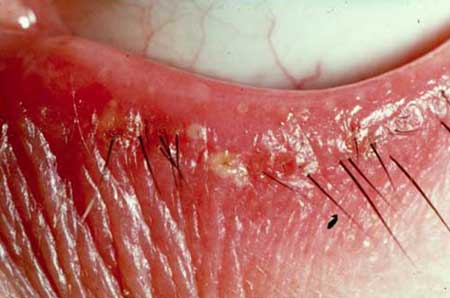
Chronic blepharitis (inflammation of the eyelid) can be classified into anterior and posterior forms.3,7 Anterior blepharitis is associated with staphylococcal infection and seborrheic dermatitis. Posterior blepharitis is associated with various disorders of the meibomian glands, which are known collectively as meibomian gland dysfunction.4-6 Meibomian gland dysfunction can disrupt the tear film in several ways (discussed below). This tear film disturbance can cause or exacerbate many of the symptoms associated with meibomian gland dysfunction (e.g., burning, irritation, itching, sudden sharp pains, foreign body sensation, and filmy vision).
The lipid layer of the tear film serves several functions. It creates a smooth optical surface, retards tear evaporation, creates a barrier to contamination by skin surface lipids, prevents tear overflow, creates a water-tight seal on closure of lids, prevents maceration of the lid margin by tears, and decreases the surface tension of the tear film, thereby increasing its stability.3,8-12 The meibomian glands that produce the lipid layer are located between the tarsal plate and the palpebral conjunctiva and their openings are at the posterior margin of the lids. The palpebral portion of the orbicularis "milks" the glands on contraction during the blinking process.13,14
Meibomian gland dysfunction has been associated with acne rosacea, seborrheic dermatitis, and aging.4,7,11,15,16 Acne rosacea is a dermal dystrophy of unknown etiology associated with intermittent flushing, abnormalities of vascular dilation and telangiectasia, and often papules and pustules. Rosacea is associated with hypertrophy and plugging of the sebaceous glands. Borrie found that nearly 100% of rosacea patients had posterior blepharitis, and ocular disease preceded cutaneous manifestation in 20%.11 Seborrheic dermatitis is characterized by hyper-keratinization of the skin in areas rich in sebaceous glands. It has been reported to be present in 36% of patients with meibomian keratoconjunctivitis.4,7,16 In this disorder, keratin deposits and desquamated epithelial cells obstruct the meibomian ducts.
There is an increased prevalence of meibomian gland dysfunction in the older population.5,15 With increasing age, some of the oils produced by the meibomian glands are discharged into the depth of the aqueous tear layer as a result of retroplacement of the meibomian orifices. Other meibomian orifice changes predisposing to glandular dysfunction, such as obliteration, pouting and narrowing, are also associated with increased age (Slide 2). Although our study population (average age 57) was not of advanced age, they were not, on average, young patients.
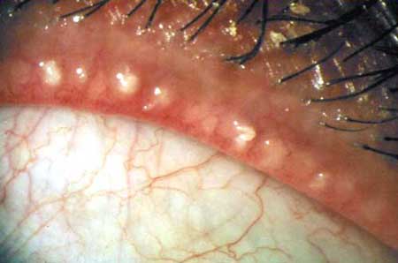
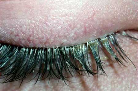
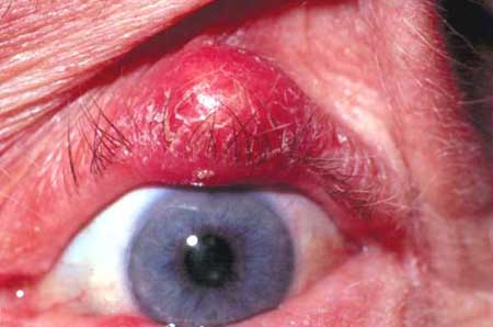
Obstruction of the meibomian ducts by any associated factors causes accumulation of meibomian gland secretions (meibum) (Slide 3). Meibum is mainly made of non-polar wax and esterol esters. Meibum is different than sebum (which is produced in the skin) because it has a lower concentration of polar triglycerides and free fatty acids. Normal meibum has a melting point equal to or lower than the ocular surface temperature.10,17 Accumulation of meibum within the meibomian gland can lead to inflammation of the gland and bacterial colonization.3,4,16,17 The colonizing bacteria have lipases that break the non-polar wax and sterol esters into triglycerides and free fatty acids (polar lipids), thus altering the normal composition of the meibum.18 The polar lipids diffuse more easily through the aqueous layer and contaminate the mucin layer, making it hydrophobic.9 This causes the tear film to become unstable, and the surface of the eye becomes non-wettable. Clinically, tear film instability may present as a decrease in the tear break-up time. In moderate to severe cases of meibomian gland dysfunction, tear film instability leads to a recalcitrant and irritating superficial punctate keratopathy.12 Also, the abnormal meibum has a melting point above the ocular surface temperature, which causes it to solidify and obstruct the ducts, which further perpetuates the problem. Chalazia can occur when pressure breaks the walls of the glands and liberates glandular contents within the lid, causing a noninfectious inflammatory reaction (Slide 4).14
Management
Although lid hygiene is currently the mainstay of treatment for meibomian gland dysfunction,2,3,4 other proven therapies for meibomian gland dysfunction include oral tetracycline, doxycycline or minocycline, topical erythromycin or bacitracin, and topical steroids.3,4,13,19 For refractory cases, topical cyclosporine can be used.20,21 However, the cost and side effects associated with these medications make them a poor choice for long-term management in patients with mild to moderate cases of meibomian gland dysfunction.
Lid hygiene is a conservative treatment often used to treat meibomian gland dysfunction. Lid hygiene has been useful in the treatment of associated eyelid disorders, such as chalazion, in which conservative management is 83% effective.13 However, to our knowledge, no study to evaluate the clinical effectiveness of conservative management for meibomian gland dysfunction has been formally attempted. To address this issue, we performed a case-cohort prospective study on the effect of lid hygiene and preservative-free artificial tears on meibomian gland dysfunction in a large inner city general ophthalmology clinic. The results of the study showed that lid hygiene and preservative-free artificial tears are 88% effective in reducing the symptoms of meibomian gland dysfunction and statistically improve the tear break-up time.22
The importance of lid hygiene has been appreciated in the management of meibomian dysfunction and blepharitis. Some techniques of lid hygiene emphasize the use of baby shampoo as essential.23,24 This at first glance appears to make sense. However, in meibomian gland dysfunction, the combination of free fatty acids and inflammation leads to saponification. Saponification creates the detergent action of the abnormal tear film. Adding baby shampoo to a disease complex characterized by too much detergent is not logical. In addition, the added step of mixing and applying baby shampoo in the correct formulation may contribute to poor patient compliance.23 We choose not to include the use of any soaps as part of the recommended treatment in our patients.
We consider the Physician-Patient Educational Conference to be an integral part of patient care (See Conference Instructions). The idea of conservative therapy is not new, but the concept of an educational discussion with the patient has often been neglected in the ophthalmic literature and most likely in clinical practice, as well.13 We believed that educated patients would be more compliant with the recommended regimen, because they would have a clearer sense of purpose in performing lid hygiene (rather than just performing it "because the doctor said so") (See Patient Instructions). We are also careful to suggest to patients that they could readily achieve positive outcomes in their disease and its symptoms. In our experience, the severity of the clinical appearance of meibomian gland dysfunction may be related to the severity of symptoms as expressed by patients. This makes meibomian gland dysfunction a good setting for taking advantage of any placebo effect that might be achieved through positive suggestions and conservative self-administered treatment.25
Our conservative treatment regimen includes warm soaks, lid cleansing, and artificial tears. The warm soaks are designed to increase the ambient temperature of the eyelids, raising the temperature of the meibomian glands above the melting point of the abnormal meibum. This may allow the meibum to be released from the ducts and may end the cycle of duct obstruction that exacerbates the condition. The use of an isotonic saline solution both prevents the excessive drying of the skin which might occur with frequent application of unsalinated wet soaks and introduces a mild, nonspecific inhibitory effect on bacterial growth.26 Lid cleansing is designed to free meibomian gland obstruction directly at the mouths of the ducts, allowing freer egress of the meibomian secretions. Unblocked meibomian glands will be less likely to sustain the environment of increased bacterial colonization and abnormal lipid formation that has been associated meibomian gland dysfunction.27 However, because the initial liberation of abnormal meibum can destabilize the tear film and because neither warm compresses nor lid cleansing will cure posterior blepharitis, preservative-free artificial tears are recommended to improve the quality of the tear film and enhance ocular surface wetting.
References
- Shimazaki J, Sakata M, Tsubota K. Ocular surface changes and discomfort in patients with meibomian gland dysfunction. Arch Ophthalmol. 1995;113:1266-1270.
- Gilbard JP. Dry eye, blepharitis and chronic eye irritation: Divide and conquer. J Ophthalmic Nurs Technol. 1999;18:109-115.
- Smith RE, Flowers CW. Chronic blepharitis: A review. CLAO J. 1995;21:200-207.
- Driver PJ, Lemp MA. Meibomian Gland Dysfunction. Surv Ophthalmol. 1996;40:343-367.
- Bron AJ, Benjamin L, Snibson GR. Meibomian gland disease. Classification and grading of lid changes. Eye. 1991;5:395-411.
- Mathers WD, Shields WJ, Sachdev MS, et al. Meibomian gland dysfunction in chronic blepharitis. Cornea. 1991;10:277-285.
- McCulley JP, Dougherty JM, Deneau DG. Classification of chronic blepharitis. Ophthalmology. 1982;89:1173-1180.
- Shimazaki J, Goto E, Ono M, et al. Meibomian gland dysfunction in patients with Sjogren syndrome. Ophthalmology 1998;105:1485-1488.
- Shine WE, McCulley JP. Keratoconjunctivitis sicca associated with meibomian secretion polar lipid abnormality. Arch Ophthalmol. 1998;116:849-852.
- Shine WE, McCulley JP. Meibomian gland triglyceride fatty acid differences in chronic blepharitis patients. Cornea. 1996;15:340-346.
- Borrie P. Rosacea with special reference to its ocular manifestations and dry eyes. Br J Dermatol. 1963;65:458-463.
- Mathers WD. Ocular evaporation in meibomian gland dysfunction and dry eye. Ophthalmology. 1993;100:347-351.
- Perry HD, Serniuk RA. Conservative treatment of chalazia. Ophthalmology. 1980;87:218-221.
- Norn MS. Meibomian orifices and Marx's line studied by triple vital staining. Acta Ophthalmol. 1985;63:698-700.
- Hykin PG, Bron AJ. Age related morphological changes in lid margin and meibomian gland anatomy. Cornea. 1992;11:334-342.
- Zengin N, et al. Meibomian gland dysfunction and tear film abnormalities in rosacea. Cornea. 1995;14:144-145.
- McCulley JP, Shine WE. Meibomian secretions in chronic blepharitis. Adv Exp Med Biol. 1998;438:319-328.
- Shine WE, Silvany R, McCulley JP. Relation of cholesterol-stimulated Staphylococcus aureus growth to chronic blepharitis. Invest Ophthalmol Vis Sci. 1993;34:2291-2296.
- Dougherty JM, McCulley JP, Silvany RE, et al. The role of tetracycline in chronic blepharitis. Inhibition of lipase production in staphylococci. Invest Ophthalmol Vis Sci. 1991;32:2970-2975.
- Stevenson OD, Tauber J, Reis BL. Efficacy and safety of cyclosporine A ophthalmic emulsion in the treatment of moderate-to-severe dry eye disease: a dose ranging, randomized trial. The cyclosporin A Phase 2 Study Group. Ophthalmology. 2000;107:967-974.
- Sall K, Stevenson OD, Mundorf TK, et al. Two multicenter, randomized studies of the efficacy and safety of cyclosporin A ophthalmic emulsion in moderate to severe dry eye disease. CsA Phase 3 Study Group. Ophthalmology. 2000;107:631-639.
- Perry HD, Romero J, Biser SA, et al. Conservative therapy for posterior blepharitis (submitted to Cornea 2002).
- Key, JE. A Comparative study of eyelid cleaning regimens in chronic blepharitis. CLAO J. 1996; 22:209-212.
- Wittpenn JR. EyeScrub: Simplifying the management of blepharitis. J Ophthalmic Nurs Technol. 1995;14:25-28.
- Staats P, Hekmat H, Staats A. Suggestion/placebo effects on pain: negative as well as positive. J Pain Symptom Manage. 1998;15(4):235-243.
- Jones EM, Bowker KE, Cooke R, et al. Salt tolerance of EMRSA-16 and its effect on the sensitivity of screening cultures. J Hosp Infect. 1997;35(1):59-62.
- Groden LR, Murphy B, Rodnite J, et al. Lid flora in blepharitis. Cornea. 1991;10(1):50-53.